
Giardia lamblia: Life Cycle, Pathogenesis, Treatment, and Laboratory Diagnosis
How does a non-invasive parasite cause fat malabsorption and greasy diarrhea without ever entering the bloodstream? Complete Giardia lamblia life cycle, the brush-border mechanism, treatment, and lab diagnosis.

On this page
A group of hikers returns from a two-week backcountry trip during which they drank untreated water directly from mountain streams, assuming it was clean because it looked clear and came from a fast-moving source. Ten days later, several develop foul-smelling, greasy diarrhea that floats in the toilet bowl, along with bloating and cramping but notably, no blood or fever. One of them has known IgA deficiency and becomes considerably more unwell than the others.
This is the classic presentation of giardiasis, popularly known as "beaver fever" for its long association with contaminated wilderness water sources. What makes Giardia lamblia pedagogically interesting is that it causes all this gastrointestinal disruption; malabsorption, weight loss, greasy stools — without ever invading tissue or entering the bloodstream. It is a purely luminal infection, and yet it is one of the most common causes of waterborne diarrheal disease worldwide. Understanding how a non-invasive organism produces such significant malabsorption is the key conceptual question this article answers.
Giardia lamblia also known as Giardia intestinalis, or Giardia duodenalisis a protozoan flagellate. It is transmitted by the fecal-oral route and causes the diarrheal illness called giardiasis (popularly known as beaver fever).
General Characteristics
- Geographical distribution: worldwide; found in the soil, water, or surfaces contaminated with feces of infected human/animal.
- Habitat: Duodenum and upper part of jejunum of humans.
Morphology: Exists in two form
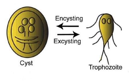 Figure: Cyst and Trophozoite of Giardia lamblia showing Encystation-excystation cycle
- Cyst: Oval cyst is thick-walled with four nuclei and several internal fibers. Each cyst gives rise to two trophozoites during excystation in the intestinal tract.
- Trophozoite: Pear-shaped with two nuclei, four pairs of flagella, and a suction disk are present.
Giardia is protected by an outer shell that allows it to survive outside the body for long periods of time and makes it tolerant of chlorine disinfection.
Life Cycle of Giardia lamblia
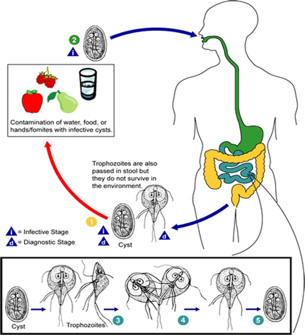 Figure: Life Cycle of Giardia lamblia (source: CDC)
- Infection occurs by the ingestion of cysts in contaminated water, food, or by the fecal-oral route (hands or fomites)
- In the small intestine, excystation releases trophozoites (each cyst produces two trophozoites).
- Trophozoites multiply by longitudinal binary fission remaining in the lumen of the proximal small bowel where they can be free or attached to the mucosa by a ventral sucking disk.
- Encystation occurs as the parasites transit toward the colon.
- Both cysts and trophozoites can be found in the feces
The cyst is the stage found most commonly in non-diarrheal feces (The cysts are hardy and can survive several months in cold water.) Trophozoites are found in diarrheal stools.
Because the cysts are infectious when passed in the stool or shortly afterward, person-to-person transmission is possible.
Pathogenesis
- Many species of mammals, as well as humans, act as reservoirs.
- Risk groups: Hikers who drink untreated stream water Male homosexuals Children in daycare centers Patients in mental hospitals IgA deficiency greatly predisposes to symptomatic infection
- The trophozoite causes inflammation of the duodenal mucosa, leading to the malabsorption of protein and fat. Mild steatorrhea (passage of yellowish greasy stools in which there is an excess of fat)
- Approximately half of those infected are asymptomatic carriers who continue to excrete the cyst for years.
Why Giardia Causes Malabsorption: The Mechanism
This is the detail most resources state without explaining, and it is worth understanding properly because it distinguishes Giardia from invasive pathogens like Entamoeba histolytica.
Giardia lamblia is a non-invasive organism, trophozoites attach to the small intestinal mucosa via their ventral sucking disk but do not penetrate the epithelium or enter tissue. Despite this, infection produces genuine malabsorption through several non-invasive mechanisms:
- Mechanical and physical disruption of the brush border, even without trophozoites entering cells, the sheer density of organisms attaching to and covering the mucosal surface (an actual "carpet" of trophozoites in heavy infections) physically interferes with normal absorption
- Shortening and disruption of intestinal microvilli, reducing the absorptive surface area available
- Disaccharidase enzyme deficiency, particularly lactase deficiency, this is why transient lactose intolerance is a recognized feature of giardiasis and may persist for some time after the infection itself has cleared
- Variable villous changes, ranging from no visible abnormality on light microscopy in many cases to partial or, in severe cases, subtotal villous atrophy
An important and somewhat counterintuitive point: the severity of diarrhea and malabsorption does not correlate reliably with the degree of visible histological damage. Some patients with significant symptoms show normal-appearing intestinal architecture under the microscope, while some asymptomatic carriers show partial villous atrophy. This tells us that much of the functional disruption happens at a level not fully visible on routine histology, likely involving direct biochemical effects of Giardia-secreted proteinases on the microvillus membrane, in addition to the mechanical effects above.
Why this matters clinically: Because the damage is functional rather than structural/invasive, treatment that clears the organism typically allows the gut to recover fully, unlike invasive parasitic diseases that can leave structural damage (such as the flask-shaped ulcers and scarring seen with Entamoeba histolytica). This is also why giardiasis, despite causing significant symptoms, is not associated with the bloody stool or systemic invasive complications (such as liver abscess) seen with truly invasive intestinal protozoa.
Clinical Findings
Giardia infection can cause a variety of intestinal symptoms, which include;
- Anorexia
- Flatulence (gas)
- Upset stomach or nausea/vomiting
- Stomach or abdominal cramps
- Watery (non-bloody), foul-smelling diarrhea
- Greasy stools that tend to float
- Dehydration (loss of fluids)
Giardiasis can cause weight loss and failure to absorb fat, lactose, vitamin A, and vitamin B12. In children, severe giardiasis might delay physical and mental growth, slow development, and cause malnutrition.
Laboratory Diagnosis
Fecal specimens containing Giardia lamblia may have an offensive odor and are pale-colored, fatty, and float in water.
- Ova and parasite (O+P) examination: Giardia cysts can be excreted intermittently, so many cases (>50%) of giardiasis will be missed with a single O+P examination, resulting in underdiagnosis. Multiple stool collections (i.e., three stool specimens collected on separate days) increase test sensitivity. Use of concentration methods increases sensitivity. Microscopical examination of freshly passed stools is used to demonstrate Giardia trophozoite and cysts.
Fresh diarrheic specimen: Try to find Giardia lamblia trophozoites. Generally difficult to detect as they attach themselves to the intestine wall. A Giemsa or Field’s stained fecal smear should be examined if giardiasis is suspected but no trophozoites are detected in a wet mount of the feces. Formed fecal specimen: Look for the Giardia lamblia cyst.
- Fecal immunoassays that are more sensitive and specific can be used: An ELISA test that detects a Giardia cyst wall antigen in the stool can be used.
- String test (Entero-Test): Swallowing a weighted piece of string until it reaches a duodenum. Trophozoites adhere to the string and can be visualized after the withdrawal of the string.
- Polymerase Chain Reaction (PCR) can be used to identify the subtypes of Giardia lamblia.
Treatment
Giardiasis is treated with antiparasitic medication, most commonly:
- Metronidazole: the most widely used first-line agent in most countries, typically given as a 5–7 day course
- Tinidazole: often preferred where available, as it can be given as a single dose with comparable or better efficacy
- Nitazoxanide: an alternative agent, useful particularly where nitroimidazole resistance or intolerance is a concern
- Albendazole: a benzimidazole alternative, sometimes used particularly in mixed helminth/protozoal infection settings
An increasingly important clinical point: treatment failure is a growing problem. Metronidazole, despite being first-line therapy, fails to cure 15–70% of cases in some reported series with a standard course, and resistance has become a documented and growing concern in recent years. Giardia metabolizes metronidazole into its active toxic form using specific parasite enzymes (nitroreductases and pyruvate-ferredoxin oxidoreductase); mutations affecting these enzymes are linked to reduced drug susceptibility. In practice, this means a patient who does not respond to a standard course should not simply receive a repeated identical course — switching drug class (e.g., to tinidazole or nitazoxanide) or extending/adjusting the regimen is a reasonable next step, and persistent treatment failure should also prompt consideration of reinfection from an ongoing environmental source rather than assuming resistance alone.
No prophylactic drug or vaccine exists for Giardia, prevention relies entirely on safe water practices (boiling, filtration, or chemical treatment of water from untreated sources, particularly relevant for the hikers/campers risk group already noted in this article) and hand hygiene, since the organism is notably tolerant of standard chlorine disinfection.
Where Students Actually Get Confused
1. "A four-nucleated cyst in stool is always Entamoeba histolytica." Both Giardia lamblia and Entamoeba histolytica cysts contain four nuclei, which is a frequently tested point of confusion. The distinguishing feature is shape: Giardia cysts are oval, while E. histolytica cysts are spherical. Nuclei count alone cannot differentiate them, shape must be assessed together with nuclei count.
2. "Giardia is an invasive parasite like Entamoeba histolytica." It is not. Giardia trophozoites attach to the intestinal mucosa via a ventral sucking disk but do not invade tissue or enter the bloodstream. This is precisely why giardiasis does not cause the bloody dysentery, flask-shaped ulcers, or extraintestinal complications (such as liver abscess) seen with E. histolytica — despite both organisms causing significant gastrointestinal symptoms.
3. "All infected individuals are symptomatic." As the article already states, approximately half of those infected are asymptomatic carriers who continue to excrete cysts for years. This has direct public health relevance: asymptomatic carriers remain infectious and contribute to ongoing transmission, particularly relevant in the daycare and institutional settings already listed among the risk groups.
4. "Treatment failure with metronidazole always means the parasite is drug-resistant." Not necessarily. While documented resistance exists and is a growing concern, treatment failure can also result from reinfection from an ongoing contaminated water or environmental source, inadequate dosing or adherence, or in rare cases, immune deficiency (such as the IgA deficiency already noted as a risk factor in this article) predisposing to more difficult-to-clear infection. A failed course should prompt evaluation of all these possibilities, not an assumption of resistance alone.
5. "A single negative stool exam excludes giardiasis." As the article already states, Giardia cysts are shed intermittently, and more than half of cases are missed with a single ova and parasite examination. Multiple specimens (three, collected on separate days) or stool antigen testing (ELISA) are both more reliable than a single microscopic examination, and the string test (Entero-Test) remains a useful option specifically when trophozoites are suspected but not found on standard stool examination.
Key Exam Facts in One Table
| Fact | Detail | Memory hook |
|---|---|---|
| Common name | "Beaver fever" | Associated with contaminated wilderness water |
| Habitat | Duodenum and upper jejunum | NOT the colon |
| Cyst | Oval, thick-walled, 4 nuclei; produces 2 trophozoites on excystation | Oval shape distinguishes from E. histolytica's spherical cyst |
| Trophozoite | Pear-shaped, 2 nuclei, 4 flagella pairs, ventral sucking disk | Sucking disk = attachment mechanism |
| Invasiveness | NON-invasive, attaches but does not penetrate tissue | No bloody stool, no liver abscess (unlike E. histolytica) |
| Key malabsorption mechanism | Brush border/microvilli disruption + disaccharidase (lactase) deficiency | Functional, not always structural, damage |
| Histology correlation | Symptom severity does NOT reliably correlate with villous damage seen | Functional disruption beyond visible histology |
| Classic stool description | Pale, fatty, foul-smelling, floats in water (steatorrhea) | Fat malabsorption signature |
| % asymptomatic carriers | ~50% | Continue excreting cysts for years |
| Risk groups | Hikers (untreated water), daycare children, institutionalized patients, IgA deficiency | IgA deficiency = predisposes to symptomatic disease |
| Person-to-person transmission | Possible, cysts infectious when/shortly after passed | Unlike Naegleria, this DOES spread person-to-person |
| Single O+P exam sensitivity | Misses >50% of cases | Use 3 specimens on separate days |
| Most sensitive stool test | ELISA antigen detection | More sensitive/specific than O+P alone |
| String test (Entero-Test) | Swallowed weighted string retrieves duodenal trophozoites | Used when trophozoites suspected but not found on stool exam |
| First-line treatment | Metronidazole (5–7 days) or tinidazole (single dose) | Tinidazole often preferred for convenience |
| Treatment failure rate | 15–70% with standard metronidazole course (reported series) | Growing resistance concern — don't assume one course always works |
| Alternative agents | Nitazoxanide, albendazole | Useful in resistance/intolerance |
| Chlorine tolerance | Resistant to standard chlorination | Boiling or filtration needed for untreated water |
References and further readings
- Sastry, A. S., & Bhat, S. (2014). Essentials of Medical Parasitology. Jaypee Brothers Medical Publishers (P) Ltd.
- Garcia, L. S. (2016). Diagnostic Medical Parasitology (6th ed.). ASM Press.
- Troeger, H., Epple, H. J., Schneider, T., et al. (2007). Effect of chronic Giardia lamblia infection on epithelial transport and barrier function in human duodenum. Gut, 56(3), 328–335. https://doi.org/10.1136/gut.2006.100198
- Lalle, M., & Hanevik, K. (2018). Treatment-refractory giardiasis: challenges and solutions. Infection and Drug Resistance, 11, 1921–1933. https://doi.org/10.2147/IDR.S141468
- Müller, J., & Müller, N. (2019). Diagnostic approaches for the detection of antigiardial drug resistance and treatment failure in humans. Pathogens, 8(4), 271. https://doi.org/10.3390/pathogens8040271
- World Health Organization. (2017). Diagnostic methods for the control and elimination of the neglected tropical diseases. WHO.
- CDC – DPDx: Giardiasis. Centers for Disease Control and Prevention. https://www.cdc.gov/dpdx/giardiasis/index.html
Frequently Asked Questions
How does Giardia lamblia cause malabsorption without invading tissue?
How does Giardia lamblia cause malabsorption without invading tissue?
Giardia lamblia is non-invasive, trophozoites attach to the small intestinal mucosa via a ventral sucking disk but do not penetrate the epithelium. Malabsorption results from mechanical disruption of the brush border by dense trophozoite attachment, shortening of intestinal microvilli, and inhibition of disaccharidase enzymes, particularly lactase. Notably, the severity of symptoms does not reliably correlate with visible histological damage — much of the functional disruption occurs at a level not fully captured by routine microscopy.
How is Giardia lamblia cyst distinguished from Entamoeba histolytica cyst?
How is Giardia lamblia cyst distinguished from Entamoeba histolytica cyst?
Both organisms have four-nucleated cysts, which is a common point of confusion. The key distinguishing feature is shape: Giardia lamblia cysts are oval, while Entamoeba histolytica cysts are spherical. Nuclei count alone is insufficient to differentiate them; shape must also be assessed.
What is the treatment for giardiasis and why does treatment sometimes fail?
Why is a single stool examination often insufficient to diagnose giardiasis?
Giardia cysts are shed intermittently in stool, so a single ova and parasite (O+P) examination misses more than 50% of giardiasis cases. Collecting three stool specimens on separate days significantly increases diagnostic sensitivity. Stool antigen ELISA testing is more sensitive and specific than microscopy alone, and the string test (Entero-Test) can be used when trophozoites are specifically suspected but not detected on stool examination.

Tankeshwar Acharya, MSc (Medical Microbiology)
Tankeshwar Acharya is an Assistant Professor in the Department of Microbiology at Patan Academy of Health Sciences (PAHS), Nepal, where he has been teaching and practicing clinical microbiology for over 14 years. He is the founder of Microbe Online, one of the leading free microbiology education resources on the web, covering bacteriology, mycology, parasitology, immunology, and clinical laboratory diagnostics written from direct experience in both the classroom and the diagnostic laboratory.
Comments
No comments yet. Be the first to share your thoughts.
Leave a comment
All comments are reviewed before they appear.