Fluorescence Microscope: Principle, Types, Applications
How a fluorescence microscope makes labeled organisms glow against total darkness, and why it's replaced ordinary staining for TB screening and several other diagnostic tests.
A high-volume TB clinic in a high-burden country needs to read dozens of sputum smears a day. Using conventional Ziehl-Neelsen staining, each slide has to be scanned field by field under 1000x oil immersion, a slow, eye-straining process, and acid-fast bacilli that are sparse or unevenly distributed can be easy to miss between fields.
Switch the same smear to auramine fluorescent staining, and the bacilli themselves start doing the work. Under a fluorescence microscope at a much lower magnification, roughly 200 to 400x, acid-fast organisms glow bright yellow-orange against total darkness, visible across a far wider field of view in a single glance. The same slide that took minutes to scan under oil immersion can now be screened in a fraction of the time, and organisms that might have been missed between narrow oil-immersion fields are far harder to overlook when they're the only bright thing on an otherwise black screen.
This is exactly why the World Health Organization recommends fluorescence microscopy over conventional staining for TB smear screening in high-burden settings: not just because it's more sensitive, but because it's dramatically faster to read, and in a busy lab, speed is itself a diagnostic advantage.
Fluorescence microscopy is a light microscope that works on the principle of fluorescence. A substance is said to be fluorescent when it absorbs the energy of invisible shorter wavelength radiation (such as UV light) and emits longer wavelength radiation of visible light (such as green or red light). This phenomenon, also called fluorescence, is widely used in clinical and diagnostic settings to detect microorganisms, antibodies, and many other substances rapidly.
Some cells fluoresce naturally under ultraviolet light because they contain fluorescent substances such as chlorophyll. If the specimen to be viewed does not naturally fluoresce, it can be stained with fluorescent dyes called fluorochromes. Commonly used fluorescent dyes are; DAPI (4′,6-diamidino-2-phenylindole), acridine orange, auramine-rhodamine, Alexa Fluors, or DyLight 488.
When fluorescence microscopy is used for the detection of antigen-antibody reaction, it is known as immunofluorescence.
Components of a Fluorescence Microscope
The fluorescence microscope has a
- Light source: Xenon arc lamps or mercury-vapor lamps are a common source of ultraviolet light; power LED and lasers are used in more advanced forms.
- A set of optical filters: Optical filters include a set of a compatible excitation filter, emission filter, and dichroic beam splitter;
An excitation filter selects the wavelengths to excite a particular dye within the specimen. A dichroic beam splitter/ dichroic mirror reflects light in the excitation band and transmits light in the emission band, enabling the classic epifluorescence incident light illumination. An emission filter provides quality control by letting only the wavelengths of interest emitted by the fluorophore pass through.
- Darkfield condenser: It provides a black background against which the fluorescent objects glow.
The filters are often plugged together in a filter cube (compound microscopes) or a flat holder (mainly stereo microscopes).
Principle
To observe the sample through a fluorescence microscope, it should first be labeled with fluorescent dyes/substances known as fluorophores.
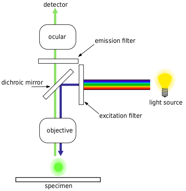 Figure: Working mechanism of Fluorescence Microscope(Photo credit: Henry Mühlpfordt)
Higher energy shorter wavelength lights (UV rays or blue light) generated from mercury vapor arc lamp pass through the excitation filter. The excitation filter allows only the short wavelength of light to pass through and removes all other non-specific wavelengths of light. The filtered light is reflected by the dichroic filter and falls on the fluorophore-labeled sample. The fluorochrome absorbs shorter wavelength rays and emits rays of longer wavelength (lower energy) that pass through the emission filter. The emission filter blocks (suppresses) any residual excitation light and passes the desired longer emission wavelengths to the detector. Thus the microscope forms glowing images of the fluorochrome-labeled microorganisms against a dark background.
To the observer, the background is dark, as there is no visible light and only the labeled specimen (cells, microorganisms, etc.) appear bright (fluoresce).
Types of Fluorescence Microscopes
There are various types of fluorescence microscopes. Some of the common types are:
Epifluorescence microscopes
It is the most common type of fluorescence microscope. In this microscope, excitation of the fluorophore and detection of the fluorescence are done through the same light path (i.e., through the objective).
Confocal microscope
In this type of fluorescence microscope, high‐resolution imaging of thick specimens (without physical sectioning) can be analyzed using fluorescent-labeled dye.
Multiphoton microscope
In this type of microscope, multiphoton fluorescence excitation captures high-resolution three-dimensional images of specimens tagged with highly specific fluorophores.
Total internal reflection fluorescence (TIRF) microscope
Total internal reflection fluorescence microscopy (TIRFM) exploits the unique properties of an induced evanescent wave or field in a limited specimen region immediately adjacent to the interface between two media having different refractive indices.
Applications of Fluorescence Microscope
Fluorescence microscopy is widely used in diagnostic microbiology and microbial ecology (for enumerating bacteria in natural environments). Some organisms, such as Pseudomonas, fluoresce naturally when irradiated with ultraviolet light. Other organisms, such as Mycobacterium tuberculosis and Treponema pallidum, are treated with fluorochrome.
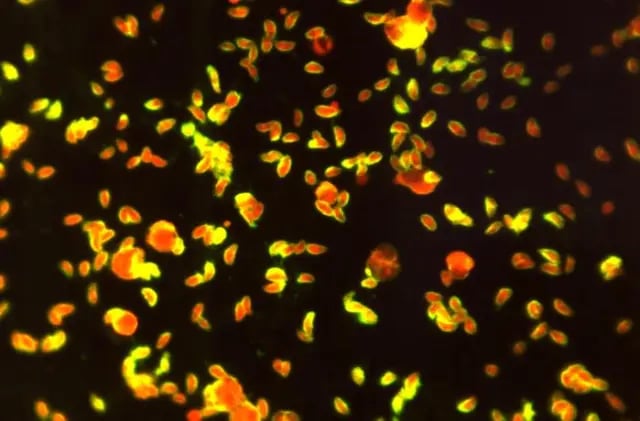 Figure: Fluorescent antibody-stained specimen showing numerous Toxoplasma sp. tachyzoite(Image credit: CDC)
- Acid-fast bacilli (AFB) in sputum or CSF are detected when stained with auramine fluorescent dye.
- Detection of Trichomonas vaginalis, intracellular gonococci, and other parasites when stained by acridine orange.
- In immunodiagnosis of infectious diseases, using both direct and indirect antibody techniques. Immunofluorescence is especially useful in diagnosing syphilis and rabies.
Limitations of Fluorescence Microscope
- Fluorophores gradually lose their ability to fluoresce as they are illuminated in photobleaching. Photobleaching can severely limit the time a sample can be observed by fluorescence microscopy. However, several techniques exist to reduce photobleaching, such as using more robust fluorophores, minimizing illumination, or using photoreactive scavenger chemicals.
- Fluorescence microscopy has enabled the analysis of live cells, but fluorescent molecules generate reactive chemical species under illumination that enhances the phototoxic effect, to which live cells are susceptible.
- Fluorescence microscopy only allows observation of the specific structures labeled for fluorescence. For example, observing a tissue sample prepared with a fluorescent DNA stain by fluorescence microscopy only reveals the organization of the DNA within the cells and reveals nothing else about the cell morphologies.
How to Remember
- What each filter does, in sequence: the excitation filter lets the right wavelength of light in; the dichroic mirror redirects it toward the sample; the emission filter lets the fluorescent glow out while blocking any leftover excitation light. Three filters, three jobs, one direction of travel.
- Why emitted light is always a different color than excitation light: the fluorophore absorbs higher-energy, shorter-wavelength light and re-emits lower-energy, longer-wavelength light. If a question describes the emitted light as shorter wavelength than the excitation light, that's backwards, energy is always lost in the process, never gained.
- Autofluorescence vs. induced fluorescence: a few organisms glow on their own under UV light without any help, Pseudomonas is the classic example. Everything else needs to be dressed in a fluorochrome "costume" before it will glow at all.
- Direct vs. indirect, in one line: direct fluorescent antibody testing uses one labeled antibody that goes straight to its target. Indirect fluorescent antibody testing uses an unlabeled primary antibody, then a labeled secondary antibody that binds to it, adding both an extra step and, often, extra signal amplification. (See the dedicated DFA and IFA articles for the full procedural detail.)
- Why fluorescence beats staining for TB screening speed, not just sensitivity: a glowing organism against total darkness is visible across a wide, low-magnification field; a stained organism has to be searched for field by field under oil immersion. The same slide takes far less time to read under fluorescence, which is exactly why WHO recommends it for high-volume TB screening.
Key exam facts in one table
| Concept | Detail | Why it's tested |
|---|---|---|
| Fluorescence, defined | Absorption of shorter-wavelength (higher-energy) light, emission of longer-wavelength (lower-energy) visible light | The direction (short to long wavelength) is the most commonly reversed detail on exams |
| Three key filters | Excitation filter (selects exciting wavelength), dichroic mirror (redirects light), emission filter (blocks excitation light, passes emitted light) | Each has a distinct, specific job in the light path |
| Autofluorescence example | Pseudomonas fluoresces naturally under UV without any stain | Distinguishes organisms that need no fluorochrome from the majority that do |
| Classic fluorochrome applications | Auramine (AFB), acridine orange (Trichomonas, gonococci), DFA/IFA (syphilis, rabies, and other pathogens) | Frequently tested organism-to-stain matching |
| Key limitation | Only labeled structures are revealed; nothing else about the specimen is shown | A common misconception is that fluorescence microscopy reveals more of the specimen overall, when it actually reveals less, just more clearly |
| Photobleaching | Fluorophores lose their ability to fluoresce with continued illumination | Limits observation time, particularly relevant for live-cell imaging |
Where Students Get Confused
- Reversing the direction of the wavelength shift. Excitation light is shorter wavelength and higher energy; emitted light is always longer wavelength and lower energy. Getting this backwards is one of the most common errors on this topic.
- Confusing autofluorescence with fluorochrome-induced fluorescence. Both make an organism glow, but only a handful of organisms (like Pseudomonas) do this naturally; everything else, including M. tuberculosis and T. pallidum, requires a fluorochrome stain first.
- Assuming fluorescence microscopy reveals more of a specimen than ordinary microscopy. It actually reveals less, only whatever was specifically labeled. A DNA-stained tissue sample under fluorescence shows DNA organization and nothing else about the cell's overall morphology.
- Mixing up direct and indirect fluorescent antibody testing. Direct testing uses one labeled antibody applied straight to the target. Indirect testing uses an unlabeled primary antibody detected by a separate labeled secondary antibody, an extra step that often improves sensitivity through signal amplification.
- Assuming fluorescence microscopy is only about sensitivity, not speed. In high-throughput settings like TB screening, the ability to scan a wide field at low magnification, rather than searching field by field under oil immersion, is itself a major practical advantage, not just a side benefit.
References and Further Readings
- Sanderson, M. J., Smith, I., Parker, I., & Bootman, M. D. (2014). Fluorescence Microscopy. Cold Spring Harbor Protocols, 2014(10), pdb.top071795. https://doi.org/10.1101/pdb.top071795
- Fluorescent Microscopy. Microscopy. Retrieved from https://serc.carleton.edu/microbelife/research_methods/microscopy/fluromic.html
- Fish, K. N. (2009). Total Internal Reflection Fluorescence (TIRF) Microscopy. Current Protocols in Cytometry, 12.18. https://doi.org/10.1002/0471142956.cy1218s50
- World Health Organization. (2014). Fluorescence Microscopy for Tuberculosis: Technical Guide. WHO Press.
Frequently Asked Questions
Why is emitted light always a longer wavelength than the excitation light in fluorescence microscopy?
What is the difference between autofluorescence and fluorochrome-induced fluorescence?
Why is fluorescence microscopy preferred over Ziehl-Neelsen staining for TB screening?
What is the difference between direct and indirect fluorescent antibody testing?
What is the main limitation of fluorescence microscopy compared to routine light microscopy?

Tankeshwar Acharya, MSc (Medical Microbiology)
Tankeshwar Acharya is an Assistant Professor in the Department of Microbiology at Patan Academy of Health Sciences (PAHS), Nepal, where he has been teaching and practicing clinical microbiology for over 14 years. He is the founder of Microbe Online, one of the leading free microbiology education resources on the web, covering bacteriology, mycology, parasitology, immunology, and clinical laboratory diagnostics written from direct experience in both the classroom and the diagnostic laboratory.