
Auramine-Rhodamine Fluorochrome Staining: Principle, Procedure, and Results

On this page
Tuberculosis kills approximately 1.5 million people annually, more than any other single infectious pathogen. In high-burden countries, the first microbiological evidence of TB comes from a sputum smear. The faster and more sensitive that smear, the sooner treatment begins.
Auramine-rhodamine fluorochrome staining was developed specifically to address a fundamental limitation of Ziehl-Neelsen staining: ZN requires examination under oil immersion at 1,000x magnification, meaning a single slide takes 15–20 minutes to screen.
Auramine-rhodamine allows screening at 250x or 400x, the entire slide area in 3–5 minutes while detecting approximately 10% more positive cases. This speed advantage is most important in high-volume laboratories processing hundreds of sputum specimens per day.
Auramine-rhodamine fluorochrome staining, also known as “Truant method of staining,” is used to visualize acid-fast bacilli (AFB). Ziehl-Neelsen (hot) and Kinyoun (cold) are still widely used in developing countries. CDC recommends fluorochrome staining for detecting AFB in primary patient specimens.
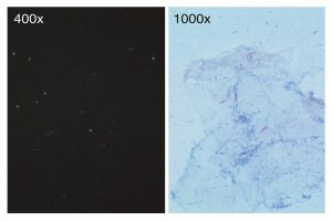 Figure: Comparison of ZN Staining and Flurochrome staining
The acid fastness of Mycobacteria is due to their thick cell wall composed of waxes and lipids with a high content of mycolic acid.
Fluorescent dyes like auramine-rhodamine bind to the mycolic acid present in them and impart bright yellow or orange fluorescence against a greenish background when viewed using a fluorescent microscope. It is also used to stain all acid-fast organisms including the sporozoan parasites.
The modified fluorochrome method (using a weaker decolorizer; 0.5% sulphuric acid rather than 3% acid-alcohol) also detects partially acid-fast organisms including Cryptosporidium parvum, Cyclospora cayetanensis, and Isospora belli oocysts in stool specimens. These coccidian parasites share the partial acid-fast property with Nocardia species and are detected by both auramine-rhodamine and the cold modified ZN method. The oocysts appear as bright yellow-orange fluorescent circles against a dark background, much easier to find at low magnification than on a standard modified ZN smear.
Principle
The fluorochrome dye, auramine-rhodamine, forms a complex with mycolic acids found in the acid-fast cell wall of organisms which resist decolorization by acid-alcohol. The counterstain, potassium permanganate, renders tissue and its debris nonfluorescent, thus reducing the possibility of artifacts. The cells visualized under ultraviolet light appear bright yellow or reddish-orange.
Materials
Reagents:
- Primary stain: auramine rhodamine solution (caution: possible carcinogen)
- Decolorizer: 0.5% acid alcohol (5 ml HCl in 995 ml 70% alcohol). (Caution: flammable, corrosive)
- Counterstain: 0.5% potassium permanganate (0.25 gm in 50 ml). (caution: corrosive)
Others:
- Slide: use only new, unscratched, and clean slides; using old, scratched, or dirty slides can lead to erroneous results.
- Identifier: Properly label each slide using graphite pencils or use a diamond or tungsten carbide stylus.
- Slide racks
- Bunsen burner
Procedure
Preparation of Smear:
For Sputum: Using a piece of stick, transfer a purulent part of the sputum (containing any yellow caseous material), to a slide and make a thin smear. An area of approximately ½ by 1 inch (or 2-cm square) is recommended. Spread the smear using circular movements. Allow to air dry.
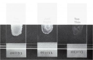 Figure: Smear Thickness
Note: Be sure to prepare smears of suitable thickness. Smears that are too thick may flake during staining and may be difficult to decolorize. Acid-fast organisms that might be present may be obscured. Smears that are too thin may not contain enough sample.
Either condition–too thick or too thin–can lead to erroneous results, particularly false negatives. Here (image 1) the smear in the center is of the proper thickness. Hold a smear about 3 to 4 inches over news-print, if you are just able to read the print, the smear is of proper thickness.
For urine: Make a smear of the deposit from three centrifuged early morning urine sediments.
Allow to air dry and heat fix the specimen. The use of an electric slide warmer is usually the preferred method for heat-fixing smears. An alternate method of heat-fixing is to pass the dried slide, smear facing upward, 2 to 3 times through the blue cone of a burner flame.
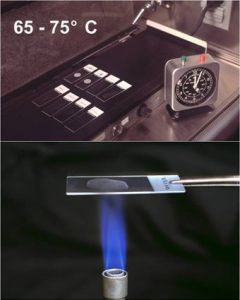 Figure: Heat Fixation of smear (Upper: using electric heater, lower: using burner)
Staining Method
- Place the fixed smear on a staining rack and flood slide with rhodamine-auramine for 15 minutes. Do not let the surface dry. (Note: Fluorochrome dyes used for acid-fast staining include Auramine O, and Auramine O in combination with another fluorochrome, Rhodamine B).
- Wash off the stain with distilled water.
- Flood slide with fluorescent decolorizer (i.e. acid-alcohol)for2-3 minutes.
- Rinse thoroughly with distilled water.
- Flood slide with potassium permanganate for 3-4 minutes. Do not allow slides to dry.
- Rinse thoroughly with distilled water and air dry.
- Examine microscopically under the same light source as used for fluorescent microscopy (i.e. a K530 excitation filter and a BG 12 barrier or G-365 excitation filter and an LP 420 barrier filter). Slides can be screened on high power (400X) and verified under oil immersion.
Result and Interpretation
- Positive Test: Acid-fast organisms fluoresce reddish-orange against a dark background.
- Negative Test: Non-acid-fast organisms will not fluoresce or may appear a pale yellow, quite distinct from the bright acid-fast organisms.
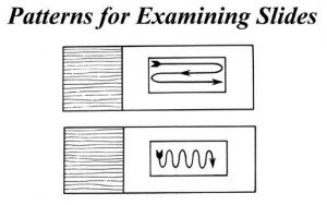 Figure: Patterns for Examining the slide
Number of Fields to Examine:
The minimum number of fields to examine before reporting a smear as negative for acid-fast organisms.
Final magnification (the objective lens magnification multiplied by the eyepiece magnification) Vs. Number of slides
- 200x: 30
- 250x: 30
- 400x: 55
- 450x: 70
Reporting of smears
- If Fluorescent AFB are seen, report the smear as AFB positive, and give an indication of the number of bacilli present in plus signs (+ to +++)
- If no fluorescent rods are seen, report the smear as NO AFB seen.
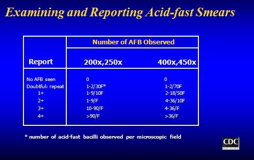 Figure: Examining and Reporting Acid fastness
Fluorochrome AFB Grading Scale (WHO/IUATLD):
| Grade | Number of AFB Seen | Magnification | Report As |
|---|---|---|---|
| No AFB | 0 in minimum fields examined | 200–450x | Negative — No AFB seen |
| Scanty | 1–9 per 100 fields | 200–250x | Report exact count; request repeat specimen |
| 1+ | 10–99 per 100 fields | 200–250x | Positive 1+ |
| 2+ | 1–10 per field (50 fields) | 200–250x | Positive 2+ |
| 3+ | >10 per field (20 fields) | 200–250x | Positive 3+ |
Note: The fluorochrome grading scale uses the same WHO/IUATLD criteria as Ziehl-Neelsen but fields are examined at lower magnification (200–250x for fluorochrome vs 1,000x oil immersion for ZN), which is why fluorochrome screening is significantly faster. For the full ZN grading discussion including clinical implications of each grade, see: Ziehl-Neelsen Staining: Principle, Procedure, Grading, and Interpretation
Advantages
- Faster screening of smears than with ZN- Smear can be examined rapidly using a 40x objective or 25x objective. This increases the chances of detecting AFB especially when they are few.
- ~10% more sensitive than ZN.
- Does not require the use of oil immersion.
- No heat is required for staining.
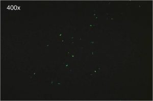 Figure: Fluorochrome stained smear showing numerous green acid-fast bacilli
Limitations of Fluorochrome Staining
A fluorescent microscope is required, which may be readily unavailable and also costly (limitation for developing countries)
A positive staining reaction provides presumptive evidence of the presence of mycobacteria. A negative staining reaction does not indicate that the specimen will be culturally negative. Therefore, cultural methods must be employed.
Reagents like auramine-rhodamine are possible carcinogens, acid –alcohol and potassium permanganate is also a strong irritant to skin, eyes, and respiratory system. Caution is required while handling and staining using such reagents.
Most strains of rapid growers may not appear fluorescent.
It is recommended that all negative fluorescent smears be confirmed with Ziehl-Neelsen stain; at least 100 fields should be examined before being reported as negative.
Key practical rule, the two-step workflow:- Positive auramine-rhodamine result → confirm with ZN staining of the same slide (fluorochrome artefacts can occasionally give false-positive results)
- Negative auramine-rhodamine result → examine minimum required fields before reporting; high clinical suspicion warrants ZN confirmation and repeat specimen
The fluorochrome result is a screen; ZN confirmation is the standard. Both together give maximum sensitivity and specificity.
Excessive exposure to the counterstain may result in a loss of brilliance of the fluorescing organism.
Stained smears should be observed within 24 hours of staining because of the possibility of fluorescence fading.
Key Exam Facts in One Table
| Feature | Detail |
|---|---|
| Also known as | Truant method of staining |
| Primary stain | Auramine O + Rhodamine B (fluorochrome dyes) |
| Decolorizer | 0.5% acid-alcohol |
| Counterstain | 0.5% potassium permanganate (renders background non-fluorescent) |
| AFB appearance | Bright yellow-orange rods against dark background |
| Screening magnification | 250x or 400x (vs 1,000x oil immersion for ZN) |
| Sensitivity vs ZN | ~10% more sensitive |
| WHO recommendation | Preferred method where fluorescence microscope available |
| Positive confirmation | Confirm with ZN staining of same slide |
| Negative confirmation | Examine minimum required fields; high suspicion → ZN + repeat specimen |
| Grading scale | Same WHO/IUATLD criteria as ZN (Scanty, 1+, 2+, 3+) |
| Modified method | 0.5% H₂SO₄ decolorizer detects Cryptosporidium, Cyclospora, Nocardia |
| Key limitation | Fluorescence microscope required; artefacts possible; results fade within 24 hours |
| Safety | Auramine-rhodamine stain is a possible carcinogen; handle in BSC with gloves |
References
- Hooja, S., Pal, N., Malhotra, B., Goyal, S., Kumar, V., & Vyas, L. (2011). Comparison of Ziehl Neelsen & Auramine O staining methods on direct and concentrated smears in clinical specimens. The Indian journal of tuberculosis, 58(2), 72–76.
- Tarhan, G., Ordulu, L., Gümüşlü, F., Ceyhan, I., & Cesur, S. (2003). Tüberküloz tanisinda auramine-rhodamine ve Erlich-Ziehl-Neelsen boyama yöntemlerinin karşilaştirilmasi [Comparison of auramine-rhodamine and Erlich-Ziehl-Neelsen staining methods for the diagnosis of tuberculosis]. Mikrobiyoloji bulteni, 37(2-3), 131–136.
- Bayot ML, Mirza TM, Sharma S. Acid Fast Bacteria. [Updated 2023 Aug 7]. In: StatPearls [Internet]. Treasure Island (FL): StatPearls Publishing; 2023 Jan-. Available from: https://www.ncbi.nlm.nih.gov/books/NBK537121/
Frequently Asked Questions
Why is auramine-rhodamine staining more sensitive than Ziehl-Neelsen for detecting acid-fast bacilli?
What is the two-step workflow for auramine-rhodamine results?
Can auramine-rhodamine staining detect organisms other than mycobacteria?

Tankeshwar Acharya, MSc (Medical Microbiology)
Tankeshwar Acharya is an Assistant Professor in the Department of Microbiology at Patan Academy of Health Sciences (PAHS), Nepal, where he has been teaching and practicing clinical microbiology for over 14 years. He is the founder of Microbe Online, one of the leading free microbiology education resources on the web, covering bacteriology, mycology, parasitology, immunology, and clinical laboratory diagnostics written from direct experience in both the classroom and the diagnostic laboratory.
Comments
No comments yet. Be the first to share your thoughts.
Leave a comment
All comments are reviewed before they appear.