Direct Fluorescent Antibody (DFA) Test: Principle, Procedure, and Clinical Uses
How the direct fluorescent antibody (DFA) test detects antigen directly in a specimen using one labeled antibody. Principle, step-by-step procedure, apple-green result, and the key clinical uses (rabies, RSV, Legionella, chlamydia)
The direct fluorescent antibody (DFA) test is a rapid microscopic procedure for detecting a specific antigen (typically a protein on the surface of a virus, bacterium, or other microbe) directly in a patient specimen, using a fluorescently labeled monoclonal antibody (mAb). It is the "direct" arrangement of immunofluorescence: a single antibody, already carrying the dye, binds the antigen in one step. Its value is speed and directness.
When you already know what you are looking for, DFA can find it in the specimen itself within about an hour, without waiting for a culture that may grow slowly or not at all. This makes it useful for visualizing organisms that are difficult or dangerous to culture, such as rabies virus in brain tissue or respiratory syncytial virus in a nasopharyngeal aspirate. For how direct immunofluorescence differs from the indirect (two-antibody) method, see indirect fluorescent antibody (IFA) test.
Principle
 Figure: Fluorescent antibody methods for detection of microbial surface antigens (Image source: Brock Biology of Microorganisms)
Fluorescent chemicals (e.g fluorescein isothiocyanate) are conjugated (attached) to the constant region of antibodies. When labeled antibodies are incubated with a test sample, the antibody will bind to an antigen, if present. Unbound antibodies are washed away. Areas, where antigens are present, are visualized as fluorescent-apple-green using a fluorescence microscope. If specific antigens are absent there will be no fluorescence.
Because the same antibody both finds the antigen and carries the dye, the reaction is complete in a single binding step. This is what makes DFA fast, and it is also why the signal is not amplified the way it is in the indirect (two-antibody) method.
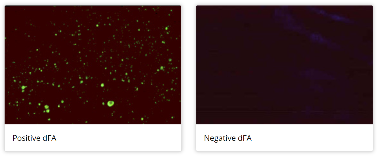 Figure: Direct fluorescent antibody (DFA) test (Image source: CDC)
Procedure
- Prepare the sample by fixing it to the glass slide.
- Apply an appropriate volume of specific fluorescein-labeled antibody to cover the fixed smear. Smear should not be allowed to dry during staining process.
- Incubate at 36 ± 2° for 25 to 65 minutes in a humidified chamber
- Decant excess antibody from the smear by rinsing with appropriate buffer (e.g. phosphate-buffered saline) or deionized water.
- Observe the stained smear in a darkened room using a fluorescence microscope. Fluorescein (FITC) is excited by blue light and emits apple-green, so the correct filter set matters. Read at the magnification appropriate to the target, commonly a 40X objective for screening and oil-immersion 100X for confirming morphology.
Result
- Positive: Specific apple-green fluorescence
- Negative: No or little nonspecific background fluorescence
Controls
Every DFA run should include a known positive and a known negative control slide. The positive control confirms the labeled antibody and microscope are working; the negative control confirms that the apple-green signal is specific and not background autofluorescence. A specimen is only interpretable when both controls behave as expected.
Uses of direct fluorescent antibody (DFA) test
 ### Uses of the direct fluorescent antibody (DFA) test
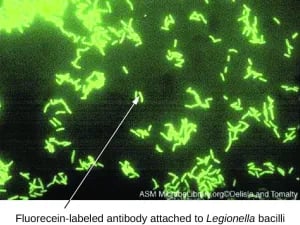
- Identification of Legionella pneumophila and other Legionella species from environmental or patient samples.
- Detection of chlamydia elementary bodies from endocervical specimens.
- Detection of respiratory syncytial virus (RSV) antigen in nasopharyngeal aspirates.
- Detection of rabies virus antigen in the brain of animals suspected of rabies. DFA is the reference (gold-standard) test for post-mortem rabies diagnosis.
- Detection and typing of herpes simplex virus (HSV-1, HSV-2) and varicella-zoster virus (VZV) directly from skin lesion scrapings.
Advantages
- Allows visual assessment of the adequacy of a specimen
- can use on microbes that can’t be easily cultured (e.g., detection of rabies virus antigen in the brain of the animals)
- is both sensitive and specific (requires a good monoclonal antibody)
- can label single cells and can view cells in natural environment
- can use different types of fluorescent-labeled antibodies, each with different dye, to see multiple cell types in one sample.
Disadvantages
- **Expensive:**Many individuals view the requirement for a fluorescent microscope as an expensive luxury.
- Fluorescence fades rapidly over time, which makes the archiving of slides difficult.
- It is often difficult to develop the monoclonal antibody that works well and cross-reactivity may be a problem.
How to Remember
One device, earning its place on the one thing students blur (direct vs the RSV/rabies use-pattern).
Direct means the dye rides along. In the direct test, the antibody that finds the antigen is the same one carrying the fluorescent dye, so there is only one antibody and one step. Tie it to the use pattern: DFA is reached for when you are hunting an antigen you can name in advance (rabies in brain, RSV in aspirate, Legionella, chlamydia), because only then can you pre-label the exact antibody you need. If you can name the target before you start, direct works.
Key exam facts in one table
| Point | What to remember |
|---|---|
| What it detects | Antigen present directly in a specimen |
| Antibody used | One monoclonal antibody, fluorophore-labeled (dye on the Fc region) |
| Steps | Single binding step (no secondary antibody) |
| Fluorophore | Fluorescein isothiocyanate (FITC); blue excitation, apple-green emission |
| Positive result | Specific apple-green fluorescence |
| Negative result | No or only faint nonspecific background fluorescence |
| Reading | Fluorescence microscope in a darkened room; 40X screen, oil 100X confirm |
| Controls | Known positive and negative control slides required each run |
| Gold-standard use | Post-mortem rabies diagnosis (brain tissue) |
| Other clinical uses | RSV (nasopharyngeal aspirate), Legionella, chlamydia, HSV/VZV in lesions |
| Main strengths | Fast; works on organisms that are hard to culture; sensitive and specific with good mAb |
| Main limitations | Fluorescence fades (photobleaching); needs a fluorescence microscope; good mAb hard to develop, cross-reactivity possible |
| Relationship to IFA | Direct = one labeled antibody; indirect (IFA) = unlabeled primary + labeled secondary |
Where students get confused
"DFA detects antibodies in the patient's blood." No. DFA detects an antigen sitting in a specimen (virus in an aspirate, bacteria in a smear, rabies antigen in brain). Detecting a patient's antibodies in serum is the job of the indirect method (IFA).
"You need a UV microscope for DFA." A common textbook slip. FITC, the standard fluorophore, is excited by blue light and emits green. You need a fluorescence microscope with the correct filter set, not specifically a UV lamp.
"DFA is more sensitive than the indirect method." Usually the reverse. DFA uses one label per target, so its signal is not amplified. The indirect method piles several labeled secondary antibodies onto each primary and is generally more sensitive. DFA's advantage is speed and low background, not raw sensitivity.
"A faded slide can be re-read later." Fluorescence photobleaches, so DFA slides do not archive well and should be read promptly. This is a practical limitation, not a minor footnote.
References and further reading
- Centers for Disease Control and Prevention. Direct fluorescent antibody test for rabies diagnosis.
- Madigan, M. T., Bender, K. S., Buckley, D. H., Sattley, W. M., & Stahl, D. A. (2018). Brock Biology of Microorganisms (15th ed.). Pearson.
- Tille, P. M. (2022). Bailey & Scott's Diagnostic Microbiology (15th ed.). Elsevier.
- Procop, G. W., et al. (2017). Koneman's Color Atlas and Textbook of Diagnostic Microbiology (7th ed.). Wolters Kluwer.
- Nowotny, A. (1979). Fluorescent-antibody techniques in diagnostic bacteriology. Bacteriological Reviews.
- United States Department of Agriculture, Center for Veterinary Biologics. Fluorescent antibody staining procedure for detection of viral antigens.
Frequently Asked Questions
What does a direct fluorescent antibody (DFA) test detect?
What does a positive DFA result look like?
Why is DFA used for rabies diagnosis?
What is the difference between DFA and IFA?
Does DFA need a UV microscope?
Why must DFA slides be read promptly?

Tankeshwar Acharya, MSc (Medical Microbiology)
Tankeshwar Acharya is an Assistant Professor in the Department of Microbiology at Patan Academy of Health Sciences (PAHS), Nepal, where he has been teaching and practicing clinical microbiology for over 14 years. He is the founder of Microbe Online, one of the leading free microbiology education resources on the web, covering bacteriology, mycology, parasitology, immunology, and clinical laboratory diagnostics written from direct experience in both the classroom and the diagnostic laboratory.