
Radioimmunoassay (RIA): The Competitive Principle (More Antigen, Less Signal)

On this page
Radioimmunoassay (RIA) is very sensitive (detecting analytes down to the picogram-to-nanogram per milliliter range) and a specific technique that is used for the quantitative detection of antigens or haptens.
RIA is one of the labeled immunoassays, a family that also includes ELISA and immunofluorescence, in which a detectable label makes an otherwise invisible antigen-antibody reaction measurable. In RIA the label is a radioisotope, and the reaction is read as radioactivity. For how it sits among the other antigen-antibody reactions, see the overview.
RIA is an extremely important tool in biomedical research and clinical practice. It is commonly used to assay microbial antigens, proteins, vitamins, hormones, or drugs in serum. A specialized RIA called radioallergosorbent test (RAST) is used to measure the amount of serum IgE antibody that reacts with a known allergen (antigen).
RAST itself has largely been replaced by non-radioactive fluorescence-based assays (such as the ImmunoCAP system) for measuring allergen-specific IgE, for the same safety reasons.
The principle of RIA is similar to that of competitive ELISA for antigen detection, except that a radioisotope (usually iodine-125 or tritium, 3H) is used as the label in place of an enzyme.
Principle of Radioimmunoassay (RIA)
Radioimmunoassay was developed by Rosalyn Yalow and Solomon Berson (1960). Yalow received the Nobel Prize in Physiology or Medicine in 1977. Berson had died in 1972 and so was not eligible, since the Nobel Prize is not awarded posthumously
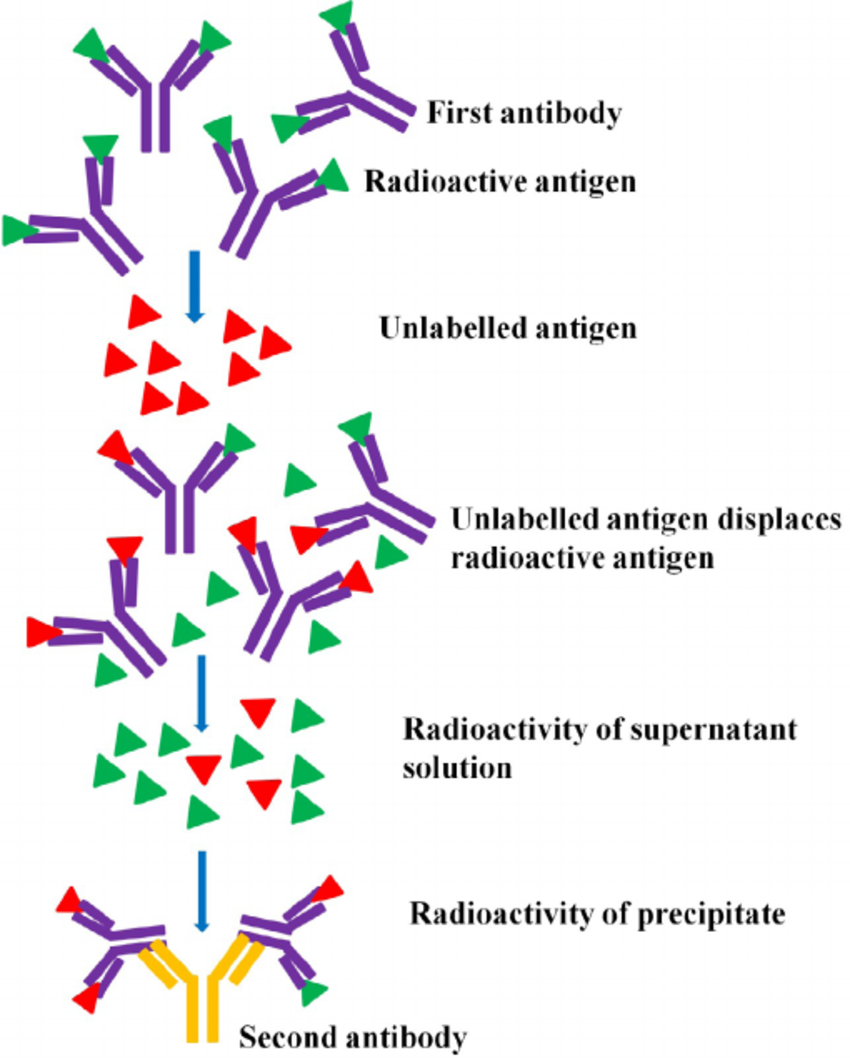 Figure: Principle of Radioimmunoassay
Radioimmunoassay uses radioisotope-labeled purified known antigen which competes with unlabeled (unknown) antigen for binding sites on a known amount of antibody.
The antigen-antibody complexes that form are then separated from unbound antigen, and the radioactivity of the bound fraction is measured with a gamma or scintillation counter.
When the sample contains a high amount of antigen, much of the antigen-binding sites of the antibodies are occupied by unlabelled antigen. So the bound complex will show little radioactivity. The radioactivity generated, in fact, is inversely proportional to the amount of antigen present in the sample.
Then the concentration of the unknown (unlabeled) antigen or hapten present in a sample is determined by plotting the value of radioactivity generated in a standard chart generated using different concentrations of same antigens against the radioactivity of the bound complex.
A crucial practical step is separating antibody-bound antigen from free antigen before counting, because only the bound fraction should be measured. This is done by precipitating the antigen-antibody complexes, classically with a second (anti-immunoglobulin) antibody, or by using a solid phase (antibody fixed to a tube or bead) so the free antigen can simply be washed away.
Why the signal is inverse
RIA works by competition, and this is the single idea that makes the whole method click. The reaction contains a fixed, limited amount of antibody and a fixed amount of radiolabeled antigen. Into this, the patient's sample adds an unknown amount of unlabeled antigen. Labeled and unlabeled antigen now compete for the same limited antibody sites.
Think of the antibody sites as a small number of chairs. The labeled antigen and the patient's unlabeled antigen are both trying to sit down. If the patient has a lot of antigen, it takes most of the chairs, and the labeled antigen is left standing (unbound, washed away), so the bound radioactivity is low. If the patient has little antigen, the labeled antigen keeps most of the chairs, and the bound radioactivity stays high.
So the bound radioactive signal is inversely proportional to the amount of antigen in the sample: more antigen means less signal. This is the opposite of what intuition suggests and is the most common point of confusion in RIA. The unknown concentration is then read off a standard curve built from known antigen concentrations.
Materials Required for Radioimmunoassay
The materials used in RIA are as follows:
- Radioactive Isotope: A radioisotope is used as a tracer to label one of the components involved in the assay. Common radioisotopes used include iodine-125 (^125I) and tritium (^3H).
- Labeled antigen: In competitive RIA, a purified, known quantity of the target antigen is labeled with the radioisotope. This labeled antigen competes with the patient's unlabeled antigen for a limited amount of specific (unlabeled) antibody. (Note: labeling the antibody instead of the antigen describes the related non-competitive method IRMA, noted below.)
- Sample: The biological sample (e.g., blood, serum, plasma, urine) containing the substance of interest must be collected and prepared for the assay.
- Standard Curve Standards: A set of standards with known concentrations of the measured substance is prepared. These standards help create a calibration curve, allowing the conversion of radioactive counts into quantitative concentrations.
- Solid Phase: Tubes or microplates coated with a material that can bind antibodies or antigens are useful to separate bound and unbound components during the assay.
- General reagents: Buffers maintain the pH and ionic strength needed for antigen-antibody binding, and wash solutions remove unbound label so that only the bound fraction is counted, which improves accuracy.
- Gamma Counter: A gamma counter measures the radioactivity in each sample. It quantifies the amount of radioactive tracer present, which is proportional to the concentration of the measured substance.
- Pipettes and Pipette Tips: Accurate and precise pipetting equipment is essential for preparing standards, samples, and reagents.
- Safety Equipment: One should wear personal protective equipment, like gloves and lab coats, when handling radioactive materials. Waste disposal procedures must also be proper.
- Calibrators and Controls: Calibrators with known concentrations and quality control samples are used to ensure the accuracy and reliability of the assay.
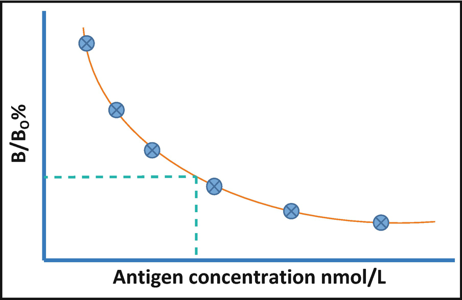 Figure: Radioactivity antigen plot
Advantages
- Extremely high sensitivity. RIA can detect analytes at very low concentrations (picogram to nanogram per milliliter), which is why it opened up measurement of hormones, drugs, and other trace substances that no earlier method could quantify.
- Quantitative and precise. Reading the unknown against a standard curve gives an actual concentration, not just positive or negative, and the method is highly reproducible.
- Specific. The antigen-antibody reaction gives specificity for the target analyte, and RIA works even for small haptens (such as drugs and steroids) that are hard to measure other ways.
- Broadly applicable. The same principle measures hormones, vitamins, drugs, microbial antigens, and allergen-specific IgE (RAST), across serum, plasma, and other fluids.
Limitations
- Radiation hazard. Handling radioisotopes requires trained staff, monitoring, shielding, and licensing, and generates radioactive waste that must be disposed of under regulation. This is the single biggest reason RIA has been displaced. This is the single biggest reason RIA has been displaced. It was displaced in two directions: by ELISA, which uses a safe enzyme label, and by chemiluminescence immunoassay (CLIA), which removed the radiation hazard while keeping the very high sensitivity that made RIA valuable.
- Short reagent shelf life. Radiolabeled reagents decay (I-125 has a half-life of about 60 days), so kits expire relatively quickly and signal strength drifts as the label ages.
- Specialized, costly equipment. A gamma or scintillation counter and a radiation-safe facility are needed, which is impractical for many routine and resource-limited laboratories.
- Not suited to point-of-care or rapid testing. The competitive binding, separation, and counting steps take time and skilled hands, unlike a lateral flow strip.
- High-dose limitation in the competitive format. At very high antigen concentrations the competitive curve flattens, so very high values are read less precisely without dilution.
RIA vs ELISA
RIA and ELISA share the same competitive logic; they differ in the label and everything that follows from it.
| Feature | RIA | ELISA |
|---|---|---|
| Label | Radioisotope (I-125, 3H) | Enzyme (with color substrate) |
| Signal read | Radioactivity (gamma/scintillation counter) | Color change (spectrophotometer) |
| Sensitivity | Very high | High, comparable in modern formats |
| Hazard | Radioactive waste, licensing, short reagent shelf life | Minimal, safer |
| Cost/equipment | Gamma counter, radiation safety | Plate reader, widely available |
| Current use | Largely replaced in routine labs | Standard in most laboratories |
The takeaway: ELISA has replaced RIA in most routine settings not because RIA is less sensitive, but because it avoids the radiation hazard, licensing, and disposal problems, while matching RIA's performance for most analytes. Where the highest sensitivity is still needed without the radiation hazard, chemiluminescence immunoassay (CLIA) has taken that role on modern automated analyzers.
How to Remember
More antigen, less signal. In RIA the patient's antigen competes with labeled antigen for limited antibody. The more the patient has, the more labeled antigen gets crowded out and washed away, so the bound radioactivity falls. High antigen, low counts. This inverse relationship is the whole trick.
Musical chairs with a limited number of chairs. The antibody sites are few chairs; labeled and unlabeled antigen both scramble for them. Whichever is more abundant wins more chairs. A patient flooded with antigen leaves the labeled version standing, so the signal drops.
Key exam facts in one table
| Point | What to remember |
|---|---|
| What it measures | Quantitative detection of antigen or hapten, extremely sensitive |
| Developed by | Berson and Yalow (1960); Yalow won the Nobel Prize (1977) |
| Principle | Competitive: labeled and unlabeled antigen compete for limited antibody |
| Key relationship | Bound radioactivity is inversely proportional to sample antigen |
| Common labels | Iodine-125, tritium (3H) |
| Separation step | Precipitate bound complex (second antibody) or use solid phase, then count |
| Read by | Gamma counter (or scintillation counter) |
| Quantitation | Unknown read off a standard curve |
| RAST | RIA variant measuring allergen-specific IgE (now mostly replaced) |
| Main limitation | Radiation hazard, licensing, waste, short reagent shelf life |
| Replaced by | ELISA in most routine labs (safety, not sensitivity) |
| Non-competitive cousin | IRMA (immunoradiometric assay): labeled antibody, direct relationship |
Where students get confused
"More antigen should give more signal." In RIA, the opposite is true. The signal comes from labeled antigen bound to antibody. The patient's unlabeled antigen competes it away, so more sample antigen means less bound label and a lower count. The inverse relationship is the defining, and most misread, feature of RIA.
"RIA measures the radioactivity of the patient's antigen." No. The patient's antigen is unlabeled and invisible. The radioactivity comes entirely from the reagent (labeled) antigen. You measure the patient's antigen indirectly, by how much labeled antigen it displaces.
"ELISA replaced RIA because RIA wasn't sensitive enough." RIA is extremely sensitive; that was never the problem. ELISA replaced it because it avoids radioactive hazard, licensing, and disposal while giving comparable results. The change was about safety and convenience.
"RIA and IRMA are the same." They are related but opposite in design. RIA is competitive (labeled antigen, inverse relationship). IRMA is non-competitive (labeled antibody sandwiching the antigen, direct relationship: more antigen, more signal). Mixing them up reverses the whole logic.
References and further reading
- Madigan, M. T., Bender, K. S., Buckley, D. H., Sattley, W. M., & Stahl, D. A. (2018). Brock Biology of Microorganisms (15th ed.). Pearson.
- Willey, J. M., Sherwood, L. M., & Woolverton, C. J. (2016). Prescott's Microbiology (10th ed.). McGraw-Hill.
- Tille, P. M. (2022). Bailey & Scott's Diagnostic Microbiology (15th ed.). Elsevier.
- Yalow, R. S., & Berson, S. A. (1960). Immunoassay of endogenous plasma insulin in man. Journal of Clinical Investigation, 39(7), 1157–1175.
Frequently Asked Questions
What is the principle of radioimmunoassay?
Why does more antigen give a lower signal in RIA?
Which radioisotopes are used in RIA?
What is the difference between RIA and ELISA?
What is the difference between RIA and IRMA?
Why has RIA been largely replaced?

Tankeshwar Acharya, MSc (Medical Microbiology)
Tankeshwar Acharya is an Assistant Professor in the Department of Microbiology at Patan Academy of Health Sciences (PAHS), Nepal, where he has been teaching and practicing clinical microbiology for over 14 years. He is the founder of Microbe Online, one of the leading free microbiology education resources on the web, covering bacteriology, mycology, parasitology, immunology, and clinical laboratory diagnostics written from direct experience in both the classroom and the diagnostic laboratory.
Comments
No comments yet. Be the first to share your thoughts.
Leave a comment
All comments are reviewed before they appear.