
Respiratory Syncytial Virus (RSV): Structure, Replication, and Pathophysiology
Respiratory syncytial virus is the top cause of bronchiolitis in infants. Learn its structure, how it replicates, why it plugs infant airways, and how it is diagnosed and prevented.

On this page
In an adult, respiratory syncytial virus is just a cold. The same virus in a two-month-old baby can fill the smallest airways with debris until breathing becomes a struggle. Nothing about the virus changes between the two patients. What changes is the size of the airway it infects. That single idea, a common virus meeting a tiny airway, explains why RSV is the leading cause of infant hospitalization for breathing problems, and why understanding its pathophysiology matters so much.
Respiratory syncytial virus (RSV) usually causes mild, cold-like symptoms, and most people recover in a week or two. But RSV can be serious, especially in infants and older adults. It is the single most common cause of bronchiolitis and pneumonia in infants and young children, and a major cause of severe respiratory illness in the elderly.
The virus takes its name from what it does to cells. When it infects them, it causes neighboring cells to fuse together into large multinucleated masses called syncytia. This cell fusion is central to how the virus spreads from cell to cell and is the origin of the word "syncytial" in its name.
Structure and classification of RSV
RSV was long classified in the family Paramyxoviridae, but it has now been reclassified into the family Pneumoviridae, in the genus Orthopneumovirus. Many textbooks still list the older classification, so it is worth knowing both. For a fuller comparison, see the article on the difference between paramyxoviruses and orthomyxoviruses
The essential structural facts, which are also the ones most often asked:
- Genome: RNA, not DNA. RSV has a single-stranded, negative-sense, non-segmented RNA genome. Because it is negative-sense, the genome cannot be read directly by the host cell and must first be copied by a viral enzyme, which the virus carries inside itself.
- Baltimore classification: Group V (negative-sense single-stranded RNA viruses).
- Enveloped. RSV has a lipid envelope, which is why it is fragile outside the body and easily destroyed by soap and disinfectants.
Three surface proteins on the envelope matter:
- G protein (attachment): binds the virus to the host cell.
- F protein (fusion): fuses the viral envelope with the host cell and, crucially, fuses infected cells to their neighbors to form the syncytia the virus is named for.
- SH protein (small hydrophobic): a smaller membrane protein whose role is less central.
One point of difference from the classic paramyxoviruses is diagnostically important: RSV has no hemagglutinin. This means the hemagglutination and hemadsorption tests used for viruses like mumps and influenza do not work for RSV, which shapes how it is diagnosed in the laboratory.
RSV has two major antigenic subtypes, A and B, which often circulate together and are distinguished mainly by differences in the G protein.
Replication (life cycle) of RSV
Because the genome is negative-sense, RSV carries its own RNA-dependent RNA polymerase inside the virion, and the entire replication cycle takes place in the cytoplasm of the host cell.
The steps of the life cycle are:
- RSV attaches to host cells via the surface glycoprotein.
- Virion envelope fuses with the cell membrane by the action of fusion glycoprotein and enters the cell.
- Release the genome RNA and RNA-dependent RNA polymerase into the cytoplasm.
- The polymerase uses the genome as a template to produce capped and polyadenylated mRNAs
- mRNAs are translated into viral proteins
- Antigenome and genome RNAs are produced
- The resulting genomes are assembled with other viral proteins and buds from the plasma membrane to produce progeny virus particles.
Pathogenesis and pathophysiology of RSV
RSV first infects and replicates in the epithelial cells lining the nasopharynx. This is the "cold" stage, and in many people the infection stays here. The danger begins when the virus spreads down into the lower respiratory tract, where it causes bronchiolitis (inflammation of the small airways) and pneumonia.
Why RSV plugs the small airways. This is the heart of RSV pathophysiology. As the virus infects the cells lining the bronchioles, several things happen at once. The infected epithelial cells die and slough off. The immune system sends in lymphocytes, which gather around the airways (peribronchiolar infiltration). The tissue beneath the lining swells with fluid (edema). And mucus production increases. The result is a plug made of dead cells, mucus, and fibrin that blocks the smaller bronchioles. Interestingly, the virus itself is often no longer detectable deep in these plugged bronchioles. Much of the damage is caused by the body's inflammatory response, not the virus directly.
Why this is an emergency in infants but not adults. The plug is the same, but the airway is not. An infant's bronchioles are tiny, only a few millimeters across. A plug of debris that an adult's wide airway would clear without trouble can completely block an infant's narrow bronchiole. When enough small airways are blocked, air gets trapped, gas exchange fails, and the baby has to work hard to breathe. This is why the same virus is a mild cold in a parent and a hospital admission in their baby. The pathophysiology is driven as much by airway size as by the virus.
Limited spread beyond the airway. Unlike many viruses, RSV rarely if ever enters the bloodstream. Viremia is uncommon. The disease is essentially confined to the respiratory tract, which is why its complications are respiratory.
Incubation and shedding. The incubation period is about 3 to 5 days. Infected infants and young children shed large amounts of virus for 1 to 3 weeks, far longer than adults, who shed for only 1 to 2 days. This prolonged, high-level shedding by young children is a major reason RSV spreads so efficiently through households and daycare settings.
The role of the immune system. A working immune system, particularly cell-mediated immunity, is needed to clear RSV. People with impaired cell-mediated immunity may become persistently infected and shed the virus for months. The larger the initial dose of virus (the inoculum), the more likely infection is to take hold.
Signs and Symptoms
- The spectrum of respiratory illness caused by RSV ranges from inapparent infection or the common cold through pneumonia in infants to bronchiolitis in very young babies
- About one-third of primary RSV infections involve the lower respiratory tract severely enough to require medical attention. The child may wheeze.
- Almost 2% of infected babies require hospitalization with peak occurrences at 2 months of age.
- Symptoms can progress rapidly in severe cases. With modern pediatric intensive care, the mortality rate in otherwise healthy infants is low. However, when RSV infection occurs on top of a pre-existing condition such as congenital heart disease or chronic lung disease of prematurity, the risk of severe outcomes is much higher.
- Infections in the elderly may cause symptoms similar to the influenza virus infection. Pneumonia may develop.
- Children who suffered from RSV bronchiolitis and pneumonia as infants often exhibit recurrent episodes of wheezing illness for many years.
- RSV is an important cause of otitis media (It is estimated that 30-50% of wintertime episodes in infants may be due to respiratory syncytial virus infection).
Laboratory Diagnosis
Specimens should be collected using flocked nylon swabs (cotton-tipped or calcium alginate swabs are not suitable) and placed immediately in the viral transport medium (VTM). Preferred samples are:
- nasal secretions,
- nasopharyngeal secretions
- nasopharyngeal swab or
- nasal wash/aspirate
Isolation of the virus and detection of viral antigens in respiratory secretions is the procedure of choice to diagnose respiratory syncytial virus infection.
Unlike the classic paramyxoviruses it was once grouped with, RSV has no hemagglutinin. Diagnostic methods therefore cannot use hemagglutination or hemadsorption assays.
Antigen (Ag) detection
- Direct identification of viral antigens in clinical samples is rapid and sensitive.
- Immunofluorescence on exfoliated cells or ELISA on nasopharyngeal secretions is commonly used.
- Large amounts of virus are present in nasal washes from young children but much less is present in specimens from adults.
- ELISA kits allow rapid diagnosis. A fast result is valuable mainly for infection control (grouping infected patients together and preventing spread on the ward) and for avoiding unnecessary antibiotics, rather than for antiviral treatment, since routine antiviral therapy for RSV is limited.
Isolation and identification of the virus
- Inoculate the sample into cell cultures immediately; freezing of clinical specimens may result in complete loss of infectivity (labile virus). Commonly used cell lines are; human heteroploid cell lines; HeLa and Hep-2.
- The presence of RSV can usually be recognized by the development of giant cells and syncytia in inoculated cultures. It may take as long as 10 days for cytopathic effects to appear.
- Definitive diagnosis can be established by detecting viral antigens in infected cells using a defined antiserum and the immunofluorescence test.
- Rapid isolation of RSV can be achieved by spin-amplified inoculation of vials containing tissue cultures growing on coverslips. Cells can be tested 24-48 hours later by immunofluorescence.
- Detection of RSV is strong evidence that the virus is involved in a current illness because it is almost never found in healthy people.
Serology
- Serum antibodies can be measured in several ways: immunofluorescence, ELISA, and neutralization tests.
- Measurements of serum antibodies are important for epidemiologic studies but play only a small role in clinical decision-making.
Nucleic Acid Detection
Detection of the genome of RSV in respiratory secretions using PCR.
- Subtyping of respiratory syncytial virus
- Analysis of genetic variation in outbreaks.
Prevention and control of RSV
For decades, RSV prevention meant only hygiene measures. That has changed significantly. There are now effective immunizations for the groups most at risk, and hygiene remains a useful additional layer.
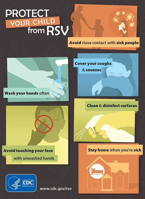 Figure: General hygiene measures that reduce RSV spread (Source: CDC).
Protecting infants. Two approaches are used, and most babies need only one of them:
- A maternal RSV vaccine given to the mother during pregnancy (around weeks 32 to 36). The mother makes antibodies that cross the placenta and protect the newborn in the first months of life.
- A long-acting monoclonal antibody given to the baby (nirsevimab, and more recently clesrovimab). This is not a vaccine; it provides ready-made antibody that protects the infant directly through their first RSV season. In real-world use, nirsevimab has reduced RSV hospital admissions in infants by roughly 80 percent.
Protecting older adults. RSV vaccines are now available for older adults, who are the other high-risk group. Health authorities recommend a single dose for all adults aged 75 and older, and for adults aged 50 to 74 who are at increased risk of severe RSV.
General hygiene measures still help reduce spread, especially within households and childcare settings:
- Wash hands often.
- Avoid close contact with people who are sick.
- Cover coughs and sneezes.
- Avoid touching the face with unwashed hands.
- Keep sick children home where possible.
Because RSV is an enveloped virus, it is readily killed by soap and standard disinfectants, which makes hand hygiene and surface cleaning genuinely effective.
How to Remember
Why the name ("cells that hold hands"): RSV makes infected cells fuse with their neighbors into giant multinucleated masses, syncytia. Picture cells joining hands into a clump. The fusion (F) protein does it, and it gives the virus its name.
Why babies, not adults ("same plug, smaller pipe"): the airway plug of dead cells and mucus is the same in everyone. The infant's bronchiole is a narrow pipe, so the same plug blocks it. Remember "small virus problem, small airway problem" and the whole pathophysiology follows.
No hemagglutinin, no hemadsorption: RSV is the paramyxovirus-like virus that broke the rule. It has no hemagglutinin, so the HA and hemadsorption tests that work for mumps and influenza do not work for RSV. If a question pairs RSV with hemadsorption, that is the trap.
Prevention has flipped ("antibody for babies, vaccine for grannies"): infants get a monoclonal antibody (nirsevimab) or protection from a maternal vaccine; older adults get their own RSV vaccine. Two ends of life, two different tools.
Key exam facts
| Fact | Detail |
|---|---|
| Current family | Pneumoviridae (formerly Paramyxoviridae) |
| Genus | Orthopneumovirus |
| Genome | Single-stranded, negative-sense, non-segmented RNA |
| Baltimore group | Group V (negative-sense ssRNA) |
| Envelope | Enveloped (fragile; killed by soap/disinfectant) |
| Key surface proteins | G (attachment), F (fusion → syncytia), SH |
| Distinctive feature | No hemagglutinin (so no HA/hemadsorption test) |
| Subtypes | A and B |
| Named for | Syncytia (fused multinucleated cells) |
| Site of replication | Cytoplasm |
| Incubation period | 3 to 5 days |
| Core disease | Bronchiolitis and pneumonia in infants |
| Key mechanism | Small-airway plugs (cells + mucus + fibrin) + narrow infant airway |
| Viremia | Rare to absent; disease confined to respiratory tract |
| Shedding | 1 to 3 weeks in infants; 1 to 2 days in adults |
| Highest-risk groups | Young infants, elderly, congenital heart/lung disease |
| Lab diagnosis | Antigen detection (IF, ELISA), culture (HeLa/Hep-2, syncytia), PCR; flocked swabs |
| Infant prevention | Maternal vaccine OR infant monoclonal antibody (nirsevimab/clesrovimab) |
| Older-adult prevention | RSV vaccine (age 75+, and 50–74 if at risk) |
Where Students Get Confused
"Is RSV a DNA or RNA virus?" RNA. Specifically single-stranded, negative-sense, non-segmented RNA (Baltimore Group V). Being negative-sense means it must carry its own polymerase to copy the genome before anything can be made.
"Is RSV a paramyxovirus?" It used to be classified in Paramyxoviridae, but it has been moved to its own family, Pneumoviridae. Many textbooks still say paramyxovirus, so know both, but the current answer is Pneumoviridae.
"Why does RSV cause severe disease in babies but only a cold in adults?" Because of airway size. RSV plugs the small airways with dead cells and mucus. An infant's bronchioles are so narrow that these plugs block them, causing bronchiolitis. An adult's wider airways clear the same plugs easily. The virus is the same; the airway is not.
"Why can't you use a hemadsorption test for RSV like for mumps?" Because RSV has no hemagglutinin. Hemadsorption and hemagglutination tests depend on that protein binding red blood cells. RSV lacks it, so these tests do not work, and diagnosis relies on antigen detection, culture, or PCR instead.
"Is there a vaccine for RSV?" Yes, now. There are RSV vaccines for older adults and for pregnant women (to protect the baby), plus a long-acting antibody (nirsevimab) given directly to infants. This is recent; older resources say there is no vaccine, which is now out of date.
"Does RSV spread through the blood like measles?" No. RSV rarely enters the bloodstream. It stays in the respiratory tract, which is why its complications are respiratory rather than systemic.
Frequently Asked Questions
What is RSV?
What is RSV?
Respiratory syncytial virus (RSV) is a common respiratory virus. It usually causes a mild cold but can cause serious lower-airway disease (bronchiolitis and pneumonia) in infants and older adults.
Is RSV a DNA or RNA virus?
Is RSV a DNA or RNA virus?
It is an RNA virus, with a single-stranded, negative-sense RNA genome (Baltimore Group V). It is enveloped.
Why is RSV so dangerous for babies?
Why is RSV so dangerous for babies?
RSV blocks the small airways with a plug of dead cells and mucus. Because a baby's airways are very narrow, these plugs can block them completely, making it hard to breathe. Adults have wider airways, so the same infection is usually just a cold.
Why is it called "syncytial"?
Why is it called "syncytial"?
Because the virus causes infected cells to fuse together into large multinucleated masses called syncytia. This cell fusion is a hallmark of the infection.
How is RSV diagnosed?
How is RSV diagnosed?
Mainly by detecting the virus in respiratory secretions using antigen tests (immunofluorescence or ELISA) or PCR. Because RSV has no hemagglutinin, hemagglutination and hemadsorption tests cannot be used.
How long is someone with RSV contagious?
How long is someone with RSV contagious?
Infants and young children can shed the virus for 1 to 3 weeks. Adults usually shed for only 1 to 2 days. This long shedding in children helps RSV spread quickly.
Is there a vaccine for RSV?
Is there a vaccine for RSV?
Yes. There are RSV vaccines for older adults and for pregnant women (to protect the newborn), and a long-acting antibody (nirsevimab) given to infants. Most babies need either the maternal vaccine or the infant antibody, not both.
Can you catch RSV more than once?
Can you catch RSV more than once?
Yes. Immunity to RSV is incomplete, so reinfections happen throughout life, though they are usually milder than the first infection.
References
- Griffiths, C., Drews, S. J., & Marchant, D. J. (2017). Respiratory syncytial virus: infection, detection, and new options for prevention and treatment. Clinical Microbiology Reviews, 30(1), 277–319. https://doi.org/10.1128/CMR.00010-16
- Battles, M. B., & McLellan, J. S. (2019). Respiratory syncytial virus entry and how to block it. Nature Reviews Microbiology, 17(4), 233–245. https://doi.org/10.1038/s41579-019-0149-x
- Centers for Disease Control and Prevention. (2026). Clinical guidance for RSV immunizations and vaccines. https://www.cdc.gov/rsv/hcp/vaccine-clinical-guidance/index.html
- Centers for Disease Control and Prevention. (2025). Respiratory syncytial virus (RSV). https://www.cdc.gov/rsv/

Tankeshwar Acharya, MSc (Medical Microbiology)
Tankeshwar Acharya is an Assistant Professor in the Department of Microbiology at Patan Academy of Health Sciences (PAHS), Nepal, where he has been teaching and practicing clinical microbiology for over 14 years. He is the founder of Microbe Online, one of the leading free microbiology education resources on the web, covering bacteriology, mycology, parasitology, immunology, and clinical laboratory diagnostics written from direct experience in both the classroom and the diagnostic laboratory.
Comments
No comments yet. Be the first to share your thoughts.
Leave a comment
All comments are reviewed before they appear.