Immunochromatography/ Lateral Flow Immunoassay: Principle and Uses
Immunochromatography/ Lateral Flow Immunoassay: Principle and Uses
Immunochromatography, also known as lateral flow immunoassay, is a combination of chromatography and immunoassay. This technology is in use for a wide array of tests for clinical, veterinary, and industrial applications.
Depending on the assay, either antigen (for detection of antibody) or antibodies (for detection of antigens) are immobilized on a nitrocellulose membrane in discreet spots in a cartridge device. Following antibody or antigen capture and a flow-through wash step, the sequential addition of an enzyme-labeled conjugate and substrate results in the appearance of a colored reaction product directly on the membrane.
A typical immunochromatographic test strip consists of
- an absorbent pad onto which specimen is applied,
- a conjugate or reagent pad that contains antibodies specific to the target analyte conjugated to colored particles (i.e., colloidal gold particles or latex microspheres),
- a reaction membrane onto which anti target analyte antibodies are immobilized in a line across the membrane as a capture zone or test line,
- a control zone containing antibodies specific for the conjugate antibodies, and
- a waste reservoir composed of another absorbent pad (wick assembly) designed to draw the sample across the reaction membrane by capillary action.
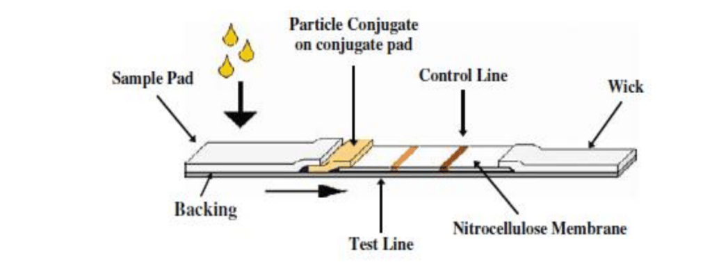 Figure: Typical configuration of a lateral flow immunoassay test strip
The components of the strip are fixed to an inert backing material and may be formatted as a simple dipstick or inside a plastic casing with a sample port and a reaction window showing the capture (test) and control zones.
Rapid diagnostic tests or RDTs are lateral flow immuno-chromatographic antigen tests, which rely on the capture of dye-labeled antibodies to produce a visible band on a strip of nitrocellulose, often encased in a plastic housing, referred to as cassettes. RDTs are also available for the detection of antibodies.
A number of variations of immunochromatographic or lateral flow immunoassays have been developed into commercial products, but they all operate according to the same basic principles.
Principle (for the detection of Antigen)
Lateral flow immunoassays used in clinical microbiology laboratories are usually double-antibody sandwich assays. For the detection of antigens, the capture zone (test line) on the membrane contains immobilized antibodies.
The specimen (e.g., serum, urine) containing the antigen to be detected is placed on the sample pad, which soaks up the specimen fluid. The fluid then migrates to the conjugate pad, which contains conjugated antibodies (conjugated with gold, colored latex, or a chromophore) directed against the antigen. Here, the antigen-antibody-conjugate complex is formed. Ag-Ab complex continues to migrate across the membrane until it reaches the capture zone where the complex will bind to immobilized antibodies. As more and more Ag-Ab complexes are captured at the “test” line, the line becomes visible on the membrane.
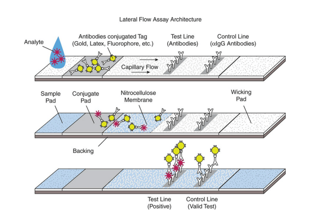 Figure: Lateral Flow Immunoassay
The sample then migrates further along the strip until it reaches the control zone where excess conjugate binds and produces a second visible line (control line) on the membrane. This control line indicates that the sample has migrated across the membrane as intended.
Antibodies that are not antigen-specific or conjugated antibodies that are not complexed with antigen are not captured in the test line and continue to migrate toward the control line. The control line is composed of immobilized antibodies directed against immunoglobulin. As more and more uncomplexed antibody passes over the “control” line, the uncomplexed antibodies are captured and become visible at the “control” line. The presence of a “control” line only indicates that the test was performed properly.
Interpretation of test results
- Positive result: A clear line in the control zone and the test area on the membrane.
- Negative result: A single line in the control zone.
- Invalid: A single line in the test area without a corresponding control line
Applications of Lateral Flow Immunoassay
 Immunochromatographic methods are widely used in clinical practice for;
- Detection of toxins
- Pregnancy tests- detection of human chorionic gonadotropin (hCG)
- Diagnosis of parasitic infections
- Malaria: Malaria- detect specific antigens (Plasmodium lactate dehydrogenase, Plasmodium aldolase, and P. falciparum histidine-rich protein-2) produced by malaria parasites in the blood of infected individuals.
- G. lamblia and Cryptosporidium parvum (ImmunoCardSTAT! Cryptosporidium/Giardia)
- G.lamblia, E. histolytica/E. dispar, and C. parvum on fecal specimen (Triage Micro Parasite Panel)
- Diagnosis of bacterial infections
- Mycoplasma pneumoniae(ImmunoCard Mycoplasma, which detects Mycoplasma pneumoniae specific IgM in serum samples)
- H. pylori antigens in stool (ImmunoCardSTAT SpSA)
- V. cholerae O1 and O139 from stool specimens
- Streptococcus pneumoniae antigen detection in CSF or in urine (Binax NOW Streptococcus pneumoniae antigen card)
- Diagnosis of viral Infections
- Antigen detection: RSV (Binax NOW RSV, Remel Xpect RSV), Rotavirus (ImmunoCardSTAT! Rotavirus), Influenza A/B (BinaxNOW Influenza A & B), Hepatitis B and Hepatitis C infection
- Antibodies detection: Detection of HIV-1 and HIV-2 antibodies (OraQuick Advance Rapid HIV-1/2 antibody test, Reveal G4 Rapid HIV-1 antibody test, Multispot HIV-1/HIV-2 Rapid Test, etc)
Advantages
- Commercially available and low cost (compared with EIA, Immunofluorescence, or RIA)
- Comparable or better sensitivity and specificity than other well-established methods
- Rapid test
- Requirement of small sample volume
- Easy to perform (no sample pre-treatment required in most of the cases)
- Simple and user-friendly (to perform as well as to interpret test results)
- Can be used in the field or rural settings: stability over a wide range of environmental conditions and a very long shelf life.
Limitations
- Mostly qualitative or semi-quantitative
- Most of the devices can detect more than one or two analytes simultaneously
Reference and further reading
- Cherian Sebastian et al. Immunochromatography: Formats and Applications.Indo American Journal of Pharmaceutical Research.2016:6(07).
- Koneman’s Color Atlas and Textbook of Diagnostic Microbiology

Tankeshwar Acharya, MSc (Medical Microbiology)
Tankeshwar Acharya is an Assistant Professor in the Department of Microbiology at Patan Academy of Health Sciences (PAHS), Nepal, where he has been teaching and practicing clinical microbiology for over 14 years. He is the founder of Microbe Online, one of the leading free microbiology education resources on the web, covering bacteriology, mycology, parasitology, immunology, and clinical laboratory diagnostics written from direct experience in both the classroom and the diagnostic laboratory.