Blood Collection Tubes: Significance of Color Coding
Blood Collection Tubes: Significance of Color Coding
Different tests and biochemical assays require varying types of sample collection tubes. In-vitro analysis of blood samples can be performed in clinical laboratories using serum or plasma.
Blood is collected in test tubes or blood collection tubes (BCTs) with air-tight closures, color-coded for practical and easy identification. These are also called Vacutainer® or evacuated tubes.
These are either made up of plastic or glass and have rubber stopper at the top. The rubber top creates a vacuum seal that helps in drawing a pre-determined volume of blood. These tubes are used in various diagnostic fields like chemical/biochemical, hematological, molecular, and serological testing.
Color of the tubes determine the types of additives added in the tubes. Thorough mixing of tubes with additives is must. All tubes with anticoagulants should be mixed gently 10-12 times to mix the additive with the blood and prevent clotting. When blood is not mixed thoroughly with the additives may result in errors in the test.
Performing more than one test is possible from one tube. You may check with the laboratory for the minimum amount of needed blood.
Components of Blood Collection Tube
The blood collection tubes are similar to the test tube in shape and size. However, these have stoppers and can be made from either plastic or glass. The following are the components of blood collection tube:
- Tube wall: The blood collection tube is of 50-150 mm in length and 10-20 mm in diameter. The glass used is borosilicate or soda-lime and the plastic used is of polyethylene terephthalate or polyethylene and polypropylene. The plastic tubes are more durable and carry less risk of cross contamination.
- Rubber stopper: The rubber stopper is colorful and easily penetrable by needle and self seal after removing the needle. Butyl rubber and halogenated butyl rubber are common materials for stopper.
- Stopper lubricant: Lubricants like silicone oils, glycerol, and fluids are applied in the stopper. These lubricants help in easy removal and insertion of stopper.
- Tube surfactant: Tube surfactant should chosen wisely as these might interfere with antibodies and disrupt the reactions required. The surfactant helps reducing non-specific adsorption, improve blood flow, and preventing absorbing of proteins, RBCs, and platelets to the tube wall.
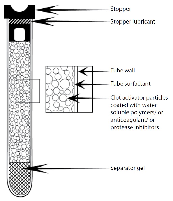 Figure: Components of an evacuated blood collection tube
Additional components
Besides the general components, additional components are present in different color caps, which are as follows:
- Separating gel: These are present in SST (serum separating tubes) and used to separate serum from clotted or whole blood. The gel used is thixotropic gel which is lodged between the packed cells and serum.
- Anticoagulants: Potassium EDTA, trisodium citrate, potassium oxalate, sodium fluoride, and heparin salts act as anticoagulants and chelating agents in blood collection tubes.
- Clot activator particles: These are present in plastic tubes and the particles activate clot either intrinsically or extrinsically. Ellagic acid, thrombin, snake venoms, and thromboplastin are activate clot extrinsically. Silica, glass, bentonite, kaolin, and diatomaceous earth activate clot intrinsically.
- Protease inhibitors: EDTA and citrate also acts protease inhibitors by limiting the activation of proteases. Aprotinin and sulfonyl halides are other protease inhibitors used in Vacutainer® tubes.
Order of Draw
Blood collection in tubes follows a principle known as “order of draw”. A specific order for transferring the drawn blood is necessary for avoiding cross-contamination of additives. The recommended order of draw for blood collection tubes are:
First draw in blood culture bottle or tube (yellow or yellow-back top)
Second draw in a coagulation tube (light blue color). Concern of contamination by tissue, fluids, or thromboplastins may arise, so one can draw in non-additive tube first followed by the light blue top.
Third draw is on a non-additive tube (red top).
Final draw in additive tube in following order:
SST (red-gray or gold top) which contains gel separator and clot activator.
PST (light green top) with lithium heparin anticoagulant and a gel separator.
Sodium heparin (dark green top)
EDTA(lavender top)
ACDA or ACDB (pale yellow top) that hs acid citrate dextrose.
Oxalate/ fluoride (light gray top)
Do not transfer a sample from one collection tube to another or mix blood from different collection tubes.
Color of the Cap and Its Purpose
As mentioned earlier, the color of the top of the tube signifies the purpose and anticoagulant added to the tube. The color of the tube also determines the clotting time required as well as the number of necessary inversions after the blood is transferred into the tubes. Standard laboratory practices use yellow, pink, blue, lavender/purple, red, green, and light blue color tubes.
The color of the tube with anticoagulant and their area of use are as follows:
Marble or Gold (SST)
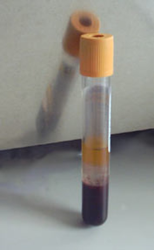 Figure: SST tube
- Additive: Plastic tubes with clot activator and gel for serum separation.
- Tube Inversions: 5 tube inversions required to ensure the mixing of clot activator with blood.
- Clotting Time Required: 30 minutes
- Commonly Associated Tests: Chemistry profiles, electrolytes, lipid panel, hepatic panel, hepatitis panel, thyroid studies, iron studies, cancer markers, lithium, alcohol, vitamin B12, vitamin D, hormone studies, cardiac markers, lidocaine, folate, therapeutic drugs (except carbamazepine), tricyclic antidepressants, salicylate, and homocysteine (ON ICE).
Plain Red
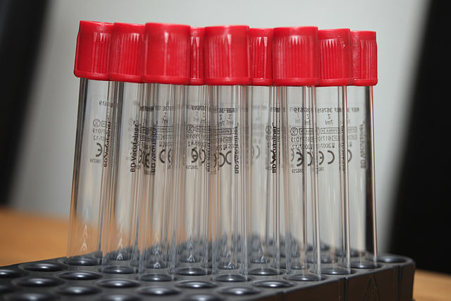 Figure: Red Vacutainer
- Additive: Silicone coated made of glass.
- Tube Inversions: No tube inversions required.
- Clotting Time Required: 60 minutes
- Commonly Associated Tests: Rheumatoid factor (RF), RPR (rapid plasma reagin test), uric acid, PTH (parathyroid hormone), insulin, prealbumin, magnesium, BhCG (beta-human chorionic gonadotropin) test, FT3/FT4 (free triiodothyronine and free thyroxine), digoxin, amylase, lipase, cortisol, CRP (C-reactive protein) test, and C-peptide.
Green
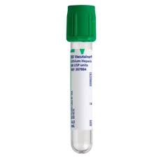 Figure: Green Vacutainer
- Additive and tube Inversions: Lithium heparin (light green tube tubes containing lithium heparin and gel for plasma separation) is the additive. Eight tube inversions to ensure mixing the anticoagulant with blood to prevent clotting.
- Clotting Time Required: No clotting time required.
- Commonly Associated Tests: Green and light green Vacutainer tubes are preferable for all STAT general chemistry requests.
Chemistry profiles Ionized calcium Lipid panel Hepatic panel Cardiac markers Rheumatoid factor (RF) test Ammonia (ON ICE) Therapeutic drugs (except for VANC and lithium). BhCG Quant
Gray
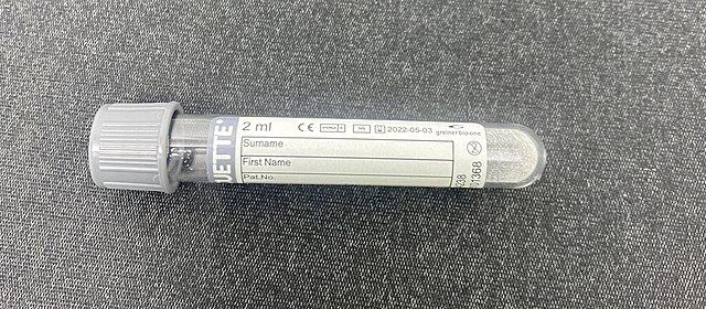 Figure: Gray Vacutainer
- Additive: Sodium Fluoride/Potassium Oxalate
- Tube Inversions: 8 tube inversions ensure proper mixing of additives with blood.
- Clotting Time Required: No clotting time required.
- Commonly Associated Tests: Lactic acid (ON ICE). Gray top tubes can be used when checking Glucose levels only. These tubes preserve glucose and are helpful when drawing blood samples a long distance from the hospital.
Purple/Lavender
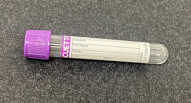 Figure: Lavender Vacutainer
- Additive: Spray-coated K2 EDTA added in a plastic tube. So, also called as EDTA tube.
- Tube Inversions: 8 tube inversions required to ensure the mixing of the anticoagulant with the blood.
- Clotting Time Required: No
- Commonly Associated Tests: CBC (Complete blood count)/PLT Count, H&H (hemoglobin and hematocrit), SED Rate (ESR- eosinophil sedimentation rate), BNP (B-type natriuretic peptide) test, HgbA1C, Cyclosporin, Sickle cell, RETIC (reticulocyte count), Path Review, Intra op PTH, Vancomycin, HIV, Direct Coombs, RBC Folate, PROGRAF, CD3/CD4.
Pink
 Figure: Pink Vacutainer
- Additive: Spray-coated K2 EDTA added in plastic tubes.
- Tube Inversions: 8 tube inversions prevent clotting.
- Clotting Time Required: No
- Commonly Associated Tests: Blood typing and RH, Blood typing and Screening, Antibody Screen, Crossmatch, RHOGAM Workup
Blue
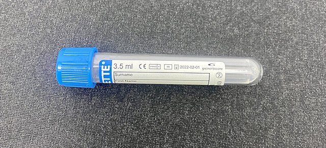 Figure: Blue Vacutainer
- Additive: Buffered sodium citrate 0.1-5 M (3.2%) glass and 0.109 M (3.2%) plastic. It is also called as PT tube.
- Tube Inversions: 3-4 tube inversions ensure proper mixing of the anticoagulant with the blood.
- Clotting Time Required: No
- Commonly Associated Tests
PT/INR (Prothrombin time/International normalized ratio) test PTT (partial thromboplastin time) test Fibrinogen D’DIMER Special Coag and Factor Assays call the lab before collection
Royal Blue
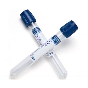 Figure: Royal blue Vacutainer
- Additive: Plastic tubes is sprayed with K2 EDTA. K2 EDTA increases the MCV (mean corpuscular volume) of RBC in higher concentrations.
- Tube Inversions: 8 tube inversions ensure proper mixing of the anticoagulant with the blood.
- Clotting Time Required: No
- Commonly Associated Tests: Toxicology testing determines the trace amount of metals like aluminum, cadmium, copper, lead, mercury, selenium, zinc, etc.
The Gold Top (SST-Serum Separator Tube) and Plain Red Top Tube should be mixed 5-6 times because they contain a clot activator. The Green Top (PST-Plasma Separator Tube) is preferred if listed.
In summary
The color of the tube and the anticoagulant added to it is as follows:
| Color of the Tube | Anticoagulant |
|---|---|
| Yellow, Pink, and Blue | Sodium polyanethole sulfonate (SPS) |
| Light Blue “citrate tube” | Sodium citrate (3.2%) |
| Red | No anticoagulant or additive inside the tube |
| Green | Heparin (sodium heparin, lithium heparin, or ammonium heparin) |
| Lavender/Purple “EDTA tubes” | Ethylene-diamine-tetra-acetic-acid |
| Gray | Potassium oxalate and sodium fluoride |
References and further readings
- MD ECK. Phlebotomy [Internet]. WebPath. [cited 2023Jan24]. Available from: https://webpath.med.utah.edu/TUTORIAL/PHLEB/PHLEB.html
- Bayot ML, Tadi P. Laboratory Tube Collection. [Updated 2022 Aug 8]. In: StatPearls [Internet]. Treasure Island (FL): StatPearls Publishing; 2022 Jan-. Available from: https://www.ncbi.nlm.nih.gov/books/NBK555991/
- BD Vacutainer Venous Blood Collection Tube Guide – Hopkins Medicine [Internet]. <www.bd.com/vacutainer>. BD Diagnostics; [cited 2023Jan24]. Available from: https://www.hopkinsmedicine.org/immunogenetics/\_docs/Tubes_chart.pdf
- Bowen RAR, Remaley AT. Interferences from blood collection tube components on clinical chemistry assays. Biochemia Medica. 2014Feb15;24(1):31–44.
- https://www.bd.com/en-us/products-and-solutions/products/product-families/bd-vacutainer-blood-collection-tubes

Tankeshwar Acharya, MSc (Medical Microbiology)
Tankeshwar Acharya is an Assistant Professor in the Department of Microbiology at Patan Academy of Health Sciences (PAHS), Nepal, where he has been teaching and practicing clinical microbiology for over 14 years. He is the founder of Microbe Online, one of the leading free microbiology education resources on the web, covering bacteriology, mycology, parasitology, immunology, and clinical laboratory diagnostics written from direct experience in both the classroom and the diagnostic laboratory.