
Bismuth Sulfite Agar (BS Agar): Composition, Principle, Uses, and Colony Morphology
Bismuth Sulfite Agar is the most sensitive medium for isolating Salmonella Typhi from stool. Learn its bismuth sulfite mechanism, characteristic black rabbit-eye colonies with metallic sheen, 2-day shelf life limitation, and how it compares to XLD and HE agar.

On this page
A 22-year-old university student returns from rural Nepal with two weeks of stepwise fever, abdominal pain, and relative bradycardia. Blood culture is sent but results will take 5–7 days. Meanwhile, the clinician also sends three consecutive stool specimens for culture, as Salmonella Typhi is shed in stool from the second week of illness onward.
In the laboratory, a key decision must be made: which solid medium will give the best chance of isolating S. Typhi from a stool specimen that contains millions of normal commensal organisms? MacConkey agar will grow coliforms prominently and may miss low numbers of S. Typhi. XLD agar is reliable, but bismuth sulfite agar is specifically regarded as the most sensitive single medium for S. Typhi isolation, producing distinctive black colonies with a metallic sheen that can be seen even at low colony counts, standing out unmistakably against the brown-black surrounding medium.
Principle
Bismuth sulfite agar achieves selectivity through two independent inhibitory mechanisms and differentiation through a unique H₂S indicator system:
Selective mechanism 1, Brilliant green dye: As in SS agar, brilliant green is incorporated at a low concentration (0.025 g/L) to inhibit the growth of Gram-positive organisms and suppress most coliforms. Salmonella species have inherent resistance to brilliant green at this concentration.
Selective mechanism 2, Bismuth sulfite: Bismuth sulfite is the primary and most distinctive selective agent in this medium. Bismuth ions (Bi³⁺) are toxic to most Gram-negative bacteria, including coliforms, because they interfere with sulphur metabolism and enzyme activity. Salmonella species possess metabolic mechanisms that allow them to tolerate bismuth toxicity while simultaneously exploiting the bismuth as part of the H₂S indicator reaction. The combination of brilliant green and bismuth sulfite creates a dual selective barrier significantly more inhibitory to coliforms than either agent alone.
Differential mechanism, the H₂S metallic sheen system: Salmonella species produce H₂S by reducing thiosulfate and other sulphur-containing compounds. On BS agar, H₂S reacts with ferrous sulfate to produce black ferrous sulfide (FeS) precipitate within the colony, giving the colony its black color. Simultaneously, H₂S and other reducing products cause the bismuth ions to be reduced to metallic bismuth (Bi⁰) in the agar immediately surrounding the colony. Metallic bismuth deposits produce the characteristic metallic sheen or luster around BS agar colonies, one of the most striking and distinctive appearances in clinical bacteriology.
The medium also contains dextrose (glucose) as the sole fermentable carbohydrate, with ferrous sulfate functioning simultaneously as an H2S indicator and iron source.
Why BS agar is considered the most sensitive medium for S. Typhi: S. Typhi is a vigorous H₂S producer and grows luxuriantly on BS agar because it handles bismuth toxicity particularly well. The high bile salt concentration and dual inhibitory agents suppress competing flora more completely than XLD or HE agar, and the metallic bismuth sheen is visible even around small or isolated colonies, making it easier to identify S. Typhi colonies at low inoculum density. This is why BS agar is the medium of choice when S. Typhi specifically is suspected and colony count may be low.
Historical note about Wilson and Blair medium: BS agar is a modification of the original Wilson and Blair medium developed by Wilson and Blair in the late 1920s (1927 to 1931) for the specific purpose of S. Typhi isolation from stool. The original medium also used bismuth sulfite as the primary indicator but required a more complex preparation procedure. The current formulation is the commercially standardized version that retains the core bismuth sulfite-ferrous sulfate indicator system while improving stability and ease of preparation.
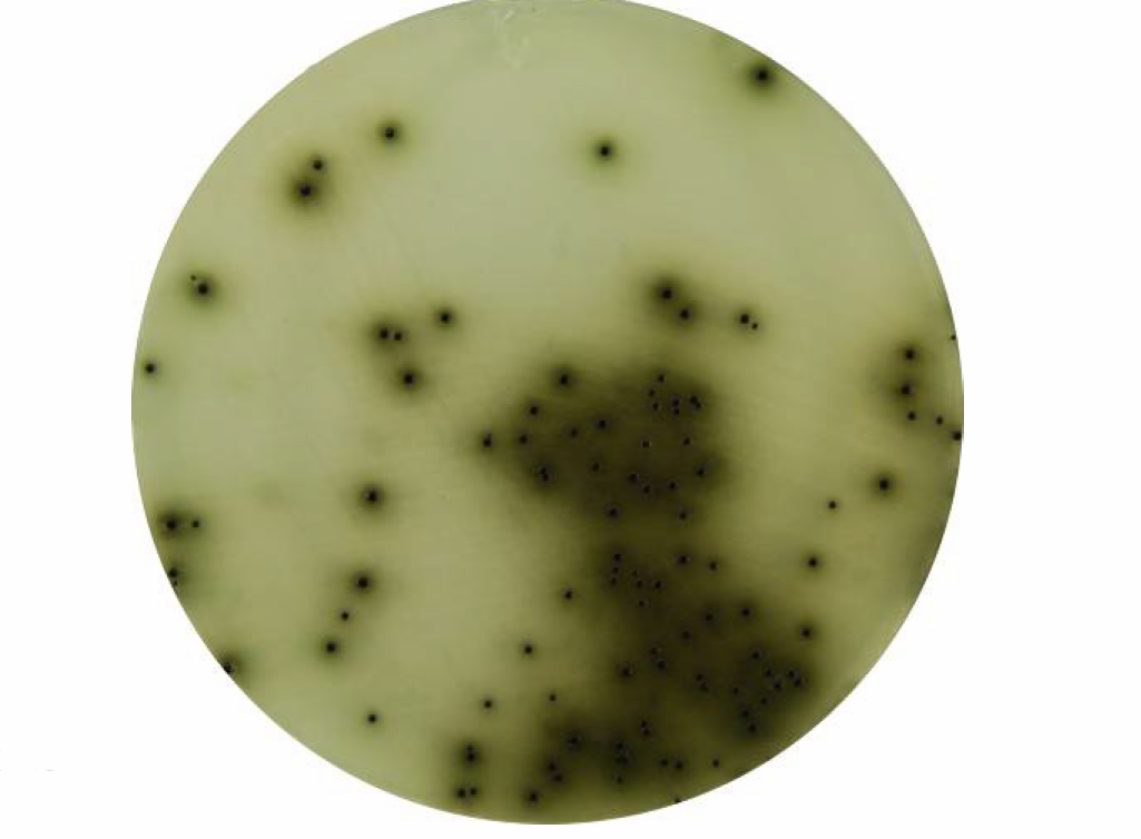 Figure: Bismuth Sulfite Agar
Uses of Bismuth Sulfite Agar
1. Primary isolation of Salmonella Typhi from clinical specimens BS agar is the preferred medium for S. Typhi isolation from:
- Stool specimens in suspected typhoid fever (second week of illness onward, the period of maximum fecal shedding)
- Stool specimens from typhoid carriers and contacts during outbreak investigation
- Urine specimens (rose spots, urine culture, S. Typhi may be shed in urine during the second and third weeks)
2. Subculture from enrichment broth For carrier specimens or specimens with low organism counts, subculture from selenite broth or Rappaport-Vassiliadis medium onto BS agar provides the optimal combination of enrichment followed by selective plating.
3. Non-typhoidal Salmonella from food and environmental samples BS agar is used in food safety microbiology for Salmonella detection in poultry, eggs, and dairy products. Its high selectivity suppresses the heavy coliform background typical of food specimens.
4. Reference and outbreak investigation laboratories BS agar is particularly valuable in reference laboratory settings where maximum S. Typhi sensitivity is required. For example, during typhoid outbreak investigations, when even a single positive culture from a carrier or contact has epidemiological significance.
Positioning relative to other enteric media: BS agar is the most selective enteric medium for Salmonella, but it is also the most inhibitory overall, including to some Salmonella variants (see Limitations). It should always be used alongside a less inhibitory medium such as XLD agar or HE agar to ensure recovery of the full range of enteric pathogens. It is never used as the sole medium.
Composition
Bismuth sulfite agar is selective due to the presence of inhibitors and is differential on the basis of hydrogen sulfide (H₂S) production. The presence of bismuth sulfite indicator and brilliant green inhibits the growth of Gram-positive bacteria and members of the coliform group while allowing Salmonella to grow luxuriantly.
| Ingredients | Gm / Litre |
|---|---|
| Peptone | 10 |
| Beef extract | 5 |
| Dextrose (Glucose) | 5 |
| Disodium phosphate | 4 |
| Ferrous sulphate | 0.3 |
| Bismuth sulphite indicator | 8 |
| Brilliant green | 0.025 |
| Agar | 20 |
| Final pH ( at 25°C) 7.7±0.2 |
Beef extract and peptones provide nitrogen, vitamins, and minerals needed for the growth of microorganisms.
Dextrose is the carbohydrate present that acts as a source of carbon. Disodium phosphate is the buffering agent which maintains the osmotic equilibrium.
Ferrous sulfate is an indicator for hydrogen sulfide production, which occurs when the H₂S produced by Salmonella reacts with the iron salt. This reaction causes a black or green metallic colony and brown or black precipitate whilst the reduction of bismuth ions to metallic bismuth produces a metallic luster around the colonies. Agar is the solidifying agent.
Note: Composition may vary according to the manufacturer. Some use enzymatic digest of casein and enzymatic digest of animal tissue instead of peptone. Glucose may be used as an alternative to dextrose. However, the inclusion purpose of each ingredient and quantity are the same.
Preparation of Media
- Suspend the required amount of dry powder (as per the manufacturer’s instruction) in 1000 mL of distilled water.
- Heat gently with frequent agitation until the medium just begins to boil and simmer for 30 seconds to dissolve the agar.
- DO NOT OVERHEAT. DO NOT AUTOCLAVE
- Cool to 50-55°C, mix well to disperse suspension, and pour plates (25 ml medium per plate).
- Allow the medium to solidify, store at 2-8°C until use and dry the prepared plates before use.
Critical shelf life limitation: Prepared bismuth sulfite agar plates must be used within 48 hours (2 days) of preparation. After 72 hours, the selective activity of the medium decreases significantly, and Salmonella recovery rates fall. BS agar cannot be prepared in large batches and stored for routine use like other media. It must be prepared fresh for each use or ordered from a manufacturer with a reliable cold chain. This is one of the most important practical differences between BS agar and other enteric media such as XLD, DCA, or MacConkey, which have 2–4 week shelf lives. If pre-prepared commercial plates are used, always check the expiry date and discard plates older than 2 days from opening.
The sensitivity of the medium depends largely upon uniform dispersion of precipitated bismuth sulphite in the final medium; therefore the medium should be mixed well in a way that there should be no sedimentation of the indicator.
Sample processing: Clinical specimens may be directly inoculated in the medium, or if the sample is considered to have a low organism load, it can be sub-cultured from an enrichment broth. For environmental samples, appropriate dilution or enrichment may be necessary before plating.
Inoculation and Incubation
- Allow the plate to reach room temperature and dry the agar surface before use.
- Inoculate directly from the specimen. Suitable specimens include stool, rectal swabs, urine, and, for food or water testing, the processed sample or its enrichment broth (for example, selenite broth subculture).
- Streak for well-isolated colonies using a four-quadrant streak. This step is not optional on BSA: some areas of the plate must be sparsely seeded, because S. Typhi in a heavily inoculated area can appear light green instead of black and be missed. The sparse quadrant is where the true black colony with metallic sheen appears.
- Incubate aerobically at 35 to 37°C.
- Read the plate at both 24 hours and 48 hours. S. Typhi often shows its black colony and sheen by 18 to 24 hours, but many non-typhi Salmonella do not blacken until 48 hours. Do not report the plate negative at 24 hours.
- BSA is highly selective and unusual in tolerating a larger inoculum than most enteric media, which improves recovery when the organism is present in low numbers, as in early typhoid or in carriers. Even so, always plate a sparse quadrant so the diagnostic colony morphology can develop.
- Read the metallic sheen by reflected light, ideally daylight, tilting the plate under the light rather than looking through it.
Colony Characteristics on Bismuth Sulfite Agar
The baseline medium color is brown to dark brown. H₂S producing organisms produce black colonies surrounded by a zone of darkened medium; metallic bismuth deposits create a distinctive metallic sheen in and around the colony.
| Organism | Colony appearance | Metallic sheen | Timing | Notes |
|---|---|---|---|---|
| Salmonella Typhi | Black "rabbit-eye" colonies surrounded by a black zone of darkened medium | Yes, pronounced metallic sheen (silver or golden) | Visible at 18–24 hrs; uniformly black at 48 hrs | "Rabbit-eye" = small black colony with a lighter ring around it resembling a rabbit's eye. Heavy inoculum warning: at very high inoculum density, S. Typhi may appear light green rather than black, do not dismiss green colonies as negative |
| Salmonella arizonae | Black colonies with brown zone | Yes | 18–24 hrs | One of only two enteric organisms producing brown zone in medium (along with S. Typhi) |
| Salmonella Paratyphi A | Variable. Green, clear, or mucoid; not typically black | Absent or faint | 18–48 hrs | Critical: S. Paratyphi A is H₂S-negative, does not produce black colonies. May appear greenish or colorless. Can be missed if only black colonies are picked |
| Salmonella Typhimurium and other non-typhi | Variable at 18 hrs (black, green, or clear mucoid); uniformly black at 48 hrs with widespread medium staining and metallic sheen | Yes at 48 hrs | Full expression at 48 hrs | Widespread darkening of surrounding medium is characteristic |
| Shigella flexneri and S. sonnei | Poor growth; if any, colorless or brown | No | — | Most Shigella inhibited; S. flexneri and S. sonnei may occasionally grow |
| Escherichia coli and other coliforms | Usually inhibited; if breakthrough occurs, dull green or brown colonies | No metallic sheen | — | Absence of metallic sheen distinguishes from Salmonella |
| Proteus spp. | Usually inhibited; occasional dull green or brown colonies | No metallic sheen | — | Distinguished from Salmonella by lack of metallic sheen and urease positivity |
| Serratia spp. | Usually inhibited; occasional dull green colonies | No | — | — |
The two-timepoint reading rule: Read BS agar plates at both 18–24 hours and 48 hours. Early reading (18–24 hrs) may show "rabbit-eye" S. Typhi colonies; 48-hour reading is required for non-typhi Salmonella, which may not fully express black pigmentation until the second day. Do not discard plates as negative after 24 hours alone.
The heavy inoculum trap: A heavy inoculum of S. Typhi produces light green rather than black colonies, this is one of the most commonly cited false-negative errors on BS agar. If a green colony appears on BS agar in a typhoid-suspected specimen, subculture it to TSI and test with Salmonella antisera before concluding it is a coliform.
Separately, some non-typhi Salmonella (Enteritidis, Cholerae-suis, Paratyphi) naturally produce green to brown colonies without the dark zone even at normal inoculum. So a green colony in a sparse, well-isolated area is more likely a genuine non-typhi Salmonella, while a green colony only in the heavy-streak area is likely S. Typhi that failed to blacken. Always read the sparse quadrant.
BS Agar in the Enteric Media Comparison
| Feature | BS Agar | XLD Agar | HE Agar | SS Agar |
|---|---|---|---|---|
| Selectivity level | Highest | Moderate-high | Moderate-high | High |
| Best for | S. Typhi specifically; low-count specimens | Broad Salmonella + Shigella | Shigella recovery | Salmonella in heavily contaminated specimens |
| S. Typhi sensitivity | Best | Good | Good | Good |
| S. Paratyphi A appearance | Greenish/colorless (no black), easy to miss | Red with no black center | Blue-green, no black center | Colorless, no black center |
| Shigella recovery | Poor, most inhibited | Good | Best | Poor |
| Shelf life | 2 days only | 2–4 weeks | 2–4 weeks | 2–4 weeks |
| Do not autoclave | Yes | Yes | Yes | Yes |
| Medium color | Dark brown | Bright red | Deep green | Pale |
| Salmonella colony | Black + metallic sheen | Red + black center | Blue-green + black center | Colorless + black center |
Practical recommendation: Use BS agar as a third plate alongside XLD and MacConkey when S. Typhi specifically is clinically suspected (typhoid fever, carrier screening, enteric fever outbreak). Its 2-day shelf life makes it unsuitable as a routine standing medium in most laboratories.
How to Remember
Two mechanisms, one goal, suppress everything except Salmonella:
- Brilliant green → inhibits Gram-positives and most coliforms (same as SS agar)
- Bismuth sulfite → additional inhibitor specific to this medium; bismuth ions are metabolically toxic to coliforms while Salmonella tolerates and even exploits them in the H₂S reaction
The metallic sheen, what it is and why it matters: The sheen is metallic bismuth (Bi⁰). H₂S produced by Salmonella reduces bismuth ions in the agar to elemental metallic bismuth, which deposits around the colony and reflects light like a metal surface. This is why BS agar colonies have a sheen reminiscent of a polished metal. No other common enteric medium produces this appearance.
The rabbit-eye analogy: S. Typhi colonies at 18–24 hours appear as a black center surrounded by a lighter ring, then a darker outer zone, resembling a cross-section of a rabbit's eye. This is one of the most visually distinctive colony descriptions in all of microbiology. If you see a "rabbit-eye" black colony with metallic sheen on a brown agar plate, think S. Typhi until proven otherwise.
The three traps:
- Heavy inoculum → S. Typhi turns green, not black. Pick green colonies too
- S. Paratyphi A → H₂S negative → no black center → appears greenish/colorless → easy to miss
- 2-day shelf life → plates lose selectivity → false growth of coliforms → always check preparation date
Where BS agar fits: BS agar answers one specific question better than any other enteric medium: "Is Salmonella Typhi present in this specimen?" For that question, it is the most sensitive tool available. For everything else (broad enteric pathogen workup, Shigella recovery, routine urine culture) other media are more appropriate.
Limitations
- Autoclaving/overheating the medium should be avoided as it destroys the selectivity of the medium.
- Due to its selectivity, Shigella species (except S. flexneri and S. sonnei) and certain Salmonella like S. Sendai, S. Berta, S. Gallinarum, S. Abortus are also inhibited.
- Incubation of bismuth sulfite agar plates at higher temperatures (i.e., 43ºC.) results in small, atypical Salmonella colonies and also reduces sensitivity showing significantly lower recovery.
- Although typical S. Typhi colonies usually develop within 24 hours, a further incubation up to 48 hours is required for growth of all typhoid strains.
- Prepared Bismuth Sulfite Agar plates should be used within 2 days. After 3 days of storage, there is a reduction in selectivity of the media, decreasing the number of Salmonella recovered.
- Colonies on bismuth sulfite agar may be contaminated with other viable organisms; therefore, isolated colonies should be subcultured onto a less selective medium (i.e., MacConkey Agar)
Where Students Get Confused
1. A green colony is not always a failure to grow. In a heavily streaked area, S. Typhi can appear light green instead of black, which is a classic false negative. But some non-typhi Salmonella naturally form green to brown colonies without a dark zone. Read the sparse, well-isolated quadrant: a green colony there is more likely a genuine non-typhi Salmonella, while a green colony only in the heavy-inoculum area is likely S. Typhi that did not blacken.
2. Do not call the plate negative at 24 hours. S. Typhi often shows its black colony and sheen by 18 to 24 hours, but non-typhi Salmonella may not blacken until 48 hours. Always hold and re-read at 48 hours before reporting negative.
3. A black colony is not proof of Salmonella. Some H₂S-producing coliforms, Proteus, and Citrobacter can form dark colonies that resemble Salmonella. The metallic sheen and brown-black zone favor S. Typhi, but confirm with TSI or KIA (these mimics ferment lactose and produce gas) and urease (Proteus is urease positive).
4. The metallic sheen needs the right light. The sheen is seen by reflected light, ideally daylight, not by looking through the plate. If you hold the plate up to the light you will miss it. Tilt the plate under a light source and look at the colony surface.
5. Never autoclave bismuth sulfite agar. Overheating destroys the bismuth sulfite indicator system and the selectivity. The medium is dissolved by boiling, cooled, and poured; it is never autoclaved. A plate that lost its brown color or selectivity was likely overheated.
Point 3 is the highest-value item because the black-colony-equals-Salmonella heuristic is the same trap that recurs across SS, DCA, HE, and now BSA, and the metallic sheen is the specific discriminator BSA adds.
References
- Gordeĭko, V. A., & Shustrova, N. M. (1989). The use of bismuth-sulfite-agar as a solid differential medium for isolating bacteria of the species Y. enterocolitica and Y. frederiksenii. Laboratornoe delo, (4), 64–65.
- Allen, S. B., Firstenberg-Eden, R., Shingler, D. A., Bartley, C. B., & Sullivan, N. M. (1993). Evaluation of Stabilized Bismuth Sulfite Agar for Detection of Salmonella in Foods. Journal of food protection, 56(8), 666–671. https://doi.org/10.4315/0362-028X-56.8.666
- Tille, P. M. (2022). Bailey and Scott's Diagnostic Microbiology (15th ed.). Elsevier.
- Cheesbrough, M. (2006). District Laboratory Practice in Tropical Countries, Part 2 (2nd ed.). Cambridge University Press.
- Wilson, W. J., & Blair, E. M. (1931). Use of a glucose-bismuth sulphite-iron medium for the isolation of B. typhosus and B. paratyphosus B. Journal of Hygiene, 31(2), 138–149.

Tankeshwar Acharya, MSc (Medical Microbiology)
Tankeshwar Acharya is an Assistant Professor in the Department of Microbiology at Patan Academy of Health Sciences (PAHS), Nepal, where he has been teaching and practicing clinical microbiology for over 14 years. He is the founder of Microbe Online, one of the leading free microbiology education resources on the web, covering bacteriology, mycology, parasitology, immunology, and clinical laboratory diagnostics written from direct experience in both the classroom and the diagnostic laboratory.
Comments
No comments yet. Be the first to share your thoughts.
Leave a comment
All comments are reviewed before they appear.