Salmonella: Properties, Disease, Lab Diagnosis
Salmonella: Properties, Disease, Lab Diagnosis
Salmonella is a Gram-negative, rod-shaped, motile bacilli that moves with the use of its peritrichous flagella. The genus Salmonella can be divided into two species (S. enterica and S. bongori), based on their phenotypic profile. The genus Salmonella is a member of the family Enterobacteriaceae. Salmonella is one of the world’s most common causes of foodborne diseases.
Most human diseases are caused by bacteria belonging to the subspecies Salmonella enterica. Salmonella lives in the intestine of many animals such as cows, dogs, pigs, and birds but Salmonella typhi only lives in humans.
Human-acquired Salmonella infection by;
- consuming contaminated meat or animal products (eg. eggs)
- direct contact with infected animals or environments
- contaminated food and water, utensils, hands of someone who handles food.
Thphoidal vs. Non-Typhoidal Salmonella
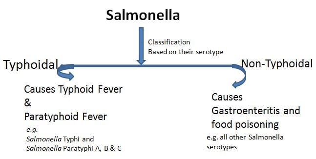 Figure: Classification of Salmonella
Salmonella bacteria are classified as either “typhoidal” or “nontyphoidal,” based on their serotype.
Main diseases caused by Salmonella
- Enteric fever (Typhoid fever and Paratyphoid fever)
- Enterocolitis
- Septicemia
- Osteomyelitis
Important properties of Salmonella species
- Gram-negative rods
- Do not ferment lactose
- Antigens of Salmonella speciesa. Cell wall O antigenb. Flagellar H antigenc. Capsular Vi (virulence) antigen)
Laboratory Diagnosis of Typhoid Fever
A. CULTURE
| Organism | Slant | Butt | H 2 S | Gas |
|---|---|---|---|---|
| S.typhi | Alk | Acid | + (weak) | -ve |
| S.paratyphi A | Alk | Acid | -ve | +ve |
| Other Salmonella spp | Alk | Acid | variable | variable |
Blood culture is the mainstay for the diagnosis of Typhoid fever. Presence of specific antibodies against Salmonella and or presence of characteristics signs & symptoms can be suggestive of typhoid fever but not definitive.
Definitive diagnosis of typhoid fever depends on the isolation of S.typhi from blood or bone marrow aspirate culture.
Sample
- Blood
- Bone marrow aspirate
- Duodenal aspirate
- Stools (especially useful for the diagnosis of typhoid carriers).
In this post, I am discussing the isolation and identification of Salmonella from blood culture only.
For blood culture
Salmonella is present in the blood of more than 80% of patients with typhoid fever. The overall volume of blood cultured is critical to increase yield (isolation rate) of the causative pathogen. Reducing the blood volume reduces the sensitivity of the blood culture.
Timing:
- Blood for culture should be taken before the patient is given antimicrobial therapy.
- Patients with a history of fever for 7 to 10 days are more likely than others to have a positive blood culture.
Volume:
- 10-15 ml from school children & adults
- 2-4 ml from toddlers and preschool children (Remember- children have a higher level of bacteremia than adults).
The optimum ratio of the volume of blood to traditional culture broth is 1:10 (e.g. 5 ml blood in 45 ml broth). For a commercial blood culture system, please read and use recommended amounts of blood.
Blood should be inoculated immediately into a blood culture bottle at the time of drawing blood using the same syringe that has been used for collection. Inoculated culture bottles should be incubated at 37°C
Do not refrigerate the sample or keep in cool places during transport.
Reading and reporting blood culture results for Typhoid fever
Check the inoculated culture bootles for turbidity, gas formation, and other evidence of growth after 1, 2, 3, and 7 days.
- For days 1, 2, and 3, only bottles showing signs of positive growth are cultured on agar plates (commonly used media for subculturing are Blood Agar, Chocolate Agar, and MacConkey Agar)
- On day 7 all bottles should be subcultured before being discarded as negative.
Subculture plates should be incubated at 37°C for 18-24 hours in an aerobic incubator. If growth is observed in the culture plates, colony morphology should be noted and biochemical tests performed to identify the isolate.
Remember, S.typhi is not the only bacterial pathogen found in the blood. Find information about organisms that are commonly isolated from blood
Colony characteristics
- Blood agar: S.typhi and S. paratyphiusually produce non-hemolytic smooth white colonies on blood agar.
- MacConkey agar: Salmonellae produce lactose non-fermenting smooth colonies on MacConeky agar.
Salmonella Typhi gives K/A with trace H2S in KIA or TSI medium.
Biochemical characteristics
Suspected colonies obtained on the above culture media are screened using the following media/tests:
Results of Salmonella spp in Triple Sugar Iron (TSI) Agar
Organism | Slant | Butt | H2S | Gas |
S.typhi | Alk | Acid | + (weak) | -ve |
S.paratyphi A | Alk | Acid | -ve | +ve |
Other Salmonella spp | Alk | Acid | variable | variable |
Results of Salmonella spp in Motility Indole Urease (MIU) medium and citrate utilization test
Organism | Motility | Indole | Urease | Citrate utilization test |
S.typhi | +ve | -ve | -ve | -ve |
S.paratyphi A | +ve | -ve | -ve | +ve |
Other Salmonella spp | +ve | -ve | -ve | variable |
Note: V= variable; Alk= Alkaline
The salmonellae that cause typhoid and paratyphoid fever have the following antigenic compositions.
Serotype | O Antigen | H Antigen | Serogroup Phase 1:2 |
S. typhi | 9, 12, (vi) | d: | Group D1 |
S. paratyphi A | 1, 2, 12 | a: (1, 5) | Group A |
S. paratyphi B | 1, 4, (5), 12 | b: 1, 2 | Group B |
S. paratyphi C | 6, 7, (vi) | C: 1, 5 | Group C1 |
Isolated Salmonellae can be characterized by detecting the presence of their somatic (O) and flagellar (H) antigens using a specific antiserum. Specific O antigen for S.typhi is O9, O2 for S.paratyphi A, O4 for S.paratyphi B, and O6/7 for S.paratyphi C.
B. Widal Test
This test has only moderate sensitivity and specificity. It can be negative in up to 30% of culture-proven cases of typhoid fever. Widal test measures agglutinating antibody levels against O and H antigens. As S.typhi shares O and H antigens with other Salmonella serotypes and has cross-reacting epitopes with other Enterobacteriaceae, false-positive Widal test results may occur in other clinical conditions such as malaria, typhus fever, bacteremia caused by other organisms as well.
References
- Forbes, S., Sahm, D. F., & Weissfeld, A. S. (2002). Bailey & Scott’s Diagnostic Microbiology. Mosby.
- Coburn, B., Grassl, G. A., & Finlay, B. B. (2007). Salmonella, the host and disease: a brief review. Immunology and cell biology, 85(2), 112–118. https://doi.org/10.1038/sj.icb.7100007
- Giannella, R. A. (1996). Salmonella. In S. Baron (Ed.), Medical Microbiology. (4th ed.). University of Texas Medical Branch at Galveston.

Tankeshwar Acharya, MSc (Medical Microbiology)
Tankeshwar Acharya is an Assistant Professor in the Department of Microbiology at Patan Academy of Health Sciences (PAHS), Nepal, where he has been teaching and practicing clinical microbiology for over 14 years. He is the founder of Microbe Online, one of the leading free microbiology education resources on the web, covering bacteriology, mycology, parasitology, immunology, and clinical laboratory diagnostics written from direct experience in both the classroom and the diagnostic laboratory.