
Total Laboratory Automation (TLA) in Clinical Microbiology: How It Works, Benefits, and Limitations

On this page
For twenty years, clinical chemistry labs ran on automated lines while microbiology stayed stubbornly manual: hands streaking plates, carrying them to incubators, pulling them out to read, carrying them back. The reason was not reluctance.
Microbiology is harder to automate because it works with living, growing organisms on solid media, not just liquid samples in tubes. But that manual workflow hides a quiet cost. Every time a plate leaves the incubator to be read or stained, its growth stalls and fastidious organisms may be lost.
Total laboratory automation changes this by never taking the plate out at all: it images the plate through the incubator wall, so growth is never interrupted. That single change, keeping plates continuously incubated, is where most of TLA's benefit comes from.
What TLA is
Total laboratory automation (TLA) is a system that automates culture-based testing in the clinical microbiology laboratory, linking specimen processing, inoculation, incubation, and imaging into one connected workflow. Its goals are to improve quality and consistency, shorten turnaround time, handle rising specimen volumes, and offset shortages of skilled staff. Because it automates work with living cultures on solid media, not just liquid samples, it arrived in microbiology decades later than in clinical chemistry.
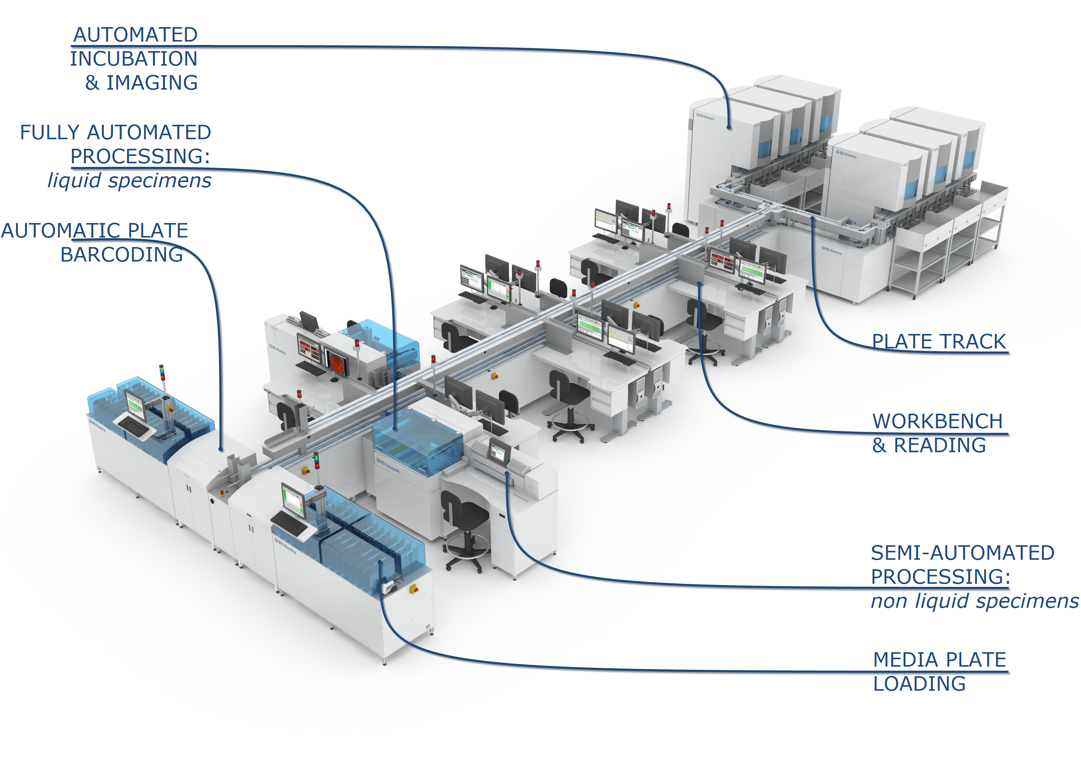 Figure: BD Kiestra (TLA in clinical Microbiology)
TLA is provided by a small number of manufacturers as conveyor-linked modular systems. The two established platforms are BD Kiestra and the bioMérieux/Copan WASPLab; both connect specimen processing, inoculation, incubation, and digital imaging along a conveyor.
Why uninterrupted incubation is the core benefit
Most of what makes TLA valuable traces back to one mechanism: the plate never leaves the incubator.
The manual problem. In a traditional workflow, a plated culture spends, on average, around three hours outside the incubator over its life in the lab, pulled out repeatedly for reading, Gram staining, and biochemical work, while the incubator door opens and closes many times. Each interruption cools the plate and shifts its atmosphere, which slows growth and can cause fastidious organisms to be missed entirely.
The TLA solution. In a TLA system, plates are incubated continuously in an optimal atmosphere and temperature, and are read as high-resolution digital images taken through the system without removing the plate. Growth is never interrupted. Because the images are enlarged and captured under several lighting conditions, even small colonies that a technologist might overlook are visible.
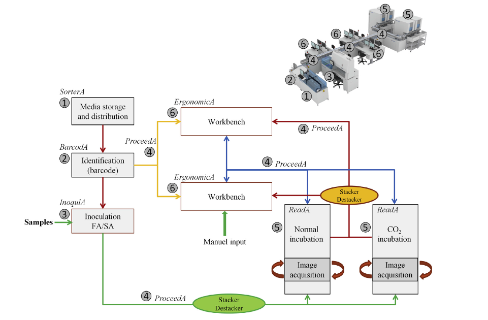 Figure: BD Kiestra TLA system workflows
The payoff, with evidence. Uninterrupted incubation improves the recovery of fastidious organisms. Laboratories implementing TLA have reported markedly improved recovery of pathogens such as Staphylococcus saprophyticus and Neisseria gonorrhoeae from urine samples.
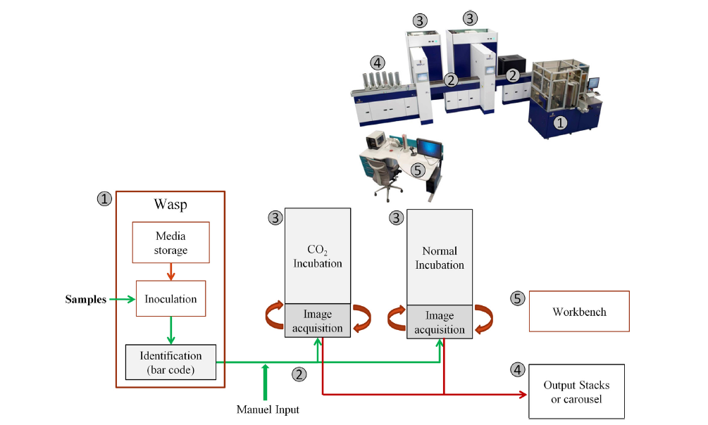 Figure: WASPLab system workflow
And because organisms grow faster and more reliably when never disturbed, TLA reduces turnaround time: the median time to a final result drops, so infections like sepsis and meningitis can be treated with the right antibiotic hours earlier. Earlier correct treatment means shorter hospital stays and lower morbidity, mortality, and cost. This turnaround-time gain is the clinical heart of the TLA case, and it flows directly from not interrupting incubation.
Other benefits
Beyond the incubation mechanism, TLA brings several efficiency and quality gains:
Frees skilled staff for skilled work. Technologists spend much of their day on tasks that need no special expertise, sorting, moving, and streaking plates, and setting up susceptibility tests. Automating these lets staff concentrate on the work that does need judgment: reading plates and working up cultures.
Uniform streaking. Streaking patterns vary widely between technologists, which affects how well colonies separate and how colony counts turn out. TLA spreads specimens across the plate uniformly, improving consistency and isolation. (BD Kiestra, for example, uses a rolling-bead streaking method that its maker reports yields more isolated colonies than loop streaking and reduces the need for subculture.)
Fewer errors. Automating steps like plate labeling cuts human errors and improves the accuracy of results.
A teaching resource. Because every plate is digitally imaged and archived, the images become a teaching library. Students can compare thousands of archived plates and even review how a colony looked at earlier time points, which is valuable where a lab is attached to a medical or technical school.
How a TLA system is built
Regardless of manufacturer, a TLA system chains the same functions along a conveyor: media storage and sorting, barcoding, automated specimen inoculation and streaking, conveyor transport, continuous incubation with digital imaging, and workbenches for the manual steps that remain (reading, identification, susceptibility testing). Plates that need follow-up work are routed automatically to those workbenches; the rest are handled digitally.
Identification can be integrated by linking MALDI-TOF (MS) into the workflow, and automated susceptibility testing and molecular diagnostics are being added to these platforms. With digital imaging plus automated MALDI-TOF identification, the proportion of plates needing any manual handling falls substantially.
The two main TLA platforms
BD Kiestra. Kiestra is a conveyor-linked, modular TLA system that connects specimen processing, automated inoculation, continuous incubation, and digital imaging. Its inoculation uses a rolling-bead streaking method, in which sterile beads roll across the plate to spread the specimen; the manufacturer reports this yields more isolated colonies than manual loop streaking and reduces the need for subculture. Plates are incubated in smart incubators and imaged through the system, and images are read at connected workstations.
bioMérieux / Copan WASPLab. WASPLab pairs the WASP (Walk-Away Specimen Processor) front end with a conveyor-linked incubation and digital-imaging line. It performs automated planting and streaking, routes plates to smart incubators, and captures high-resolution images that can be read on screen, with software that can pre-sort plates into "growth" and "no growth" to reduce the reading workload.
Both platforms automate the same core sequence, and the choice between them turns on laboratory layout, specimen mix, throughput, and the identification and susceptibility systems a lab wants to connect. The underlying principle, continuous incubation with imaging so plates are never removed, is identical.
Digital imaging
Digital imaging is the component that makes uninterrupted incubation possible, so it deserves its own note. A high-resolution camera photographs each plate at intervals the lab sets, without removing it from the incubator. The images can be read and annotated on screen for downstream identification or susceptibility testing.
Because the software captures several exposures and angles, it can often make simple calls automatically, most importantly growth versus no growth, and flag only the plates that need a human. All of a patient's specimens (for example urine, sputum, and resistance screening) can be reviewed together, and every image is archived for later teaching or quality control.
Limitations
TLA is powerful but not universally appropriate, and the trade-offs are real.
Not for all specimen types. Automated systems handle liquid specimens (such as urine) most efficiently. Specimens needing special processing, like tissue or organ biopsies, do not fit an automated line easily. Liquid-based transport systems help by converting swabs into liquid specimens the system can process, which extends TLA's reach.
High cost, not for every lab size. The initial investment is very high, and ongoing costs for supplies, space, and infrastructure add to it. For a small laboratory, these costs may outweigh the benefits. And the more complex the system, the greater the risk and impact of a failure.
Consequences of system failure. Because TLA centralizes the workflow, a breakdown can be crippling. Service and maintenance contracts with fast response are essential, and a lab should keep a minimal manual backup (conventional incubators and test systems) so that a major failure does not shut down testing entirely.
Dependency and skill loss. Relying on automation for routine work can erode staff proficiency in manual methods over time. During downtime, urgent samples must be processed manually, and if those skills have faded, patient care can suffer. Maintaining manual competency alongside automation matters.
How to Remember
The plate never leaves the box. The one idea that carries most of TLA's benefit. Continuous incubation with imaging through the system means growth is never interrupted, which is why fastidious organisms are recovered better and results come faster. Manual workflow pulls plates out repeatedly; TLA never does.
Three hours out equals organisms lost. The manual cost in one line. A plate spends around three hours outside the incubator over its life in a manual lab, and that stalled growth is what loses fastidious organisms like N. gonorrhoeae. TLA closes that gap to zero.
Faster growth, faster treatment. Uninterrupted incubation shortens turnaround time, so sepsis and meningitis get the right antibiotic hours earlier. The mechanism (no interruption) and the clinical payoff (earlier treatment) are directly linked.
The catch is cost and dependency. TLA is expensive and centralizes everything, so a failure hurts, and staff can lose manual skills. Big benefits, but only for high-volume labs that keep a manual backup.
Key exam facts in one table
| Concept | Fact to remember |
|---|---|
| What TLA is | Conveyor-linked automation of culture-based microbiology: processing, inoculation, incubation, imaging |
| Why it came late to micro | It automates work with living cultures on solid media, harder than automating liquid samples |
| Core mechanism | Continuous incubation with digital imaging; plates are never removed, so growth is never interrupted |
| Key recovery benefit | Better recovery of fastidious organisms (e.g. Staphylococcus saprophyticus, Neisseria gonorrhoeae) |
| Key clinical benefit | Reduced turnaround time, earlier correct treatment of sepsis and meningitis |
| Uniform streaking | Automated streaking is more consistent than manual, improving colony isolation |
| Identification integration | MALDI-TOF can be linked for organism identification |
| Main platforms | BD Kiestra and bioMérieux/Copan WASPLab |
| Main limitations | High cost, poor fit for biopsy specimens, risk if the system fails, and staff skill loss |
Where Students Get Confused
The main benefit is uninterrupted incubation, not just speed of handling. TLA's biggest gain comes from never removing plates from the incubator, which improves growth and pathogen recovery. Faster plate handling matters, but the continuous-incubation effect is the core.
TLA does not replace the microbiologist. It automates sorting, streaking, moving, and imaging, and can auto-call simple growth/no-growth decisions, but reading complex plates, identification, and interpretation still need trained staff. It redirects expertise, it does not remove it.
Why fastidious organisms benefit most. Organisms like N. gonorrhoeae are sensitive to cooling and atmosphere changes. Every time a plate leaves a manual incubator, these organisms suffer. Continuous incubation is exactly what they need, which is why their recovery improves most under TLA.
TLA is not for every lab or every specimen. It suits high-volume labs and liquid specimens. It fits poorly with biopsy and tissue specimens, and its high cost makes it hard to justify for small laboratories.
Automation creates a new risk: dependency. Centralizing the workflow means a system failure can halt testing, and over-reliance can erode manual skills. Labs must keep a manual backup and maintain staff competency, which is why dependency is listed as a genuine limitation.
References
- Croxatto A, Prod'hom G, Faverjon F, Rochais Y, Greub G. Laboratory automation in clinical bacteriology: what system to choose? Clin Microbiol Infect. 2016;22(3):217-235. doi:10.1016/j.cmi.2015.09.030
- Croxatto A, Dijkstra K, Prod'hom G, Greub G. Comparison of inoculation with the InoqulA and WASP automated systems with manual inoculation. J Clin Microbiol. 2015;53(7):2298-2307. doi:10.1128/JCM.03076-14
- Lippi G, Da Rin G. Advantages and limitations of total laboratory automation: a personal overview. Clin Chem Lab Med. 2019;57(6):802-811. doi:10.1515/cclm-2018-1323
- Lainhart W, Burnham CA. Enhanced recovery of fastidious organisms from urine culture in the setting of total laboratory automation. J Clin Microbiol. 2018;56(8):e00546-18. doi:10.1128/JCM.00546-18
- Zhang W, Wu S, Deng J, et al. Total laboratory automation and three shifts reduce turnaround time of cerebrospinal fluid culture results in the Chinese clinical microbiology laboratory. Front Cell Infect Microbiol. 2021;11:765504. doi:10.3389/fcimb.2021.765504
Frequently Asked Questions
What is total laboratory automation (TLA) in microbiology?
What is total laboratory automation (TLA) in microbiology?
TLA is an integrated system that connects the individual steps of the microbiology workflow, specimen processing, plating, incubation, digital imaging, and colony reading, into one automated, track-linked line. It is not a diagnostic test; it is the infrastructure that moves and processes specimens through the laboratory with minimal manual handling.
Why does TLA improve the recovery of fastidious organisms?
Why does TLA improve the recovery of fastidious organisms?
Because plates are incubated continuously and imaged without being removed, growth is never interrupted. In a manual workflow, plates are repeatedly taken out to be read and stained, cooling them and changing their atmosphere, which can cause delicate organisms like Neisseria gonorrhoeae to be missed. Uninterrupted incubation preserves these organisms, improving recovery.
How does TLA reduce turnaround time?
How does TLA reduce turnaround time?
Uninterrupted incubation lets organisms grow faster and more reliably, and digital imaging allows plates to be read as soon as growth appears without waiting to physically retrieve them. This shortens the median time to a final result, so serious infections can be treated with the correct antibiotic hours earlier.
How does digital imaging work in TLA?
How does digital imaging work in TLA?
A high-resolution camera photographs each plate at set intervals without removing it from the incubator. Staff read and annotate the images on screen, and the software can make simple calls such as growth versus no growth automatically, flagging only the plates that need human review. Images are archived for teaching and quality control.
What are the main limitations of TLA?
What are the main limitations of TLA?
It requires a very high initial investment, so it suits high-volume labs more than small ones. It handles liquid specimens well but not tissue or biopsy specimens. A system failure can halt testing, so a manual backup and service contract are essential. And reliance on automation can erode staff skills in manual methods over time.
What are the main TLA systems used in microbiology?
What are the main TLA systems used in microbiology?
The two most widely known are BD Kiestra and COPAN WASPLab. Both connect automated specimen processing and plating with smart incubation and digital plate imaging, so colonies can be read on a screen rather than by handling each plate.
Why is automation in microbiology different from other laboratory disciplines?
Why is automation in microbiology different from other laboratory disciplines?
Microbiology works with living cultures that need incubation over hours to days, with plate handling and visual colony reading, rather than the liquid samples and immediate instrument readings of chemistry or hematology. TLA is built around that difference, its key advantage is uninterrupted incubation and digital imaging, which manual workflows interrupt every time a plate is removed to be examined.
Is TLA the same as an automated blood culture system?
Is TLA the same as an automated blood culture system?
No. An automated blood culture system (such as BACTEC) monitors blood culture bottles for growth. TLA is the broader workflow automation that handles plated specimens through processing, incubation, and reading. They automate different parts of the laboratory.

Tankeshwar Acharya, MSc (Medical Microbiology)
Tankeshwar Acharya is an Assistant Professor in the Department of Microbiology at Patan Academy of Health Sciences (PAHS), Nepal, where he has been teaching and practicing clinical microbiology for over 14 years. He is the founder of Microbe Online, one of the leading free microbiology education resources on the web, covering bacteriology, mycology, parasitology, immunology, and clinical laboratory diagnostics written from direct experience in both the classroom and the diagnostic laboratory.
Comments
No comments yet. Be the first to share your thoughts.
Leave a comment
All comments are reviewed before they appear.