
RCUT for Neisseria: The Maltose Result That Separates Gonorrhoeae from Meningitidis

On this page
Why It Matters
Two oxidase-positive, Gram-negative diplococci land on a bench looking almost identical: Neisseria gonorrhoeae and Neisseria meningitidis. Under the microscope they are indistinguishable. On chocolate agar they can look the same. But the two organisms could hardly be more different clinically. One is a sexually transmitted pathogen that triggers partner notification and contact tracing. The other is a cause of life-threatening meningitis and septicemia, sometimes carried harmlessly in the throat, that occasionally turns up at a genital site and must not be misreported as gonorrhoea.
The test that separates them is sugar utilization, and the single most important result is maltose: N. gonorrhoeae uses glucose only, while N. meningitidis uses glucose and maltose. One letter of the pattern changes the diagnosis, the treatment, and the public-health response.
The rapid carbohydrate utilization test (RCUT) is the modern way to read that pattern. It replaced the old cystine trypticase agar (CTA) sugars because CTA was slow (24 to 72 hours) and prone to a specific failure: Neisseria produce very little acid, and the peptone in CTA generates alkaline ammonia that can neutralize what little acid there is, giving false negatives. RCUT fixes this by using a buffered, peptone-free saline so the small amount of acid actually registers, and it delivers a result in 4 hours.
This article covers how RCUT works, how to read the sugar panel that separates the pathogenic and commensal Neisseria, and the traps that produce a wrong answer.
Rapid carbohydrate utilization tests is also done for the identification of Aggregatibacter aphrophilus (formerly called Haemophilus aphrophilus, one of the HACEK organisms),A. actinomycetemcomitans and Gardnerella vaginalis.
Traditionally, cystine trypticase agar (CTA) supplemented with a 1% carbohydrate solution was used to determine acid production by Neisseria and related species. This test is no longer recommended now and has been replaced by the rapid carbohydrate utilization test (RCUT). In this test, patterns of acid production from the carbohydrates – glucose, maltose, lactose, sucrose, (and fructose) – are used to identify Neisseria and related species. Production of acid from carbohydrates is indicated by a change in the color of the phenol red indicator from red to yellow.
Principle
Small (0.10 mL) aliquots of phosphate-buffered saline solution containing phenol red as the pH indicator are distributed in a series of tubes. These tubes are labelled as glucose, maltose, sucrose, and lactose.
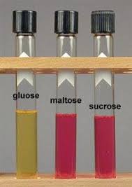 Figure: Neisseria gonorrhoeae test result
Single drops of the individual carbohydrates (20% w/v) are added to individual tubes according to the labelling e.g. 20% w/v of glucose is added in the tube labelled as glucose.
A heavy suspension of the test isolate is prepared in the buffer without carbohydrates. One drop of this suspension is added to each of the labelled tubes.
Neisseria species utilize the carbohydrate source present in the tubes and produce acid by an oxidative pathway (which is in contrast to other organisms which produces acid by fermentative pathway). The acid thus produced when exceeds the buffering capacity of the solution, the color of the medium changes from red (alkaline) to yellow (acid).
This oxidative (not fermentative) acid production has two practical consequences that shaped the test:
- Neisseria make very little acid. Oxidation yields far less acid than fermentation. So the medium must be sensitive: a low buffering capacity and a peptone-free base, so that the small amount of acid produced is enough to tip the phenol red from red to yellow. This is why RCUT uses buffered saline with the carbohydrate, not a rich peptone medium.
- Peptone would sabotage the result. Many Neisseria produce ammonia from peptone, which is alkaline and neutralizes the weak acid. A peptone-containing medium can therefore give a false negative. Removing the peptone removes that neutralizing effect. This is the core reason RCUT replaced the older CTA sugars.
A related trap involves CO₂: the test must not be incubated in a CO₂ atmosphere, because dissolved CO₂ forms carbonic acid and can turn the indicator yellow with no carbohydrate utilization at all, a false positive. (The article's procedure note already flags this; it follows directly from the low buffering capacity that makes the test sensitive.)
Media and Reagents
A. Phosphate Buffered Saline Solution (PBS) or Balanced Salt Solution (BSS) (a 10× stock solution having the following formula)
- Dipotassium phosphate (K2HPO4): 0.40 g
- Monopotassium phosphate (KH2PO4): 0.10 g
- Potassium chloride (KCl): 8.0 g
- Distilled water: 100 mL
Filter-sterilize and store at 4°C–8°C
B. Working Solution
Add 10 mL of the 10× BSS to 90 mL of distilled water. After this, 0.5–0.8 mL of a 1% aqueous solution of phenol red is added to the solution, so that the final product is “cherry red.” This working solution is then filter-sterilized.
To ensure that the distilled water has the proper pH, the use of pharmacy-grade sterile distilled water is recommended.
C. Stock Carbohydrate Solutions
Weigh out 10 g each of glucose, maltose, sucrose, and lactose individually. Each is dissolved in 50 mL of distilled water. The solutions are filter-sterilized, dispensed into sterile vials, and frozen at −20°C.
It is important that “reagent-grade” carbohydrates be used, because the maltose from some bacteriologic media vendors may be contaminated with glucose.
Sample: Pure culture (18h-24h) of Gram-negative, oxidase-positive, catalase-positive diplococci grown on nonselective (chocolate agar or equivalent) medium.
Procedure
Carbohydrate tube preparation
- Label a series of sterile 12 × 75-mm tubes with the carbohydrate to be tested (i.e. glucose, maltose, sucrose, and lactose) and add 0.10 mL of working BSS in each tube.
- Add a single drop of each of the carbohydrates to the appropriately labeled tube with a Pasteur pipette.
Inoculum Preparation
- Take a sterile tube and label it with the isolate or specimen number.
- Dispense 0.30–0.40 mL of BSS in it.
- Transfer few isolated colonies of the test organism (18-24 hour culture from chocolate agar) with a sterile bacteriologic loop.
- Vortex the tube carefully to obtain a uniform, heavy suspension.
Mixing and Incubation
- Add a single drop of inoculum suspension to each of the carbohydrate-containing tubes.
- Agitate the tubes briefly to ensure thorough mixing.
- Place the tubes in an aerobic incubator or water bath at 35°C for 4 hours.
Note: Carbohydrate tubes should NOT be incubated in an incubator with carbon dioxide-supplemented atmosphere. Carbon dioxide may diffuse into the medium and form carbonic acid which may cause a false-positive acid reaction.
Results and Interpretations
The development of yellow color in any of the carbohydrate-containing tubes indicates utilization of that carbohydrate by the organism. Negative carbohydrate-utilization tests remain red or red-orange.
| Organism | Glucose | Lactose | Maltose | Sucrose |
|---|---|---|---|---|
| Neisseria gonorrhoeae | Yes | No | No | No |
| Neisseria meningitidis | Yes | No | Yes | No |
| N. lactamica | Yes | Yes | Yes | No |
| N. mucosa | Yes | No | Yes | Yes |
| M. catarrhalis | No | No | No | No |
Acid production patterns of Neisseria species and M. catarrhalis
Commercial Application
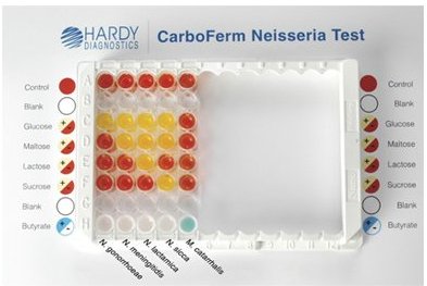 Figure: CarboFerm Neisseria test result
CarboFerm Neisseria Kit from Hardy Diagnostics is a rapid method for the identification of N. gonorrhoeae, N. meningitidis, N. sicca, N. lactamica and Moraxella catarrhalis isolates. CarboFerm Neisseria kit consists of an 8-well reagent cuvette that comes packaged as 12 strips snapped into a plastic frame. Based on the principle of the rapid carbohydrate utilization test, this kit gives results within 4 hours.
Where students get confused
Incubating in a CO₂ incubator. The single most common error. Neisseria love CO₂ for primary growth, so the instinct is to put everything in the CO₂ incubator. But dissolved CO₂ forms carbonic acid, which turns the phenol red yellow with no sugar utilization, a false positive across every tube. RCUT is incubated in a plain aerobic incubator or water bath, no CO₂.
The overoxidation trap (N. cinerea looks glucose-negative). Some Neisseria, notably N. cinerea, do use glucose but then rapidly over-oxidize the acid all the way to CO₂, so acid never accumulates in the tube. The result reads glucose-negative even though the organism used the glucose. N. cinerea can therefore be mistaken for a non-saccharolytic organism or misidentified. If a presumptive Neisseria reads negative on everything but does not fit M. catarrhalis, consider overoxidation.
Maltose contaminated with glucose. Reagent-grade carbohydrates matter. Maltose from some suppliers is contaminated with free glucose, which can cause a N. gonorrhoeae (glucose-only) isolate to read falsely maltose-positive, mimicking N. meningitidis. Since the maltose result is the critical gonorrhoeae/meningitidis discriminator, this contamination directly causes misidentification. Use reagent-grade maltose.
Reading a weak acid producer as negative. Some N. gonorrhoeae strains are weak acid producers and can appear glucose-negative in a poorly buffered or peptone-containing medium. This is exactly what RCUT's peptone-free, low-buffer design is meant to prevent, but a too-light inoculum can reproduce the problem. Use a heavy suspension.
Skipping the prerequisite tests. RCUT interpretation assumes the organism is already known to be an oxidase-positive, Gram-negative diplococcus. Running it on an unconfirmed isolate is meaningless. Gram stain and oxidase come first.
Confusing utilization with fermentation. The reason this is called "carbohydrate utilization" and not "fermentation" is that Neisseria oxidize the sugar aerobically rather than ferment it anaerobically. The result looks the same (acid, yellow) but the mechanism is different, which is why the medium and incubation conditions differ from a standard sugar fermentation tube.
See the carbohydrate fermentation test for the fermentative version.
How to remember
Maltose is the meningococcus letter. The one result that matters most: *N. meningitidis* uses maltose; N. gonorrhoeae does not. One M matches the other. Glucose is positive for both, so glucose alone tells you nothing; maltose is the discriminator.
Lactamica adds lactose. Walk up the ladder: gonorrhoeae = glucose only; meningitidis = glucose + maltose; lactamica = glucose + maltose + lactose (the name tells you). N. lactamica is the non-pathogenic throat commensal that would otherwise be mistaken for meningococcus; lactose (or a positive ONPG) separates them.
No sugars at all = Moraxella catarrhalis. M. catarrhalis is asaccharolytic: red across the board. If every tube stays red on an oxidase-positive Gram-negative diplococcus, think M. catarrhalis (confirm with DNase and butyrate esterase).
No CO₂ in the incubator. Neisseria grow in CO₂, but RCUT must not, because CO₂ makes carbonic acid and false-positive yellow. Grow in CO₂, test out of it.
References and further reading
- Procop GW, Church DL, Hall GS, Janda WM, Koneman EW, Schreckenberger PC, Woods GL. Koneman's Color Atlas and Textbook of Diagnostic Microbiology. 7th ed. Philadelphia: Wolters Kluwer; 2017.
- Kellogg DS, Holmes KK, Hill GA. Cumitech 4. In: Marcus S, Sherris JC, eds. Laboratory Diagnosis of Gonorrhea. Washington, DC: American Society for Microbiology, 1976.
- Acid detection test, Centers for Disease Control and Preventions.
- Kulkarni, S., Bala, M., & Risbud, A. (2015). Performance of tests for identification of Neisseria gonorrhoeae. The Indian journal of medical research, 141(6), 833–835. doi:10.4103/0971-5916.160721
- CarboFerm Neisseria kit. Hardy Diagnostics
- Centers for Disease Control and Prevention. Acid detection test for Neisseria identification.
Frequently Asked Questions
Why is maltose the key sugar in identifying Neisseria?
Why must RCUT not be incubated in a CO2 incubator?
Why did RCUT replace the older CTA sugar test?
What does it mean if all RCUT sugars stay red?
How is Neisseria lactamica distinguished from Neisseria meningitidis?
Why can contaminated maltose cause a Neisseria misidentification?

Tankeshwar Acharya, MSc (Medical Microbiology)
Tankeshwar Acharya is an Assistant Professor in the Department of Microbiology at Patan Academy of Health Sciences (PAHS), Nepal, where he has been teaching and practicing clinical microbiology for over 14 years. He is the founder of Microbe Online, one of the leading free microbiology education resources on the web, covering bacteriology, mycology, parasitology, immunology, and clinical laboratory diagnostics written from direct experience in both the classroom and the diagnostic laboratory.
Comments
No comments yet. Be the first to share your thoughts.
Leave a comment
All comments are reviewed before they appear.