Nocardia: Properties, Pathogenesis, Lab Diagnosis
Nocardia: Properties, Pathogenesis, Lab Diagnosis
Nocardia species are gram-positive, variably acid-fast, filamentous, and strictly aerobic organisms belonging to the actinomycetes group. Nocardia spp forms branched filaments that extend along the surface (substrate hyphae) and into the air (aerial hyphae).
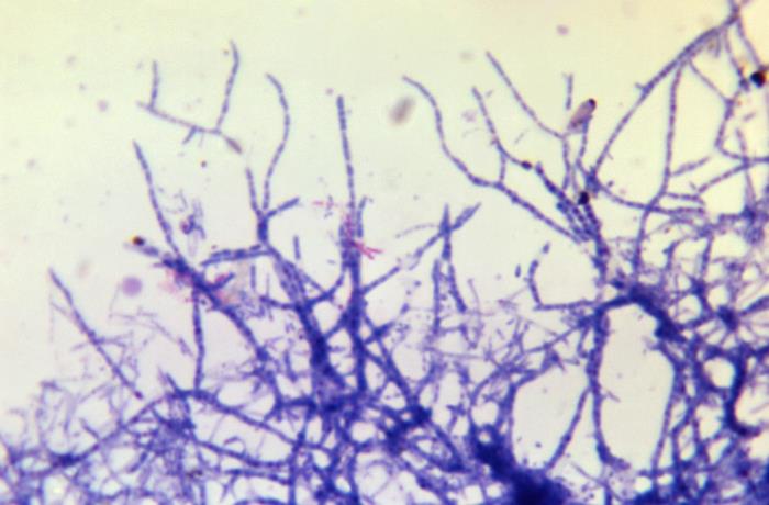 Figure: Filamentous Nocardia spp. in Kinyoun acid fast stain(Image source; CDC/Phil Health)
They are normal inhabitants of soil and water but can cause infections both in immunocompetent and immunocompromised individuals. Among various species, Nocardia asteroides and Nocardia brasiliensis are associated with clinical infections and cause a disease called nocardiosis.
N. asteroides alone causes >80% of infections.
Pathogenesis
Mode of Transmission
People acquire Nocardia infections either by traumatic inoculation or inhalation.
Clinical infections
Nocardia spp. specially Nocardia brasiliensis cause three types of skin infections in immunocompetent individuals:
- Mycetoma: A chronic, localized, painless, subcutaneous infection
- Lymphocutaneous infections
- Skin abscesses or cellulitis
In individuals, who are immunocompromised (especially those with reduced cell-mediated immunity), Nocardia spp. can cause invasive pulmonary and disseminated infections. Nocardia asteroides typically causes pneumonia, lung abscess with cavity formation, lung nodules, or empyema.
From the lung, the organism can spread hematogenously to various organs, notably the brain, where it causes brain abscess. Disseminated nocardiosis has a very poor prognosis.
Laboratory Diagnosis
Sample
When nocardiosis is clinically suspected, multiple specimens should be submitted for culture, because smears and cultures are simultaneously positive in only a third of the cases. The choice of the sample depends on the site affected. Depending on the clinical presentations, samples include sputum, pus from an abscess, and granules.
As Nocardia spp. are ubiquitous, isolation do not necessarily mean infection. It can possibly be the colonization of skin or upper respiratory tract or rarely laboratory contamination.
Direct Microscopy
If granules are seen in the actinomycetomas sample, the granules should be washed in saline, emulsified in 10% KOH, or crushed between two slides and gram stained to check for the presence of gram-positive filamentous bacilli.
Gram-staining (Brown-Brenn modification)
Presence of gram-positive branching or partially branching, filamentous bacilli often with beaded appearance suggest the presence of actinomycetes.
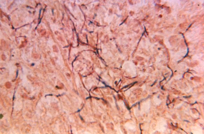 Figure: Nocardiaspp in tissue section stained using the Brown & Brenn method(Image source: CD/Phil Health)
Modified acid-fast staining (Kinyoun method)
Nocardiaspp. are partially acid-fast and appear as branching and filamentous red-colored acid-fast bacilli when stained by a modified acid-fast staining method (using 1% sulfuric acid as decolorizer).
Histopathology (H and E stain)
H and E staining of the granules show multilobulated with sunray appearance.
Culture
Nocardia spp. do not have complex growth requirements and can grow on various media such as brain heart infusion agar, sheep blood agar, chocolate agar, and Sabouraud dextrose agar (SDA). Colonies will be visible only after 2-3 days of incubation at 37°C. Because of slow growth contaminating flora may be overgrown. To overcome this problem various selective media have been in use to isolate Nocardia which are:
- Buffered yeast extract containing polymyxin and vancomycin.
- Sabouraud dextrose agar with chloramphenicol.
- Paraffin bait technique: Media using paraffin as the sole carbon source
- Buffered charcoal yeast extract agar with polymyxin, anisomycin, and vancomycin.
- Brain-heart infusion agar with chloramphenicol and cycloheximide.
- Lowenstein-Jensen medium: Produces moist glabrous colonies (differentiates from mycobacteria)
Colonies are creamy, wrinkled, pigmented (orange or pink-colored due to carotenoid-like pigments), and adhere firmly to the medium. Some colonies possess abundant aerial growth and have a cotton wool ball appearance.
Biochemical Identification
Nocardia species are non-motile, catalase-positive, and utilize a number of sugars oxidatively. Various biochemical tests are done for species identification such as:
- Decomposition of casein, hypoxanthine, tyrosine
- Growth in lysozyme
- Acetamide utilization
- Growth at 45°C for 3 days
- Acid from rhamnose
- Gelatin hydrolysis
- Opacification of Middlebrook agar
Serology
Currently, no serological tests are available to diagnose nocardiosis.
References and further readings
- Tille, P. (2017). Bailey & Scott’s Diagnostic Microbiology (14 edition). Mosby.
- Procop, G. W., & Koneman, E. W. (2016). Koneman’s Color Atlas and Textbook of Diagnostic Microbiology(Seventh, International edition). Lippincott Williams and Wilkins.

Tankeshwar Acharya, MSc (Medical Microbiology)
Tankeshwar Acharya is an Assistant Professor in the Department of Microbiology at Patan Academy of Health Sciences (PAHS), Nepal, where he has been teaching and practicing clinical microbiology for over 14 years. He is the founder of Microbe Online, one of the leading free microbiology education resources on the web, covering bacteriology, mycology, parasitology, immunology, and clinical laboratory diagnostics written from direct experience in both the classroom and the diagnostic laboratory.