
Mitochondria: Structure, Function, Location, and Why It Has Its Own DNA
Mitochondria structure (cristae, matrix, double membrane), function in ATP production, location and number in the cell, mitochondrial DNA and the endosymbiotic theory, plus Janus Green B vital staining. Foundation-level notes for biology and pre-medical students.

On this page
Every living cell needs energy to survive and do its work, and in eukaryotic cells most of that energy is produced in the mitochondria. Mitochondria are double-membraned organelles found in the cytoplasm of almost all eukaryotic cells, from a plant cell to a human muscle cell. They are often called the powerhouse of the cell because they generate most of its usable energy in the form of ATP.
 The word is often confused: one is a mitochondrion, two or more are mitochondria. "Mitochondria" is the plural. A single cell may contain anywhere from one to several thousand of them, depending on how much energy that cell needs.
Mitochondria are unusual among organelles in two ways that this article will keep returning to: they have their own DNA, separate from the DNA in the nucleus, and they are strikingly similar in size to a bacterium. Both facts point to the same remarkable idea about where mitochondria came from, which we cover below.
Where are mitochondria located in the cell?
Mitochondria are found in the cytoplasm of eukaryotic cells, suspended in the cytosol outside the nucleus. They are not fixed in one spot: they move through the cytoplasm and cluster wherever the cell's demand for energy is highest.
The number of mitochondria varies enormously from one cell type to another, and it tracks how much energy the cell uses:
- Heart muscle cells are packed with them. Mitochondria can occupy up to 40% of the cytoplasm, because cardiac muscle never stops contracting and has a huge, constant energy demand.
- Liver cells contain some of the highest numbers, on the order of 1,000 to 2,000 mitochondria per cell, reflecting the liver's intense metabolic workload.
- Mature red blood cells have no mitochondria at all. They lose them during maturation, which leaves more room for hemoglobin and forces them to rely entirely on anaerobic glycolysis for energy. This is a favorite exam point.
The rule to carry away: the more energy a cell needs, the more mitochondria it has. A cell's mitochondrial count is a direct clue to how metabolically active it is.
Structure of Mitochondria
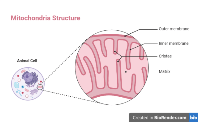
A mitochondrion is a rod-shaped or oval organelle, roughly 0.75 to 3 micrometers in size, which is about the same size as a typical bacterial cell. What makes it distinctive among organelles is that it is bounded by two membranes, not one, and each membrane does a different job.
Outer membrane. A smooth, continuous membrane that surrounds the whole organelle. It contains many copies of a barrel-shaped protein called porin, which forms channels wide enough to let small molecules and ions pass freely. Because of porins, the outer membrane is fairly permeable.
Intermembrane space. The narrow gap between the outer and inner membranes. Its contents matter for energy production, because this is the compartment into which protons are pumped during respiration, and it is where cytochrome c sits (important for the apoptosis role below).
Inner membrane. This is the working surface of the mitochondrion, and it is very different from the outer one. It is impermeable to most ions and molecules, so substances can only cross using specific transport proteins. This tight control is what allows the mitochondrion to build up the proton gradient that drives ATP synthesis. The inner membrane holds the electron transport chain and the enzyme ATP synthase.

Cristae. The inner membrane is thrown into deep folds called cristae. These folds dramatically increase the membrane's surface area, and since the machinery that makes ATP sits on the inner membrane, more surface area means more ATP-producing capacity. A cell that needs a lot of energy has mitochondria with many, tightly packed cristae.
Matrix. The gel-like space enclosed by the inner membrane. It contains the enzymes of the Krebs cycle (citric acid cycle), the mitochondrion's own DNA, and its own ribosomes. This is where the early steps of energy extraction from food take place.
Mitochondrial DNA and ribosomes. Inside the matrix, the mitochondrion carries a small loop of its own DNA and its own ribosomes, which are covered in the next section because they are the key to understanding where mitochondria came from.
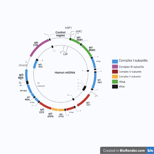 Figure: Structure of human mitochondrial DNA
Functions of mitochondria in the cell
Energy (ATP) production
The main job of the mitochondrion is to make ATP, the energy currency the cell spends on nearly everything. This happens in two connected stages:
- In the matrix, the Krebs cycle breaks down products of digested food and captures their energy in electron carriers (NADH and FADH2). The Krebs cycle itself makes only a small amount of ATP directly.
- Those electron carriers then feed the electron transport chain on the inner membrane. As electrons pass along the chain, protons are pumped into the intermembrane space, and the resulting gradient drives ATP synthase to produce the bulk of the cell's ATP. This oxygen-dependent process is called oxidative phosphorylation.
So the division of labor is worth memorizing: Krebs cycle in the matrix prepares the fuel; the electron transport chain on the inner membrane makes most of the ATP. Confusing these two is one of the most common mistakes students make.
Other functions
Mitochondria do more than make energy:
- Calcium storage. Mitochondria take up and store calcium ions and release them when needed. Calcium is a signal for many processes, including muscle contraction, nerve signaling, and blood clotting.
- Heat production. In brown fat, mitochondria deliberately "leak" the proton gradient instead of using it for ATP, releasing the energy as heat. This is why brown fat, abundant in newborns, is important for keeping babies warm.
- Controlling cell death (apoptosis). When a cell is damaged or no longer needed, its mitochondria release cytochrome c, which triggers the controlled self-destruction of the cell. This makes mitochondria central to how the body removes old or faulty cells.
Why do mitochondria have their own DNA?
Most organelles have no DNA of their own; they rely entirely on the instructions in the cell's nucleus. Mitochondria are different. Each one carries a small, circular loop of DNA (called mtDNA), about 16,569 base pairs in humans, along with its own ribosomes to read it. This lets the mitochondrion make some of its own proteins.
Two more clues make mitochondria look surprisingly bacterial:
- Their DNA is circular, like a bacterium's, not linear like the DNA in the nucleus.
- Their ribosomes are the 70S type found in bacteria, not the 80S type found elsewhere in the eukaryotic cell.
- They are about the same size as a bacterium, and they divide by splitting in two, much as bacteria do.
These observations are the basis of the endosymbiotic theory: the idea that mitochondria descend from a free-living bacterium that was engulfed by an early host cell billions of years ago. Instead of being digested, the bacterium stayed on, providing energy in exchange for a safe home, and over time became a permanent organelle. It is a striking idea, and a nineteenth-century biologist noticed the resemblance long before the theory was accepted: in 1890 Robert Altmann saw that these granules were about the size of bacteria and called them "bioblasts."
Inheritance note: because sperm contribute virtually no mitochondria to the egg, you inherit your mtDNA almost entirely from your mother. This maternal inheritance is why mitochondrial DNA is used to trace maternal ancestry.
Do bacteria have mitochondria?
No. Bacteria do not have mitochondria, or any other membrane-bound organelle, because bacteria are prokaryotic cells. This raises an obvious question that appears often in exams: if a bacterium has no mitochondria, how does it produce energy?
The answer is that the bacterial cell membrane (plasma membrane) does the job the mitochondrion does in a eukaryotic cell. The electron transport chain and the machinery of respiration are built into the bacterial cell membrane itself, so the membrane, rather than a separate organelle, is where ATP is generated. In some bacteria the membrane folds inward to increase this surface area, and these folds are sometimes called mesosomes.
This connects directly to the endosymbiotic theory above: a mitochondrion behaves like a respiring bacterium tucked inside a eukaryotic cell precisely because that is, in evolutionary terms, what it once was. For students moving from biology into microbiology, this is one of the clearest links between the two subjects.
Vital staining of mitochondria (Janus Green B)
Mitochondria are hard to see in a living, unstained cell. A classic laboratory exercise makes them visible using a supravital stain called Janus Green B, a dye that stains mitochondria in living cells without killing them.
Principle. Janus Green B is blue-green when oxidized and colorless (or pink) when reduced. Inside an active mitochondrion, the enzyme cytochrome oxidase keeps the dye in its oxidized, blue-green form, so the mitochondria stand out. Everywhere else in the cell, the dye is reduced and loses its color. The result is that only the mitochondria are stained blue-green. This is elegant because the stain is not just marking a structure, it is reporting on a function: only mitochondria with a working electron transport chain hold the color.
The buccal epithelium preparation. The commonest version of this practical uses cells scraped gently from the inside of the cheek (buccal epithelial cells), because they are easy to obtain and lie flat on a slide. A drop of Janus Green B is added, left for a few minutes, and the mitochondria appear as blue-green granules scattered through the cytoplasm.
Why it is taught. It is a simple, memorable demonstration that ties an organelle's structure to its function: the cells stain only where respiration is happening. For students, it is often the first time an invisible organelle becomes something you can actually see down a microscope.
How to Remember
Two membranes, two jobs. The outer membrane is the leaky wall (porins let things through); the inner membrane is the sealed, folded workbench where ATP is actually made. Leaky outside, tight and folded inside.
Matrix prepares, inner membrane pays. Krebs cycle in the matrix gets the fuel ready; the electron transport chain on the inner membrane produces most of the ATP. Do not credit the Krebs cycle with the big ATP payout, that belongs to the chain on the folds.
Cristae = capacity. More folds, more surface, more ATP. A hard-working cell has mitochondria stuffed with cristae.
Bacterial fingerprints. Circular DNA, 70S ribosomes, bacterial size, divides by splitting. Four clues, one conclusion: the mitochondrion was once a bacterium (endosymbiotic theory).
Janus Green stays green where the cell breathes. Cytochrome oxidase in active mitochondria keeps the dye oxidized and colored; everywhere else it fades. The stain follows the respiration.
Key exam facts
| Question | Answer |
|---|---|
| Singular and plural of mitochondria? | One mitochondrion; two or more mitochondria |
| Why is the mitochondrion called the powerhouse of the cell? | It produces most of the cell's ATP by oxidative phosphorylation |
| How many membranes does a mitochondrion have? | Two (outer and inner) |
| What are cristae? | Folds of the inner membrane that increase surface area for ATP production |
| Where does the Krebs cycle occur? | In the matrix |
| Where is most ATP made? | On the inner membrane, by the electron transport chain and ATP synthase |
| Size of human mitochondrial DNA? | About 16,569 base pairs, circular |
| What type of ribosomes do mitochondria have? | 70S (bacterial type) |
| Which cell has no mitochondria? | Mature red blood cell |
| Which cells have the most mitochondria? | High-energy cells such as heart muscle and liver cells |
| From which parent do you inherit mtDNA? | The mother (maternal inheritance) |
| Do bacteria have mitochondria? | No; the cell membrane carries out respiration instead |
| Which dye is used for vital staining of mitochondria? | Janus Green B |
Where Students Get Confused
The Krebs cycle makes most of the ATP. It does not. The Krebs cycle in the matrix produces only a little ATP directly; it mainly loads electron carriers. Most ATP comes from the electron transport chain on the inner membrane. This is the single most common mitochondrial error.
Mitochondria are found only in animal cells. No. Mitochondria are in almost all eukaryotic cells, plant and animal alike. Plant cells have both mitochondria (to release energy) and chloroplasts (to capture it). Chloroplasts do not replace mitochondria.
Bacteria have tiny mitochondria. Bacteria have none. They are prokaryotes; respiration happens at the cell membrane. A mitochondrion resembles a bacterium because of shared ancestry, but a bacterium does not contain one.
"Mitochondria" is singular. It is plural. One is a mitochondrion.
All cells have the same number of mitochondria. The number varies from zero (mature red blood cells) to thousands (liver, heart), depending on energy demand.
References
- Alberts B, Heald R, Johnson A, et al. Molecular Biology of the Cell. 7th ed. New York: W. W. Norton; 2022.
- Iwasa J, Marshall W. Karp's Cell and Molecular Biology. 8th ed. Hoboken: Wiley; 2016.
- Reece JB, Urry LA, Cain ML, et al. Campbell Biology. 12th ed. New York: Pearson; 2021.
- Cooperstein SJ, Lazarow A. Studies on the mechanism of Janus Green B staining of mitochondria. J Biol Chem.
Frequently Asked Questions
Where are mitochondria located in the cell?
Where are mitochondria located in the cell?
In the cytoplasm of eukaryotic cells, outside the nucleus. They move around and gather in the parts of the cell that need the most energy. The number ranges from none in mature red blood cells to a few thousand in liver and heart muscle cells.
What is the main function of mitochondria in an animal cell?
What is the main function of mitochondria in an animal cell?
To produce ATP, the cell's usable energy, by oxidative phosphorylation. Mitochondria also store calcium, generate heat in brown fat, and help trigger controlled cell death (apoptosis).
Is mitochondria singular or plural?
Is mitochondria singular or plural?
Plural. One is a mitochondrion; two or more are mitochondria.
Why do mitochondria have their own DNA?
Why do mitochondria have their own DNA?
Because they descend from a free-living bacterium that was taken into an early cell (the endosymbiotic theory). They kept a small loop of their own circular DNA and their own 70S ribosomes, which is why they can make some of their own proteins and why mtDNA looks bacterial.
Do bacteria have mitochondria?
Do bacteria have mitochondria?
No. Bacteria are prokaryotes and have no organelles. Their cell membrane carries out respiration and makes ATP, doing the job that mitochondria do in eukaryotic cells.
Which structure performs the function of mitochondria in bacteria?
Which structure performs the function of mitochondria in bacteria?
The bacterial cell membrane (plasma membrane). The respiratory electron transport chain is built into the membrane, so it produces ATP in place of a mitochondrion.
What is Janus Green B used for?
What is Janus Green B used for?
It is a vital stain that makes mitochondria visible in living cells. Active mitochondria keep the dye in its blue-green oxidised form (through the enzyme cytochrome oxidase), so they show up as blue-green granules while the rest of the cell stays colourless.
Which parent do you inherit mitochondrial DNA from?
Which parent do you inherit mitochondrial DNA from?
Your mother. Sperm contribute almost no mitochondria to the egg, so mtDNA is passed down the maternal line.

Tankeshwar Acharya, MSc (Medical Microbiology)
Tankeshwar Acharya is an Assistant Professor in the Department of Microbiology at Patan Academy of Health Sciences (PAHS), Nepal, where he has been teaching and practicing clinical microbiology for over 14 years. He is the founder of Microbe Online, one of the leading free microbiology education resources on the web, covering bacteriology, mycology, parasitology, immunology, and clinical laboratory diagnostics written from direct experience in both the classroom and the diagnostic laboratory.
Comments
No comments yet. Be the first to share your thoughts.
Leave a comment
All comments are reviewed before they appear.