
Urine Culture: Sample Collection, Transport and Lab Diagnosis
The organisms that cause UTI, how to collect and culture urine correctly, and how to read colony counts to separate real infection from contamination.

On this page
A young woman's urine culture grows three different organisms at 10⁴ CFU/mL each. The intern is ready to start antibiotics. The bench technologist reads the same plate and reports it as a contaminated specimen, no significant growth, please recollect. Both are looking at the same plate. The difference between them is not the organism list, it is knowing what a urine culture can and cannot tell you. That judgment is what this article is about.
Bacteria can invade and cause UTI via three major routes: ascending, hematogenous, and lymphatic. The ascending route is the most common in females. Hematogenous spread accounts for fewer than 5% of UTIs and rarely occurs with gram-negative bacilli.
Polymicrobial UTI in the absence of anatomic abnormalities, foreign bodies, or trauma is rare. Three or more organisms in large quantities usually indicates a contaminated specimen, a point that becomes central when you interpret the culture later in this article.
Etiology
Escherichia coli: Escherichia coli is the most common cause of community-acquired symptomatic UTIs, especially cystitis. Uropathogenic E. coli (UPEC) has remained the predominant uropathogen (80%) isolated in acute community-acquired uncomplicated UTIs. Escherichia coli can adhere to the urethral and bladder mucosa via pili.
Staphylococcus aureus : Staphylococcus aureus can cause pyelonephritis (infection of the renal parenchyma). In the case of bacteremic patients, S. aureus reaches the kidney via hematogenous spread or the descending route.
Because S. aureus typically reaches the kidney hematogenously, S. aureus in urine should prompt a question about bloodstream infection rather than a simple ascending UTI. This is why any count of S. aureus from a suprapubic or catheter specimen can be significant (see interpretation below).
Staphylococcus saprophyticus: It is common in young women. Approximately 10% to 15% of cases of UTIs in women of reproductive age groups are caused by S. saprophyticus. S. saprophyticus is the one coagulase-negative staphylococcus you do not dismiss as skin contaminant in a symptomatic young woman.
Mycobacteria: Mycobacteria can cause UTI in HIV-positive patients.
Pseudomonas aeruginosa: Pseudomonas aeruginosa can cause urinary tract infections, most common in patients staying in healthcare settings for longer. Patients with anatomic or neurologic abnormalities affecting their urinary tract or heavily antibiotic-experienced patients are also predisposed to UTIs from Pseudomonas aeruginosa.
Enterococci: Infrequently cause uncomplicated cystitis and pyelonephritis.
Other bacteria: Other bacteria commonly isolated from patients with UTIs are Klebsiella spp., Proteus spp. Enterobacter spp. Acinetobacter, Citrobacter, beta-hemolytic streptococci etc.
Candida: Candida species can cause UTI in patients with extensive prior antibiotic use and indwelling Foley catheters. Other high-risk groups are patients with diabetes, immunocompromised patients, and those on immunosuppressive therapy.
Viruses: Viruses rarely cause UTIs. Adenovirus, BK virus, and cytomegalovirus can cause hemorrhagic cystitis. These viruses almost exclusively cause cystitis in immunocompromised hosts such as those who have undergone stem cell transplants.
Factors affecting prevalence of etiological agents
The etiology of UTI is also affected by underlying host factors that complicate UTI, such as
- Age: The most common organisms isolated in children with uncomplicated UTI are Enterobacteriaceae. Gram-positive organisms are common, and polymicrobial infections account for up to 1 in 3 infections in the elderly.
- Gender: Women are affected far more often than men because the female urethra is much shorter (about 4 cm) than the male urethra (about 18 to 20 cm), so bacteria reach the bladder more easily. The urethral opening in women also lies close to the vaginal and anal flora.
- Diabetes: Etiologic pathogens associated with UTI among patients with diabetes include Klebsiella spp., Group B streptococci, and Enterococcus spp., as well as E. coli.
- Spinal cord injury or urinary catheterization: Patients with spinal cord injuries commonly have E. coli infections.
Complicated vs. Uncomplicated UTI
Complicated UTI has a more diverse etiology than uncomplicated UTI, and organisms that rarely cause disease in healthy patients can cause significant disease in hosts with anatomic, metabolic, or immunologic underlying diseases.
When to send urine for culture
Send urine for culture only when there is strong clinical suspicion of UTI, to avoid detecting asymptomatic bacteriuria. Culture is specifically indicated in suspected pyelonephritis (loin pain and fever), suspected UTI in men, recurrent UTI, pregnancy, and failed empiric treatment or persistent symptoms.
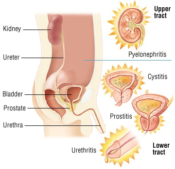 Common symptoms are frequency, urgency, dysuria, suprapubic pain, cloudy or bloody or strong-smelling urine, and flank pain.
Urine Sample Collection
Both noninvasive and invasive methods for collecting urine samples are available. Samples obtained from invasive methods are reliable as they are less likely to be contaminated and easy to interpret.
Urine collected by noninvasive methods passes through the contaminated milieu, so semi-quantitative culture is used to diagnose urinary tract infections (UTI) and to discriminate between contamination, colonization (asymptomatic bacteriuria), and clinical infection.
The methods form a ladder from cleanest to most contamination-prone. You step down the ladder to the least invasive method that will still answer the clinical question.
| Rank | Method | Best for | Contamination risk | When to use it |
|---|---|---|---|---|
| 1 (cleanest) | Suprapubic aspiration | Infants, young children, or when other methods fail | Lowest (bypasses the urethra) | Rarely needed; reference standard |
| 2 | Straight (in-and-out) catheter | Patients who cannot void a clean sample | Low | When clean-catch is unreliable and a clean result is essential |
| 3 (routine) | Clean-catch midstream | Most cooperative adults | Moderate (passes distal urethra) | The default for routine adult collection |
| 4 | Indwelling catheter port | Patients already catheterized | Moderate to high | Aspirate from the port, never the drainage bag |
| Not acceptable | Urine from a drainage bag | Never | Very high | Do not culture; organisms multiply in the bag |
Children are the main reason the ladder exists. Infants and young children cannot give a reliable clean-catch, so suprapubic aspiration or catheterization is used when a trustworthy result is needed. Bag specimens may be used for screening only: a negative bag specimen helps rule out infection, but a positive one must be confirmed by a cleaner method before treatment, because bag samples are heavily contaminated by skin and perineal flora.
Suprapubic aspiration
Suprapubic aspiration is the reference method for avoiding contamination, because the needle enters the bladder directly through the abdominal wall and bypasses the urethra entirely. Urine is withdrawn into a syringe through a percutaneously inserted needle.
Use: Reserved for situations where a clean specimen cannot be obtained any other way, most often infants and young children who cannot give a clean-catch sample, and occasionally adults when other methods have failed or given equivocal results. It is invasive and resource-intensive, so it is not used for routine adult collection.
Straight catheter technique
Collection of urine by use of a single catheter (straight catheter technique) is the next-best technique for obtaining urine specimens with minimal contamination. It is an invasive technique with added disadvantages because inserting a catheter through the urethra can introduce bacteria into the bladder (and thereby cause UTI), and rare complications have been reported.
Use: It gives a low-contamination sample and is the next-best option after suprapubic aspiration, but it is too labor-intensive and costly for routine use, and passing a catheter can itself introduce bacteria. Reserve it for patients who cannot provide a reliable clean-catch when a clean result is essential.
Clean Catch Midstream Urine
It is the least invasive technique and is used widely. It has an obvious disadvantage compared to the techniques mentioned above. The chances of contamination from normal vaginal, perineal, and anterior urethral flora are high. The urine sample passes through the distal urethra and can become contaminated with commensal bacteria.
Urine is one of the most commonly collected microbiology specimens. For how it fits within the general principles of collection, transport, and rejection, see Specimen Collection and Transport in Microbiology.
Commensal flora (resident flora) found in the urine samples are
- Anaerobic cocci
- Anaerobic gram-negative bacilli
- Coagulase-negative staphylococci (excluding S. saprophyticus)
- Commensal Mycobacterium spp.
- Commensal Mycoplasma spp.
- Diphtheroids (Corynebacterium spp.)
- Lactobacilli
- Nonpathogenic Neisseria spp.
- Propionibacterium spp.
- Viridans and non-hemolytic streptococci
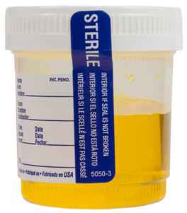 Use: Most urine specimens are obtained from adult patients via the clean-catch midstream technique
How to collect Midstream Specimen of Urine (MSU)?
Instruction for male patients
- Remove undergarments.
- Wash hands.
- Retract the foreskin completely.
- Wipe the head of the penis in a single motion with the first towelette. Repeat with a second towelette. If not circumcised, hold foreskin back before cleansing.
- Void 20 to 25 ml into the toilet and catch a portion of the remaining urine in the cup without stopping the stream. Do not touch the cup with the penis.
- Place the lid on the cup securely
- Immediately transfer to the microbiology laboratory or follow the procedure as indicated by the Hospital personnel.
Instructions for female patients
- Wash your hands.
- Sit on the toilet with your legs spread apart. Separate the labia (the folds of skin around the urinary opening) with the thumb and forefinger of one hand, and keep them held apart through the whole collection.
- Using the first towelette, wipe the inner folds from front to back in a single motion, then discard it. Repeat on the other side with a second towelette, and down the center over the urinary opening with a third.
- Keeping the labia held apart, void 20 to 25 mL into the toilet, then, without stopping the stream, catch a portion of the remaining urine in the cup. Do not touch the cup to the skin.
- Place the lid on the cup securely.
- Transfer to the laboratory promptly, or refrigerate if there will be a delay.
Front-to-back wiping and holding the labia apart are the two steps that most reduce contamination in women. Skipping them is the commonest reason a female clean-catch grows mixed perineal flora and has to be recollected.
Indwelling Catheter
Specimen collection from patients with indwelling catheters requires a scrupulous aseptic technique. The catheter tubing should be clamped off above the port to allow the collection of freshly voided urine. The catheter port or wall of the tubing should then be cleaned vigorously with 70% ethanol, and urine aspirated via a needle or syringe; the integrity of the closed drainage system must be maintained to prevent the introduction of organisms into the bladder.
Do not culture the Foley catheter tip. It is colonized by urethral and bag flora regardless of infection, so it does not reflect what is in the bladder. Sample fresh urine aspirated from the catheter port instead.
Transporting the urine sample
Urine is an excellent growth medium, so bacteria multiply in it at room temperature and inflate the colony count. Deliver the sample to the laboratory within 1 to 2 hours of collection. If that is not possible, refrigerate at 2 to 8 degrees C for up to 24 hours, or use a boric acid preservative tube, which holds the bacterial count stable during transport.
A sample left at room temperature for hours will overgrow and can turn a contaminant or a low count into a falsely significant one. This is also why the collection time must be recorded on the request form: without it the laboratory cannot judge the transport interval or flag a delayed specimen, one of several fields whose absence quietly changes the result, covered in the laboratory requisition form.
Processing of Urine Sample
Calibrated Loop: Semi-Quantitative Urine Culture
The calibrated loop is the most clinically significant type for diagnostic microbiology. Unlike an ordinary inoculating loop, which is not calibrated for a fixed volume, calibrated loops deliver a precise, reproducible volume, which allows semi-quantitative counting without a full serial dilution.
Standard volumes and their applications:
| Loop size | Volume delivered | Colony count interpretation | Clinical use |
|---|---|---|---|
| 1 µL (0.001 mL) | 1 µL | Colonies × 1000 = CFU/mL | Routine urine culture |
| 10 µL (0.01 mL) | 10 µL | Colonies x 100 = CFU/mL | Higher sensitivity; low-count infections |
How the calibrated loop urine culture works:
- Mix the urine specimen thoroughly (invert tube gently 5–10 times).
- Hold the calibrated loop vertically: the loop must be perpendicular to the surface of the urine to pick up a consistent volume by surface tension. Tilting the loop reduces the volume picked up.
- Inoculate CLED agar (or blood agar + MacConkey) using a continuous back-and-forth streak across the full plate diameter, then streaking perpendicular lines across the primary streak.
- Incubate at 37°C for 18–24 hours.
- Count colonies on the primary streak area for the semi-quantitative result.
The key technique point: Vertical loop angle is everything. A loop held at 45° will underdeliver volume. A loop dipped more than 2–3 mm into the urine will overdeliver. Practice with water and a calibrated volume until the technique is consistent; a reproducible result depends entirely on reproducible loop angle and immersion depth.
Interpretation:
Colony count (with 1 µL loop) | Estimated CFU/mL | Interpretation |
|---|---|---|
Confluent growth on primary streak | >10⁵ | Significant bacteriuria (likely infection) |
Isolated colonies on primary streak only | 10⁴ to 10⁵ | Borderline; repeat or interpret with clinical context |
Few colonies, secondary and tertiary areas only | <10⁴ | Likely contamination in most clinical contexts |
No growth | <10³ | No significant growth |
NOTE:
In certain conditions, even growth between 10³–10⁵ CFU/mL is considered significant. Those conditions are:
- Patient is on diuretics
- Patients being on antimicrobial
In certain conditions, even any count is considered significant. Those conditions are
- Specimen obtained from catheter tubing
- Suprapubic aspirate
- Suspected hematogenously acquired infection (e.g., S. aureus)
Culture
The standard protocol plates 1 µL of well-mixed urine onto 5% sheep blood agar and MacConkey agar (or CLED as a single-plate alternative), incubated aerobically at 35–37°C for 18–24 hours. Blood agar supports most uropathogens, MacConkey differentiates lactose fermenters and suppresses swarming Proteus, and CLED prevents Proteus swarming while allowing colony counting on one plate.
The next day, read the plates for colony count (against the interpretation table above), colony morphology, and any sign of contamination. Only specimens with significant growth are processed further for identification and susceptibility testing.
Treatment of Urinary Tract Infections
Choice of the antibiotics for the treatment of any infections depends on patients’ specific factors (age, underlying diseases/abnormalities) local resistance patterns of the etiological agents, and cost & availability of the drugs).
Several national and international guidelines help choose an empiric regimen. The widely cited IDSA/ESCMID guidance (published 2011, and titled a "2010 update") established nitrofurantoin, trimethoprim-sulfamethoxazole (co-trimoxazole), fosfomycin, and pivmecillinam as first-line options for uncomplicated cystitis in women. That guidance is now more than a decade old, so always check current national guidance and your local hospital antibiogram before choosing a regimen, since resistance patterns and recommendations have shifted since then.
Drugs used for the empiric treatment of uncomplicated UTIs are:
- Ciprofloxacin: Avoid fluoroquinolones for uncomplicated UTIs when alternative antibiotics are possible. Fluoroquinolones should not be used if the local prevalence of resistance of the uropathogen exceeds 10%.
- Nitrofurantoin: In patients with reduced renal function, nitrofurantoin should only be used if local resistance data suggests a high resistance to alternative agents. Due to lower drug levels in the renal parenchyma, nitrofurantoin and fosfomycin should be avoided in cases of suspected pyelonephritis.
- Trimethoprim-sulfamethoxazole (co-trimoxazole): should not be used if the local prevalence of resistance of uropathogen (E.coli) exceeds 20%.
How to Remember
- The three C's of a positive urine culture: Contamination, Colonization, and Clinical infection. A positive culture never means "UTI" by itself. It means one of three things: organisms from outside the bladder (contamination), organisms living in the bladder without disease (colonization, i.e., asymptomatic bacteriuria), or organisms causing disease (clinical infection). The count narrows it; the patient's symptoms decide it. The word "clinical" in the third C is the whole point: symptoms are what separate infection from colonization.
- "10⁵ or it didn't happen." For a clean-catch midstream specimen, ≥10⁵ CFU/mL is the significance threshold. Every exception loosens it, and each loosens it for a reason. The specimen is cleaner (suprapubic aspirate or catheter tube, so any count matters), or the patient is primed so organisms are washed out (diuretics or prior antimicrobials, so 10³ to 10⁵ counts matter), or the route bypasses the urethra entirely (hematogenous S. aureus, so any count matters).
Key exam facts
| Fact | Detail |
|---|---|
| Most common uropathogen | E. coli (UPEC), ~80% of acute uncomplicated community-acquired UTI |
| Second in young women | S. saprophyticus, ~10–15% of reproductive-age female UTI |
| Reaches kidney by blood, not ascent | S. aureus (think bloodstream source) |
| Healthcare-associated / catheter / prior antibiotics | Pseudomonas aeruginosa, Candida |
| Why women affected more than men | Female urethra much shorter (about 4 cm) vs male (about 18 to 20 cm) |
| Best low-contamination collection | Suprapubic aspiration (rarely needed clinically) |
| Most widely used collection | Clean-catch midstream urine |
| Significance threshold (clean-catch) | ≥10⁵ CFU/mL |
| Doubtful, consider repeat | 10⁴–10⁵ CFU/mL |
| Any count significant | Suprapubic aspirate, catheter-tube specimen, suspected hematogenous S. aureus |
| Standard culture media | Blood agar + MacConkey, or CLED |
| Routine calibrated loop | 1 µL (Colonies x 1000 = CFU/mL) |
| Avoid in suspected pyelonephritis | Nitrofurantoin, fosfomycin (low renal-parenchyma levels) |
| Empiric first-line (uncomplicated cystitis) | Nitrofurantoin, co-trimoxazole, fosfomycin, pivmecillinam |
| Co-trimoxazole resistance cutoff | Avoid if local E. coli resistance >20% |
| Fluoroquinolone resistance cutoff | Avoid if local resistance >10% |
Where Students Get Confused
- A positive culture means the patient has a UTI. No. It means one of three things: contamination, colonization (asymptomatic bacteriuria), or clinical infection. The count and the symptoms decide which.
- Three organisms must be a bad polymicrobial infection. Usually the opposite: three organisms in a clean-catch specimen almost always signals contamination, not severe infection.
- Any coagulase-negative staph in urine is skin contaminant. S. saprophyticus is the exception: significant in a symptomatic young woman.
- The 10⁵ threshold is absolute. It's the default for clean-catch. It loosens (to any count, or to 10³) for cleaner specimens (suprapubic, catheter) and primed patients (diuretics, prior antibiotics).
- Loop angle is a detail. It's the whole result. A loop held at 45° or dipped too deep delivers the wrong volume and invalidates the count.
- Nitrofurantoin treats everything. It reaches poor levels in the renal parenchyma, making it the wrong choice for pyelonephritis.
References
- Tille PM. Bailey & Scott's Diagnostic Microbiology. 15th ed. St. Louis: Elsevier; 2022.
- Leber AL, editor. Clinical Microbiology Procedures Handbook. 4th ed. Washington, DC: ASM Press; 2016. DOI: 10.1128/9781683670438.CMPH
- Minardi, D., d'Anzeo, G., Cantoro, D., Conti, A., & Muzzonigro, G. (2011). Urinary tract infections in women: etiology and treatment options. International Journal of General Medicine, 4, 333–343. https://doi.org/10.2147/IJGM.S11767
- Chu, C. M., & Lowder, J. L. (2018). Diagnosis and treatment of urinary tract infections across age groups. American Journal of Obstetrics and Gynecology, 219(1), 40–51. https://doi.org/10.1016/j.ajog.2017.12.231
- Kaur, R., & Kaur, R. (2021). Symptoms, risk factors, diagnosis and treatment of urinary tract infections. Postgraduate Medical Journal, 97(1154), 803–812. https://doi.org/10.1136/postgradmedj-2020-139090
- Llor, C., Moragas, A., Aguilar-Sánchez, M., García-Sangenís, A., Monfà, R., & Morros, R. (2023). Best methods for urine sample collection for diagnostic accuracy in women with urinary tract infection symptoms: a systematic review. Family Practice, 40(1), 176–182. https://doi.org/10.1093/fampra/cmac058
- Karacan, C., Erkek, N., Senel, S., Akin Gunduz, S., Catli, G., & Tavil, B. (2010). Evaluation of urine collection methods for the diagnosis of urinary tract infection in children. Medical Principles and Practice, 19(3), 188–191. https://doi.org/10.1159/000273068
- Sinawe, H., & Casadesus, D. Urine Culture. In: StatPearls [Internet]. Treasure Island (FL): StatPearls Publishing; 2023. https://www.ncbi.nlm.nih.gov/books/NBK557569/
- Gupta K, Hooton TM, Naber KG, et al. International Clinical Practice Guidelines for the Treatment of Acute Uncomplicated Cystitis and Pyelonephritis in Women: A 2010 Update by the Infectious Diseases Society of America and the European Society for Microbiology and Infectious Diseases. Clin Infect Dis. 2011;52(5):e103–e120. https://doi.org/10.1093/cid/ciq257
Frequently Asked Questions
When should urine be sent for culture, and when should it not?
When should urine be sent for culture, and when should it not?
Send it only when there is real clinical suspicion of UTI: suspected pyelonephritis (loin pain and fever), UTI in men, recurrent UTI, pregnancy, or failed empiric treatment. Culturing every patient risks detecting asymptomatic bacteriuria, which usually does not need treatment.
Which collection method gives the least contaminated sample?
Which collection method gives the least contaminated sample?
Suprapubic aspiration, because the needle enters the bladder directly and bypasses the urethra. It is reserved for infants, young children, or cases where other methods fail. For routine adults, the clean-catch midstream sample is the practical standard despite its higher contamination risk.
Why can urine never be cultured from a catheter drainage bag?
Why can urine never be cultured from a catheter drainage bag?
Bacteria multiply in the bag, so the count no longer reflects what is in the bladder. For a catheterized patient, clamp the tubing above the port, disinfect the port, and aspirate fresh urine through it.
What does the calibrated loop actually do, and why does the angle matter so much?
What does the calibrated loop actually do, and why does the angle matter so much?
It delivers a fixed, reproducible volume of urine (commonly 1 microlitre) so that counting colonies gives an estimate of CFU per mL. The result depends entirely on picking up that exact volume, so the loop must be held vertical and dipped only a few millimetres. A tilted or over-dipped loop delivers the wrong volume and invalidates the count.
Why is Staphylococcus aureus in the urine treated differently from other organisms?
Why is Staphylococcus aureus in the urine treated differently from other organisms?
S. aureus usually reaches the kidney through the bloodstream rather than by ascending the urethra. So S. aureus in urine raises the question of a bloodstream infection, and any count from a suprapubic or catheter specimen can be significant rather than dismissed as a low count.
Is Staphylococcus saprophyticus a contaminant like other coagulase-negative staphylococci?
Is Staphylococcus saprophyticus a contaminant like other coagulase-negative staphylococci?
No. Most coagulase-negative staphylococci in urine are skin contaminants, but S. saprophyticus is a genuine uropathogen in young women and should not be dismissed in a symptomatic patient.
How quickly must urine reach the laboratory?
How quickly must urine reach the laboratory?
Within 1 to 2 hours at room temperature. If delayed, refrigerate at 2 to 8 degrees C for up to 24 hours, or use a boric acid preservative tube. Urine left warm overgrows and can turn a contaminant or a low count into a falsely significant result.
Why is nitrofurantoin a poor choice for pyelonephritis?
Why is nitrofurantoin a poor choice for pyelonephritis?
It reaches good concentrations in the bladder urine but poor levels in the kidney tissue (renal parenchyma). That makes it effective for lower UTI (cystitis) but inadequate for a kidney infection. Fosfomycin is avoided in pyelonephritis for the same reason.

Tankeshwar Acharya, MSc (Medical Microbiology)
Tankeshwar Acharya is an Assistant Professor in the Department of Microbiology at Patan Academy of Health Sciences (PAHS), Nepal, where he has been teaching and practicing clinical microbiology for over 14 years. He is the founder of Microbe Online, one of the leading free microbiology education resources on the web, covering bacteriology, mycology, parasitology, immunology, and clinical laboratory diagnostics written from direct experience in both the classroom and the diagnostic laboratory.
Comments
No comments yet. Be the first to share your thoughts.
Leave a comment
All comments are reviewed before they appear.