Klebsiella pneumoniae: Properties, Diseases, Lab Diagnosis
Klebsiella pneumoniae: Properties, Diseases, Lab Diagnosis
Klebsiella pneumoniae is a Gram-negative rod-shaped bacteria of the genus Klebsiella and the family Enterobacteriaceae. They are members of the normal intestinal flora of humans and animals and may be isolated from a variety of environmental sources.
K. pneumoniae was first isolated in the late 19th century and was initially known as Friedlander’s bacterium. Classic cases of pneumonia, characterized by production of brick-red or “currant jelly” sputum, were known to be caused by Friedlander’s bacillus (Klebsiella pneumoniae).
Klebsiella pneumoniae causes infections in people of all age groups, especially in infants, the elderly, immunocompromised, and alcoholics. It is one of the leading causes of hospital-acquired (nosocomial) infections. The range of infections includes pneumonia (it is a frequent cause of ventilator-associated pneumonia), urinary tract infection, bloodstream infection (BSI), and liver abscesses. Though Klebsiella pneumoniae accounts for a small percentage of pneumonia cases, the case fatality rates are high (up to 90% in untreated cases).
Klebsiella pneumoniae is a major threat to public health with the emergence of multidrug-resistant strains, rendering infection by these strains very challenging to treat. Strains of K.pneumoniae producing extended-spectrum beta-lactamases (ESBLs)or carbapenemases are considered global priority pathogens.
General Properties of Klebsiella pneumoniae
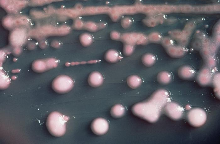 Figure: mucoid colonies
- Gram–negative
- Non-spore-forming rods
- Facultative anaerobes
- Catalase Test: Positive
- Oxidase Test: Negative
- Lactose fermenter (forms pink-colored colonies on MacConkey Agar).
- Presence of polysaccharide capsule (in the culture plate mucoid colonies are seen).
- Non-motile (Klebsiella species are nonmotile and non-flagellated and thus have no H antigens).
Virulence Factors of Klebsiella pneumoniae
- Capsule
- Cell wall receptors
- Lipopolysaccharide (endotoxin)
- Fimbriae
- Siderophores
Virulence factors of Klebsiella pneumoniae will be discussed in-depth in another post.
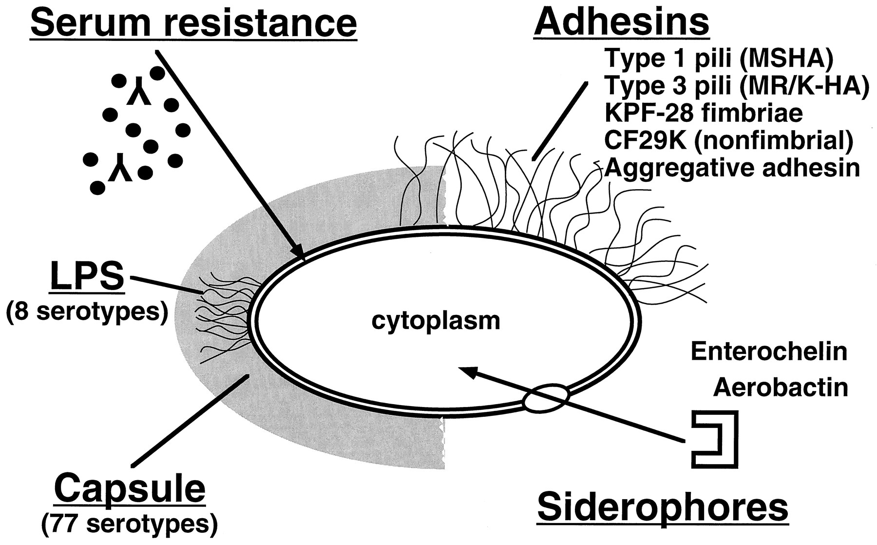 Figure: Virulence factors of K. pneumoniae
Laboratory diagnosis of K. pneumoniae infection
Sample: Sputum (Red currant-jelly sputum may be seen in a patient infected with K. pneumoniae), mid-stream urine, or blood (depending on the suspected illness/clinical presentation).
Colony characteristics of Klebsiella pneumoniae
- Blood Agar: Mucoid, non-hemolytic colonies
- MacConkey Agar: Mucoid, lactose-fermenting (pink colored) colonies
| Test Name | Results |
|---|---|
| Catalase test | Positive |
| Oxidase test | Negative |
| Indole Production Test | Negative (K. oxytoca is indole positive) |
| Methyl-Red Test | Negative |
| Voges-Proskauer Test | Positive |
| Citrate Utilization Test | Positive |
| Hydrogen Sulfide (H2S) Production | Negative |
| TSI test | Acid/Acid, Gas (++), No H₂SH₂S |
| Urea Hydrolysis Test | Positive |
| Lysine Decarboxylase Test | Positive |
| Arginine Dihydrolase Test | Negative |
| Ornithine decarboxylase test | Negative |
| Motility at 36 °C | Non-motile |
| D-Glucose (acid/gas) | Positive/Positive |
| D-mannitol fermentation | Positive |
| Sucrose fermentation | Positive |
| Lactose fermentation | Positive |
| D-sorbitol fermentation | Positive |
| Cellobiose | Positive |
| Esculin hydrolysis | Positive |
| Acetate Utilization Test | Positive |
| ONPG Test | Positive |
Various biochemical tests using conventional methods or miniature commercial system (API-20E or Enterotube test) is done to identify the suspected colony as Klebsiella pneumoniae. Some of the commonly used tests for the identification of Klebsiella pneumoniae are given below.
Note: To minimize the cost and effort, some diagnostic/hospital laboratories perform only certain tests such as Triple Sugar Iron Agar (TSI), sulfite indole motility (SIM) / urease indole motility (UIM), and citrate utilization test to identify isolates of Enterobacteriaceae family, full panel/commercially available miniature test system (API-20E and Enterotube test) is used only when the test results are inconclusive.
References
- Bailey & Scott’s Diagnostic Microbiology, Forbes, 11th edition
- Ashurst JV, Dawson A. Klebsiella Pneumonia. [Updated 2023 Jul 20]. In: StatPearls [Internet]. Treasure Island (FL): StatPearls Publishing; 2023 Jan-. Available from: https://www.ncbi.nlm.nih.gov/books/NBK519004/
- Qu, T. T., Zhou, J. C., Jiang, Y., Shi, K. R., Li, B., Shen, P., Wei, Z. Q., & Yu, Y. S. (2015). Clinical and microbiological characteristics of Klebsiella pneumoniae liver abscess in East China. BMC infectious diseases, 15, 161. https://doi.org/10.1186/s12879-015-0899-7
- Madigan Michael T, Bender, Kelly S, Buckley, Daniel H, Sattley, W. Matthew, & Stahl, David A. (2018). Brock Biology of Microorganisms (15th Edition). Pearson.

Tankeshwar Acharya, MSc (Medical Microbiology)
Tankeshwar Acharya is an Assistant Professor in the Department of Microbiology at Patan Academy of Health Sciences (PAHS), Nepal, where he has been teaching and practicing clinical microbiology for over 14 years. He is the founder of Microbe Online, one of the leading free microbiology education resources on the web, covering bacteriology, mycology, parasitology, immunology, and clinical laboratory diagnostics written from direct experience in both the classroom and the diagnostic laboratory.