
Hektoen Enteric (HE) Agar: Composition, Principle, Colony Characteristics, and Uses
Hektoen Enteric (HE) Agar is a selective and differential medium for Salmonella and Shigella with better Shigella recovery than SS agar. Learn its green-medium principle, three-carbohydrate differentiation, H₂S indicator system, and colony colors.

On this page
In 1968, microbiologists at the Hektoen Institute in Chicago were trying to solve a specific problem: Shigella strains were being missed in stool cultures because the highly selective media available at the time (particularly SS agar) were too inhibitory. Brilliant green dye, the primary selective agent in SS agar, suppressed not just coliforms but also the Shigella the medium was supposed to isolate.
Sylvia King and William Metzger's solution was to reformulate the medium from the ground up: use lower-toxicity dyes (bromothymol blue and acid fuchsin instead of brilliant green), add extra carbohydrates and peptones to offset bile salt inhibition, and build in a three-carbohydrate differentiation system that could distinguish Salmonella and Shigella from each other and from coliforms in a single incubation.
The result was HE agar; a medium specifically engineered for better enteric pathogen recovery, with Shigella isolation in mind from the outset.
Hektoen Enteric (HE) Agar is currently applicable as both direct and indirect plating medium for fecal specimens to enhance the recovery of species of Salmonella and Shigella from heavy numbers of mixed normal fecal flora.
Uses of Hektoen Enteric Agar
1. Primary plating of stool specimens for enteric pathogen isolation. HE agar is used as a primary selective/differential plating medium for fecal specimens from:
- Acute diarrheal illness (watery or bloody)
- Suspected typhoid fever or non-typhoidal salmonellosis
- Suspected bacillary dysentery, particularly where Shigella recovery is a priority
- Gastroenteritis outbreak investigation
2. Subculture from enrichment broth. After overnight incubation in selenite broth or GN (gram-negative) broth, subculture onto HE agar provides selective solid medium particularly suited for low-count Salmonella specimens or carrier detection, where the combination of enrichment and HE plating maximizes recovery.
3. Food and water microbiology. HE agar is used for Salmonella and Shigella detection in food samples (particularly poultry, eggs, raw vegetables) and water samples during public health investigations.
4. Preferred medium when Shigella recovery is the clinical priority. Because HE agar uses lower-toxicity dyes than SS agar, it is the preferred enteric selective medium when Shigella dysenteriae or other Shigella species are clinically suspected. For example, in pediatric bloody diarrhea, during epidemic dysentery outbreaks, or in regions where Shigella is endemic.
Principle
Hektoen enteric agar is a selective as well as differential media for the isolation and differentiation of enteric pathogens from clinical specimens.
The presence of the bile salts and dyes inhibits most gram-positive organisms allowing only gram-negative rods to grow on HE agar. The high concentration of bile salts partially or fully inhibits most of the nonpathogenic coliform flora of the intestinal tract. Since the enteric pathogens, Salmonella and Shigella can tolerate these inhibitory substances they generally grow faster and larger than the coliforms.
Why HE agar is green and what color changes mean:
HE agar appears deep green when freshly prepared. This color comes from bromothymol blue at its neutral-to-alkaline pH range (green at pH 6.0–7.6). Organisms that ferment any of the three carbohydrates (lactose, sucrose, salicin) produce acid, lowering the pH and shifting bromothymol blue from green toward yellow, while acid fuchsin contributes orange-red tones — resulting in the characteristic orange-yellow to salmon-pink colonies of fermenters. Organisms that do not ferment any carbohydrate maintain the alkaline or neutral local environment, producing blue-green to teal colonies that stand out against the orange background of coliform growth.
Why lower-toxicity dyes improve Shigella recovery:
SS agar uses brilliant green as its primary selective dye. Brilliant green is highly effective at suppressing coliforms but also inhibits most Shigella strains because they share a susceptibility to this compound.
HE agar replaces brilliant green with bromothymol blue and acid fuchsin, dyes with less inhibitory effect on Shigella. The reformulation also reduced the total bile salt content and added extra peptones and carbohydrates to offset the inhibition that bile salts still cause. These changes work together: the milder dyes and the compensating nutrients are both needed to recover Shigella dysenteriae, S. sonnei, and S. flexneri more reliably than SS agar, while keeping good selectivity against normal coliform flora.
Composition of Hektoen Enteric Agar
| Ingredient | Amount (g/L) | Function |
|---|---|---|
| Protease peptone | 12.0 | Nitrogen, amino acids, growth factors |
| Yeast extract | 3.0 | Vitamins, nitrogen, carbon |
| Lactose | 12.0 | Fermentable carbohydrate, most coliforms ferment this |
| Sucrose | 12.0 | Fermentable carbohydrate, additional fermenter discrimination |
| Salicin | 2.0 | Fermentable carbohydrate. Some organisms ferment salicin but not lactose/sucrose; helps discriminate further |
| Sodium chloride | 5.0 | Osmotic balance |
| Bile salts | 9.0 | Selective agent. Inhibits most Gram-positive organisms and partially inhibits coliforms. Can also be mildly inhibitory to some Gram-negative strains, which is why extra peptone is added to compensate. |
| Sodium thiosulfate | 5.0 | Sulphur source; H₂S indicator (reacts with ferric ammonium citrate) |
| Ferric ammonium citrate | 1.5 | H₂S indicator; reacts with H₂S → black iron sulfide precipitate |
| Bromothymol blue | 0.064 | pH indicator; green at neutral/alkaline; yellow-orange when acid |
| Acid fuchsin | 0.1 | pH indicator; adds red-orange component to fermenter colonies; low toxicity (cf. brilliant green in SS agar) |
| Agar | 13.5 | Solidifying agent |
Final pH: 7.5 ± 0.2 at 25°C. Medium appears deep green at baseline
Three carbohydrates for finer discrimination: the lactose, sucrose, and salicin system is the reason HE agar separates fermenters from non-fermenters more reliably than lactose-only media. The full color logic is explained in the Principle section above. In short, Salmonella and Shigella ferment none of the three and stay blue-green, while an organism that ferments even one turns orange-yellow.
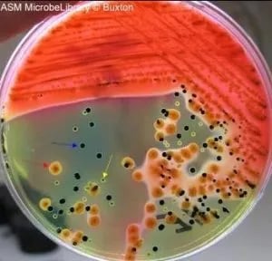 Figure: Stool culture on Hektoen enteric agar: mixed flora including Escherichia coli (red arrow), Salmonella (blue arrow), and Proteus vulgaris (yellow arrow).
Preparation of HE Agar
- Suspend 76 grams of dehydrated HE agar powder in one liter of distilled or deionized water. (Follow manufacturer instructions: formulations vary between Oxoid, BD Difco, and HiMedia.)
- Mix well and heat with frequent agitation, bringing to the boil.
- Do not autoclave. Autoclaving degrades the carbohydrates, dyes, and bile salts, reducing both selectivity and differential capacity. Boiling for one minute only is sufficient for sterilization of this medium.
- Cool to 50°C and pour into sterile Petri dishes (approximately 20 mL per plate).
- Allow to solidify on a level surface. The set medium should appear deep blue-green.
- Store inverted at 2–8°C. Use within two weeks.
- Plates should be used immediately after warming to room temperature. Do not allow plates to remain at room temperature for extended periods before inoculation, as the medium continues to acidify slightly with time.
Quality check: Uninoculated HE agar should be deep green to blue-green after solidification. A yellow or orange tint before inoculation indicates acid degradation during preparation; discard and remake. A black precipitate forming before inoculation indicates thiosulfate-ferric citrate reaction has already occurred; discard the batch.
Inoculation and Incubation
- Warm the plate to room temperature and dry the agar surface before use.
- Inoculate directly with fresh feces or a rectal swab. For a swab, roll it over a small area of the agar first. For a fecal suspension, apply a loopful.
- Streak for isolation with a sterile loop so that well-separated colonies form on part of the plate.
- Incubate aerobically at 35 to 37°C for 18 to 24 hours.
- If growth is light or no non-fermenting (blue-green) colonies are seen, reincubate for a further 18 to 24 hours. Extended incubation also improves the color separation between Salmonella and Shigella.
- HE agar is used alongside other media, not alone. Plate the same specimen on a less inhibitory medium (MacConkey or blood agar) so that any strain suppressed on HE is still recovered.
Colony Characteristics on HE Agar
The deep green background of HE agar makes colony colors particularly vivid. Reading the plate requires noting both colony color and the presence or absence of a black center.
| Organism | Colony color | Black center | Mechanism | Notes |
|---|---|---|---|---|
| Salmonella Typhi | Blue-green to teal | Yes (variable; may be small) | No carbohydrate fermentation; H₂S positive | Small black center can be faint, always pick blue-green colonies regardless |
| Salmonella typhimurium (non-typhi) | Blue-green to teal | Yes (prominent) | No carbohydrate fermentation; H₂S positive | Classic HE agar Salmonella appearance |
| Salmonella paratyphi A | Blue-green | No black center | No carbohydrate fermentation; H₂S negative | Critical: mimics Shigella — differentiate by serology and TSI |
| Shigella sonnei | Greener than Salmonella; blue-green fading at colony edge | No black center | No carbohydrate fermentation; H₂S negative | Color fades toward periphery, useful distinguishing feature from Salmonella |
| Shigella flexneri | Blue-green | No black center | No carbohydrate fermentation; H₂S negative | Similar to S. sonnei; serology for speciation |
| Shigella dysenteriae | Blue-green (grows better on HE than on SS agar) | No | No carbohydrate fermentation | Better recovery on HE than SS agar, key advantage of this medium |
| Escherichia coli | Orange to salmon-pink | No | Rapid lactose fermenter; acid production | Moderately inhibited but may produce visible orange colonies in direct plating |
| Klebsiella pneumoniae | Orange-yellow, mucoid | No | Lactose + sucrose fermenter; mucoid capsule | Very mucoid; may be hard to pick individual colonies |
| Proteus mirabilis | Small, transparent, glistening; orange-yellow with black center (variable) | Variable | H₂S positive; may ferment salicin | Proteus mimicry warning: orange-yellow colony + black center; distinguish from Salmonella (blue-green + black center) by colony color and urease test |
| Citrobacter freundii | Orange-yellow with black center | Yes | H₂S positive; lactose fermenter (variable) | Another H₂S positive mimic; bile salts usually suppress it; if it grows, orange-yellow color distinguishes it from Salmonella |
| Yersinia enterocolitica | Small, blue-green, 0.5–1 mm | No | Non-fermenter; H₂S negative | Small colony size; better recovered at 25°C; important in pediatric diarrhea |
The key visual rule for HE agar: Blue-green = non-fermenter = potential pathogen (Salmonella or Shigella). Orange-yellow = fermenter = likely coliform. The exception, orange-yellow with a black center, usually means Proteus or Citrobacter, not Salmonella (which would be blue-green with a black center). When in doubt: urease test immediately differentiates Proteus (strongly positive) from Salmonella (negative).
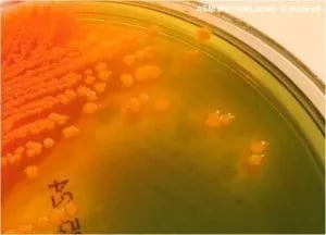 Figure: Yellow-orange colonies of Proteus vulgaris in Hektoen Enteric Agar
HE Agar vs Other Enteric Media
| Feature | HE Agar | XLD Agar | SS Agar | DCA |
|---|---|---|---|---|
| Primary selective agents | Bile salts + bromothymol blue + acid fuchsin | Sodium deoxycholate | Bile salts + sodium citrate + brilliant green | Sodium deoxycholate + sodium citrate |
| Selectivity level | Moderate-high | Moderate-high | High | Moderate |
| Shigella dysenteriae recovery | Good | Good | Poor (inhibited by brilliant green) | Moderate |
| Salmonella colony appearance | Blue-green with black center | Red-pink with black center | Colorless with black center | Colorless with black center |
| Shigella colony appearance | Blue-green, fading at edge, no black center | Red-pink, no black center | Colorless (if grows) | Colorless |
| Coliform appearance | Orange-yellow | Yellow | Pink-red | Pink |
| Baseline medium color | Deep green | Bright red | Pale | Pale pink |
| H₂S indicator system | Thiosulfate + ferric ammonium citrate | Thiosulfate + ferric ammonium citrate | Thiosulfate + ferric citrate | Thiosulfate + ferric ammonium citrate |
| Do not autoclave | Yes | Yes | Yes | Yes |
| Best for | Shigella recovery; when brilliant green toxicity a concern | Three-step Salmonella differentiation; best single enteric medium | Maximum coliform suppression; Salmonella specialist | Standard stool culture; Salmonella + Shigella |
Practical recommendation: For the most complete enteric pathogen recovery, use XLD + MacConkey as the standard two-medium battery, with HE agar substituted for XLD when Shigella epidemic dysentery is the primary clinical concern. HE agar and XLD together in a two-plate battery provides excellent coverage of both Salmonella and Shigella across all species.
How to Remember
HE agar was designed to fix a specific problem and the name of that problem is brilliant green.
SS agar uses brilliant green, which works brilliantly for suppressing coliforms but also kills Shigella. HE agar replaced brilliant green with two milder dyes (bromothymol blue + acid fuchsin) and added extra carbohydrates to compensate. The result: better Shigella recovery without losing Salmonella selectivity.
The green medium rule:
Green plate → blue-green colony = non-fermenter = pick it (Salmonella or Shigella suspect) Green plate → orange-yellow colony = fermenter = ignore it (coliform) Orange-yellow + black center = Proteus/Citrobacter, not Salmonella (which is blue-green + black center)
Three carbohydrates, one rule: Lactose, sucrose, salicin: Salmonella and Shigella ferment none of them. Any organism that ferments even one of these three turns orange-yellow. This three-carbohydrate system reduces false negatives from organisms that might escape detection with lactose alone.
Historical anchor: King and Metzger, Hektoen Institute, 1968. The medium is named after the institute, not a person. The goal was to recover more Shigella — and the key innovation was replacing brilliant green. This historical context helps remember why HE agar exists in a world that already had SS agar, XLD, and DCA: it was a deliberate improvement in Shigella sensitivity.
Where Students Get Confused
The article has excellent reading rules scattered through it (the green-medium rule, the Proteus warning), but no consolidated confusion section. Add after How to Remember, as normal markdown:
1. Blue-green means non-fermenter, which means possible pathogen. The color logic on HE agar is the reverse of what students expect. On most media, the pathogen "stands out"; here the pathogen is the quiet blue-green colony that looks like the background, while the harmless coliforms turn bright orange-yellow. Pick the blue-green colonies, not the colorful ones.
2. A black center means H₂S, not automatically Salmonella. Proteus and Citrobacter also produce H₂S and can form black-centered colonies. The distinguishing feature is the base color: Salmonella is blue-green with a black center, while Proteus and Citrobacter are orange-yellow with a black center, because they ferment one of the three sugars. When a black-centered colony is orange-yellow rather than blue-green, suspect Proteus or Citrobacter and confirm with a urease test.
3. S. Paratyphi A has no black center and mimics Shigella. Most Salmonella produce a black center from H₂S. S. Paratyphi A is H₂S negative, so it is blue-green with no black center, the same as Shigella. A missing black center does not rule out Salmonella. Confirm blue-green colonies with serology and TSI regardless of the black center.
4. Shigella sonnei color fades toward the colony edge. S. sonnei colonies are blue-green but the color fades toward the periphery, which can help distinguish them from the more uniformly colored Salmonella. This is a subtle visual cue, not a definitive test; confirm by serology.
5. Never autoclave HE agar, and read the plate color before you inoculate. Autoclaving degrades the carbohydrates, dyes, and bile salts, destroying both selectivity and differentiation. A properly made plate is deep blue-green. If the uninoculated plate is yellow or orange, it acidified during preparation and must be discarded, because the color reactions will no longer be reliable.
References
- Goo VY, Ching GQ, Gooch JM. Comparison of brilliant green agar and Hektoen enteric agar media in the isolation of salmonellae from food products. Applied Microbiology. 1973;26(3):288-292. https://doi.org/10.1128/am.26.3.288-292.1973
- King S, Metzger WI. A new plating medium for the isolation of enteric pathogens. I. Hektoen enteric agar. Applied Microbiology. 1968;16(4):577-578. https://doi.org/10.1128/am.16.4.577-578.1968
- Tille PM. Bailey and Scott's Diagnostic Microbiology. 15th ed. St. Louis: Elsevier; 2022.
- Cheesbrough M. District Laboratory Practice in Tropical Countries, Part 2. 2nd ed. Cambridge: Cambridge University Press; 2006.

Tankeshwar Acharya, MSc (Medical Microbiology)
Tankeshwar Acharya is an Assistant Professor in the Department of Microbiology at Patan Academy of Health Sciences (PAHS), Nepal, where he has been teaching and practicing clinical microbiology for over 14 years. He is the founder of Microbe Online, one of the leading free microbiology education resources on the web, covering bacteriology, mycology, parasitology, immunology, and clinical laboratory diagnostics written from direct experience in both the classroom and the diagnostic laboratory.
Comments
No comments yet. Be the first to share your thoughts.
Leave a comment
All comments are reviewed before they appear.