
Formal-Ether (Formalin-Ethyl Acetate) Sedimentation Technique: Principle, Procedure, and Results
Step-by-step guide to the formal-ether (formalin-ethyl acetate) sedimentation technique, or FECT: principle, the four layers and which one holds the parasites, quality control, and when to choose it over flotation or Kato-Katz.

On this page
A district laboratory in rural Ethiopia receives a stool specimen from a child with chronic intermittent diarrhea. A direct saline wet mount is prepared but turns out to be negative. The clinician is not convinced. The technician then runs a formal-ether sedimentation: after centrifugation, a single Giardia cyst is visible in the sediment. Treatment is started.
This scenario is the reason concentration techniques exist. Direct wet mount examines roughly 2 mg of stool from a 10 to 20 g sample. Concentration techniques like formal-ether sedimentation, often abbreviated FECT (formal-ether concentration technique), process a much larger volume, physically accumulate parasites into a small sediment, and increase detection sensitivity by 3 to 5 times.
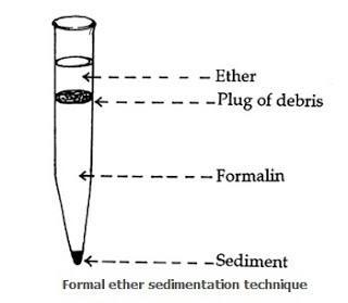 Figure: Fig-1: Formal Ether Sedimentation Technique
The parasitic forms of protozoa and helminths in a fecal specimen are often too few to be seen microscopically in direct wet mounts or in stained smear preparation. In such cases, the use of the concentration technique increases the chances of detecting parasitic organisms, thus increasing the sensitivity of copromicroscopic technique.
The two most commonly used stool concentration techniques are sedimentation and flotation. Sedimentation techniques are performed commonly in general diagnostic laboratories because they are easier to perform and less prone to technical errors.
When to Use Formal-Ether Sedimentation
| Scenario | Best method |
|---|---|
| Suspected protozoa + helminths (mixed) | Formal-ether as it detects cysts, eggs, larvae |
| Quantifying helminth egg burden (EPG) | Kato-Katz as it gives eggs per gram |
| Surveillance survey for soil-transmitted helminths | Kato-Katz |
| Suspected Cryptosporidium, Giardia (single method) | Formal-ether + modified acid-fast for Cryptosporidium |
| Light-density infection, single sensitive method needed | Formal-ether |
| Flotation with zinc sulfate | Used for Giardia cysts and some nematode eggs; not for heavy eggs (Fasciola, Taenia, unfertilized Ascaris) |
Key advantage over flotation: Formal-ether detects all organisms regardless of density: heavy eggs (Fasciola hepatica, Schistosoma, unfertilized Ascaris) that do not float in zinc sulfate still sediment and are recovered. This is why sedimentation is the workhorse method in general diagnostic laboratories.
Principle of Formal Ether Sedimentation Technique
Sedimentation techniques use solutions of lower specific gravity than the parasitic organisms, thus concentrating the latter in the sediment.
It takes advantage of the high specific gravity of protozoan cysts and helminth eggs compared to water. Their natural tendency to settle out in aqueous solutions can be accelerated by light centrifugation.
Formalin fixes the eggs, larvae, oocysts, and spores, so that they are no longer infectious, as well as preserves their morphology. Fecal debris is extracted into the ethyl acetate phase of the solution. Parasitic elements are sedimented at the bottom.
Materials required
- Glass container
- Gauze
- Funnel
- Centrifuge tube (15ml capacity)
- Centrifuge
- Physiological saline (0.85% w/v NaCl)
- 10% buffered formalin
- Ether (ethyl acetate)
- Test tubes with stopper
- Glass rod
- Iodine
- Microscope
Procedure for Formal Ether Sedimentation Technique
- Wear gloves when handling stool specimens.
- In a suitable container, thoroughly mix a portion of stool specimen about the size of a walnut into 10mL of saline solution. Mix thoroughly.
- Filter the emulsion through fine mesh gauze into a conical centrifuge tube.
- Centrifuge the suspension at a relative centrifugal force (RCF) of 600 g (about 2000 rpm) for no less than 10 minutes. The suspension should yield about 0.75mL of sediment for fresh specimens and 0.5 mL for formalinized feces.
- Decant the supernatant and wash the sediment with 10 mL of saline solution. Centrifuge again and repeat washing until supernatant is clear.
- After the last wash, decant the supernatant and add 10 mL of 10% formalin to the sediment. Mix and let stand for 5 minutes to effect fixation.
- Add 1 to 2 mL of ethyl acetate. Stopper the tube and shake vigorously.
- Centrifuge at 450 g RCF (about 1500 rpm) for 10 minutes. Four layers should result as follows: a top layer of ethyl acetate; plug of debris; layer of formalin; and sediment
- Free the plug of debris from the side of the tube by ringing with an applicator stick. Carefully decant the top three layers.
- With a pipette, mix the remaining sediment with the small amount or remaining fluid and transfer one drop each to a drop of saline and iodine on a glass slide. Cover with a coverslip and examine microscopically for the presence of parasitic forms.
After the final centrifugation, four layers form. From top to bottom they are: ethyl acetate, a plug of debris, formalin, and sediment. The parasites are in the bottom sediment layer. Every other layer is discarded. A useful way to hold the order is that the two useful things sit at the extremes: the fat solvent works at the top, the parasites collect at the bottom, and the two waste layers sit in the middle.
What each layer contains and why it forms:
| Layer | Contents | Why it forms |
|---|---|---|
| Top: ethyl acetate | Dissolved fats, lipids, and fat-soluble debris | Ethyl acetate (density < water) floats; extracts lipids away from parasites |
| Plug: debris | Non-lipid organic debris (plant fibers, undigested food) | Concentrated at interface by defatting |
| Layer: formalin | Aqueous phase with preserved parasite elements | Water-miscible formalin solution |
| Bottom: sediment | Parasitic cysts, eggs, larvae | Heaviest elements; gravity + centrifugation pellets them |
Clinical point: The sediment is the only layer examined. The debris plug must be freed from the tube wall and discarded cleanly. If it falls back into the sediment before decanting, it obscures the parasites below.
Quality Control
- Check solutions with each use to be sure they are clear and free of any bacterial contamination.
- Run known positive specimens through the procedure to verify organism recovery. This should be done at least two times per year.
Observation and Results
Systematically examine the entire surface of each coverslip with the 10x objective or, if needed for identification, higher power objectives of the microscope in a systematic manner so that the entire coverslip area is observed. When organisms or suspicious objects are seen, switch to higher magnification (40X) to see the more detailed morphology of the object in question.
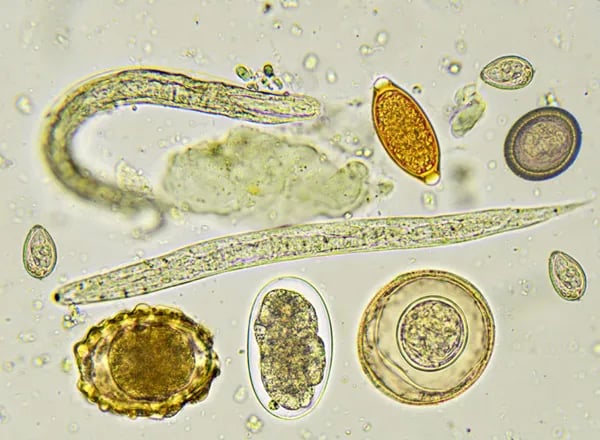 Figure: Eggs (ova) of various intestinal parasites
Procedure Notes-sedimentation technique
- The sedimentation procedure can also be used to process polyvinyl alcohol (PVA) fixed specimens. Fill one half of a tube with the stool-PVA mixture and add 0.85% NaCl almost to the top of the tube. Then filter the mixture through wet gauze into a 15 mL centrifuge tube and follow the remaining standard procedure.
- If ethyl acetate is used, swab the insides of the tube with a cotton-tipped applicator stick after the plug of debris is rimmed and the excess fluid is decanted. Excess ethyl acetate in the sediment at the time smears are prepared will lead to bubbles that may obscure parasitic forms one is attempting to observe.
- Errors in interpretation may occur if too much or too little feces is used in the sedimentation procedure. Adhere to the recommended formula of 0.75 mL of sediment for fresh specimens and 0.5 mL for formalinized feces.
- Allow the centrifuge to reach maximum speed before you start timing. If the centrifugation time is too little, certain smaller parasitic forms, such as the oocysts of Cryptosporidium species, may not reach the sediment.
Limitation of Sedimentation Technique
- Certain parasites, such as Giardia lamblia, hookworm eggs, and Trichuris eggs may not concentrate well from PVA-preserved specimens. The oocysts of Isospora belli do not routinely appear in concentrates. Therefore, examination of permanently stained smear is highly recommended.
- With both the sedimentation and the flotation techniques, species identification may not be possible in all cases, depending on the clarity of the forms observed. Permanently stained smears are usually required to make the final identification, particularly when attempting to confirm the identity of the Entamoeba histolytica.
- Cryptosporidium oocysts: Cryptosporidium parvum oocysts are very small (4–6 μm) and may not sediment reliably if centrifugation time or speed is insufficient. Additionally, even when present in the sediment, oocysts are not reliably identified by standard wet mount, they require a modified acid-fast stain applied to the sediment smear. If Cryptosporidium is suspected (HIV/AIDS patient, traveler's diarrhea, young child), specifically request modified acid-fast staining of the formal-ether sediment. Read more about Cryptosporidium in this article.
Sedimentation vs. Flotation: Which to Choose
Both are concentration methods, and they work by opposite logic.
Sedimentation uses a solution of lower specific gravity than the parasites, so cysts, eggs, and larvae settle into the sediment while lighter debris stays suspended. Because everything denser than the solution sinks, sedimentation recovers parasites of every density, including heavy eggs.
Flotation uses a solution of higher specific gravity than the parasites, typically zinc sulfate or a saturated salt or sugar solution. The parasites rise to the surface film and are lifted off with a coverslip, while heavier debris sinks. This gives a cleaner preparation but only for the eggs and cysts light enough to float.
The practical rule: sedimentation is the general-purpose method because it misses nothing on the basis of density, though the preparation contains more debris. Flotation gives a cleaner field but fails to recover dense eggs such as those of Fasciola, Schistosoma, operculated eggs, and unfertilized Ascaris, which stay at the bottom. When a single method must cover a mixed or unknown infection, sedimentation is the safer choice.
| Feature | Sedimentation (formal-ether) | Flotation (zinc sulfate) |
|---|---|---|
| Solution specific gravity | Lower than parasites | Higher than parasites |
| Parasites collect | In the bottom sediment | In the top surface film |
| Density coverage | All densities, nothing excluded | Only low-density eggs and cysts |
| Heavy eggs (Fasciola, Schistosoma, unfertilized Ascaris) | Recovered | Missed |
| Preparation clarity | More debris | Cleaner field |
| Best use | General-purpose, mixed or unknown infections | Clean recovery of light eggs and cysts |
For a fuller comparison of all the stool concentration methods, including quantitative techniques, see the overview of stool concentration techniques.
How to Remember
The two extremes rule (layer order). In the four-layer tube, the two things you care about sit at the two ends: the fat solvent floats at the very top, the parasites pack at the very bottom. The middle is waste. If you can remember "useful at the edges, waste in the middle," you never confuse which layer to examine.
Sink vs. float logic. Sedimentation makes parasites sink; flotation makes them float. The trick is what the solution does: a light solution lets heavy parasites fall (sediment), a heavy solution pushes light parasites up (flotation). The method is named for what the parasite does, not what the solution does.
Why heavy eggs betray flotation. Picture Fasciola and unfertilized Ascaris as the "heavy" eggs. They are too dense to float, so a flotation method leaves them stranded at the bottom and misses them. Sedimentation, which collects everything that sinks, catches them. Heavy eggs are the reason sedimentation is the workhorse.
Formalin fixes, so no movement. Formalin fixes and kills. Fixed things do not move. That is the one-line reason formal-ether cannot show trophozoites: they are dead and motionless. If you need to see a moving trophozoite, you need a fresh saline mount, not this method.
Where Students Actually Get Confused
1. "Formal-ether detects trophozoites." No, formalin fixation kills trophozoites and destroys motility. Formal-ether concentration is for cysts, eggs, and larvae only. To detect trophozoites, a direct saline wet mount from fresh stool processed within 30 minutes is required.
2. "The debris plug is part of the sediment to examine." The debris plug must be cleanly discarded with the other upper layers. Allowing it to fall back into the sediment obscures the parasites. The procedure note about "ringing with an applicator stick" is specifically to free the plug before decanting.
3. "Ethyl acetate and diethyl ether are the same." The original procedure used diethyl ether, which is highly flammable and explosive. Ethyl acetate (ethyl acetate concentration technique) replaced diethyl ether in modern laboratories for safety reasons. The principle is identical, both extract fats. The current standard name is "formalin-ethyl acetate sedimentation" (FEAS); "formal-ether" is the legacy name retained because of its historical prevalence in curricula.
4. "Centrifugation time doesn't matter." Too short a centrifugation fails to pellet small oocysts (Cryptosporidium). The recommended minimum is 10 minutes at 450–600g. Always wait for the centrifuge to reach full speed before starting the timer.
5. "PVA-preserved specimens cannot be processed by formal-ether." PVA-preserved specimens can be processed by formal-ether sedimentation using the modification described in the procedure notes. However, Giardia cysts, hookworm eggs, and Trichuris eggs may not concentrate well from PVA, fresh or formalin-preserved specimens give better results.
6. "Flotation is more sensitive because the field is cleaner." A cleaner field is not the same as higher sensitivity. Flotation gives a clean preparation, but only for eggs and cysts light enough to rise in the solution. Dense eggs (Fasciola, Schistosoma, unfertilized Ascaris) never reach the surface film and are missed entirely. Sedimentation looks messier but recovers parasites of every density, which is why it is preferred when the infection is mixed or unknown.
Key Exam Facts
| Fact | Detail | Memory hook |
|---|---|---|
| Principle | Differential specific gravity: parasites sediment, fats float into ethyl acetate | Heavy parasites sink; fat floats |
| Fixative used | 10% buffered formalin | Preserves morphology; kills organisms (non-infectious) |
| Fat solvent | Ethyl acetate (replaces diethyl ether due to safety concerns) | Extracts fecal fats into top layer |
| Four layers after centrifugation | Ethyl acetate / debris plug / formalin / sediment | Examine only the sediment |
| Centrifugation | 600g × 10 min (first spin); 450g × 10 min (concentration spin) | Two-spin protocol |
| Detects | Cysts + eggs + larvae | NOT trophozoites (killed by formalin) |
| Does NOT detect | Trophozoites, Cryptosporidium reliably | Add modified acid-fast for Cryptosporidium |
| Advantage over flotation | Recovers heavy eggs (Fasciola, unfertilized Ascaris, Schistosoma) | Sedimentation = all density parasites |
| Sensitivity increase vs direct smear | 3–5× | Processes larger volume |
| Safety note | Ethyl acetate replaced ether | Ether = fire/explosion risk |
Frequently Asked Questions
What is the principle of the formal-ether sedimentation technique?
What does formal-ether sedimentation detect and what does it miss?
Formal-ether sedimentation detects protozoan cysts, helminth eggs, and larvae with high sensitivity: approximately 3 to 5 times more sensitive than direct wet mount. It does not detect trophozoites (killed by formalin) and has unreliable sensitivity for Cryptosporidium oocysts, which require a modified acid-fast stain applied to the sediment smear.
Why is ethyl acetate used instead of ether in the modern technique?
Diethyl ether, used in the original procedure, is highly flammable and has explosive vapor risk, a significant laboratory safety hazard especially near centrifuges and electrical equipment. Ethyl acetate performs the same function (dissolving fecal lipids into the top layer) without the explosion risk and has become the standard reagent in modern formalin-ethyl acetate concentration (FEAS) protocols.
What does FECT mean in parasitology?
What does FECT mean in parasitology?
FECT stands for formal-ether concentration technique, also written as the formalin-ethyl acetate concentration technique. It is the stool concentration method described on this page. "FECT" is the abbreviation commonly used in laboratory request forms and curricula for the same procedure.
Which layer contains the parasites in the formal-ether technique?
Which layer contains the parasites in the formal-ether technique?
The parasites are in the bottom sediment layer. After the final centrifugation, four layers form: ethyl acetate at the top, a plug of debris, a formalin layer, and the sediment at the bottom. Only the sediment is examined. The top three layers are discarded.
What is the difference between sedimentation and flotation for stool examination?
What is the difference between sedimentation and flotation for stool examination?
Sedimentation uses a solution lighter than the parasites, so all cysts, eggs, and larvae settle into the sediment regardless of density. Flotation uses a solution heavier than the parasites, so only light eggs and cysts rise to the surface film to be collected. Sedimentation recovers heavy eggs (Fasciola, Schistosoma, unfertilized Ascaris) that flotation misses, which is why it is the general-purpose method. Flotation gives a cleaner preparation but only for the parasites light enough to float.
References
1. Garcia, L. S. (2016). Diagnostic Medical Parasitology (6th ed.). ASM Press.
2. Procop, G. W., Church, D. L., Hall, G. S., Janda, W. M., Koneman, E. W., Schreckenberger, P. C., & Woods, G. L. (2017). Koneman's Color Atlas and Textbook of Diagnostic Microbiology (7th ed.). Wolters Kluwer.
3. Cheesbrough, M. (2006). District Laboratory Practice in Tropical Countries (2nd ed., Part 1). Cambridge University Press.
4. World Health Organization. (2012). Bench aids for the diagnosis of intestinal parasites (2nd ed.). WHO. https://apps.who.int/iris/bitstream/handle/10665/37323/9789241544764_eng.pdf
5. Ritchie, L. S. (1948). An ether sedimentation technique for routine stool examinations. Bulletin of the US Army Medical Department, 8(4), 326.
6. Allen, A. V. H., & Ridley, D. S. (1970). Further observations on the formol-ether concentration technique for faecal parasites. Journal of Clinical Pathology, 23(6), 545–546. https://doi.org/10.1136/jcp.23.6.545
7. CDC – DPDx: Laboratory Identification of Parasites of Public Health Concern. https://www.cdc.gov/dpdx/index.html

Tankeshwar Acharya, MSc (Medical Microbiology)
Tankeshwar Acharya is an Assistant Professor in the Department of Microbiology at Patan Academy of Health Sciences (PAHS), Nepal, where he has been teaching and practicing clinical microbiology for over 14 years. He is the founder of Microbe Online, one of the leading free microbiology education resources on the web, covering bacteriology, mycology, parasitology, immunology, and clinical laboratory diagnostics written from direct experience in both the classroom and the diagnostic laboratory.
Comments
No comments yet. Be the first to share your thoughts.
Leave a comment
All comments are reviewed before they appear.