
Enzymes Involved in DNA Replication: Roles at the Fork, with Diagram

On this page
Copying DNA looks like it should need one machine, but no single enzyme can do the whole job. Unwinding the helix, starting a new strand, adding thousands of bases accurately, and joining the pieces are different chemical problems, and each needs a different specialist. This is why DNA replication runs on a team of enzymes, each with one job, arriving at the replication fork in a set order.
DNA replication copies the parental DNA into two identical daughter molecules before a cell divides. It is the basis of biological inheritance and happens in all living organisms. The copying is not done by one enzyme but by a team, and each member has one job at the replication fork.
The order matters, and the lagging strand is where most students lose the thread. This article walks through each enzyme by the job it does, in the order the fork needs it: helicase, gyrase (topoisomerase), primase, DNA polymerase, and ligase.
Full process of replication (the modes, the step-by-step fork mechanism, and termination) is covered in the companion article on DNA replication. This article focuses on the enzymes themselves.

How many enzymes are involved in DNA replication?
There is no single number, because it depends on how you count and on the organism. The core team that does the main work is small, usually given as five: helicase, primase, DNA polymerase, ligase, and topoisomerase (gyrase).
But replication also uses helper proteins that are not always counted as "enzymes," such as single-stranded binding proteins, and in bacteria the polymerase job is split across several DNA polymerases (I, II, III, IV, and V), while eukaryotes use a different set (α, β, γ, δ, ε). So the honest answer is: about five core enzymes carry the process, with several supporting proteins alongside them.
A quick guide to who does what, before the detail below:
| The job | The enzyme |
|---|---|
| Relieve the twist ahead of the fork | Topoisomerase / gyrase |
| Unwind the two strands | Helicase |
| Start a new strand (lay a primer) | Primase |
| Add nucleotides to build the new strand | DNA polymerase III (main) |
| Remove the RNA primer and fill the gap | DNA polymerase I |
| Seal the joins between pieces | DNA ligase |
The enzyme that "positions nucleotides," is DNA polymerase: it reads the template and places each matching nucleotide onto the growing strand.
Helicase
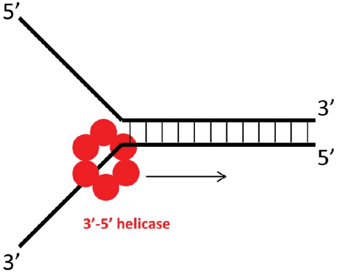 Helicase is the class of enzyme that separates the double strands of nucleic acids into single strands. It is also known as the helix destabilizing enzyme. Helicases require the energy which ATP provides. They are of two types, i.e., DNA helicase and RNA helicase. Helicase is involved in the different DNA modification processes like DNA replication, repair, recombination, transcription, translation, etc.
Helicase plays a crucial role in the DNA replication process. In bacteria, the main replicative helicase is DnaB, a ring-shaped hexamer of six identical subunits. It loads onto the DNA and moves along one strand, unwinding the double helix ahead of it. Replication usually proceeds in both directions from an origin, and the unwound region forms the Y-shaped replication fork.
DNA Topoisomerase
DNA topoisomerase is the enzyme that cuts and resolves the supercoils formed during the unwinding process. There are two types of supercoiling, positive and negative supercoiling.
A positive supercoil is twisted more and has many turns than negative supercoiling. It is faced towards the tightening of the coil, whereas the negative supercoil is faced towards losing the coils. Based on the twists and turns, topoisomerase acts on the strands. Topoisomerase forms the ester bond between a tyrosine residue of the enzyme and the DNA molecule. Then it reseals the cuts by forming a phosphodiester bond, and the enzyme gets released from the DNA.
Type I topoisomerase
Type I topoisomerase relaxes supercoils by cutting a single strand of the double helix, letting it turn, and resealing it. It needs no ATP for this. In bacteria, topoisomerase I mainly relaxes negative supercoils.
Type II topoisomerase
Type II topoisomerase cuts both strands of the helix, passes another segment of DNA through the break, and reseals it. This needs ATP. In bacteria, the type II enzyme is DNA gyrase, which removes the positive supercoils that build up ahead of the fork as helicase unwinds the DNA. Without gyrase, the DNA ahead of the fork would become too tightly wound for replication to continue.
Clinical note: DNA gyrase is the target of the fluoroquinolone antibiotics (for example, ciprofloxacin and levofloxacin). By blocking gyrase, these drugs stop the bacterium from relieving the strain ahead of its replication fork, and replication stalls. This is covered in full in the exam table at the end.
Primase
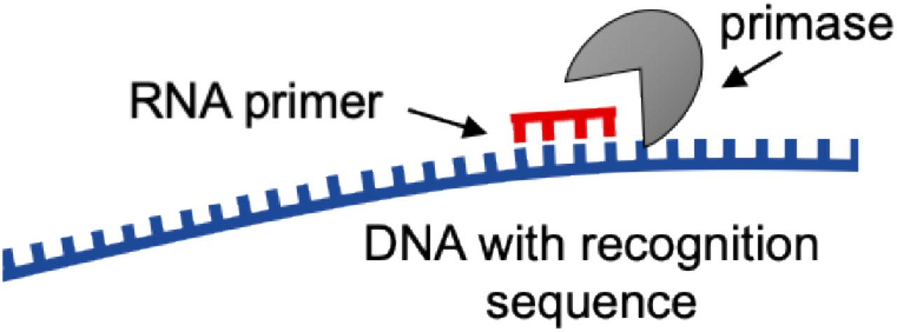 Primase is a type of RNA polymerase that creates the RNA primer. Primase binds to the DNA helicase in bacteria in the form of primosomes. Helicase activates the primase then it synthesizes the primer. Primers are short, roughly 10 to 12 nucleotides long. There are two different types of primase, i.e., Dna G and AEP. Dna G is in bacteria, and AEP is in eukaryotes and archaea.
Primase makes the short RNA primers on the single-stranded DNA template. These RNA primers are the short stretches of RNA complementary and antiparallel to the DNA template. This produces a short RNA-DNA hybrid, where the RNA primer base-pairs with the DNA template. One primer is enough for the leading strand, but the lagging strand needs a new primer for every Okazaki fragment. Then DNA polymerase adds the nucleotides to elongate DNA during the replication process.
DNA Polymerase
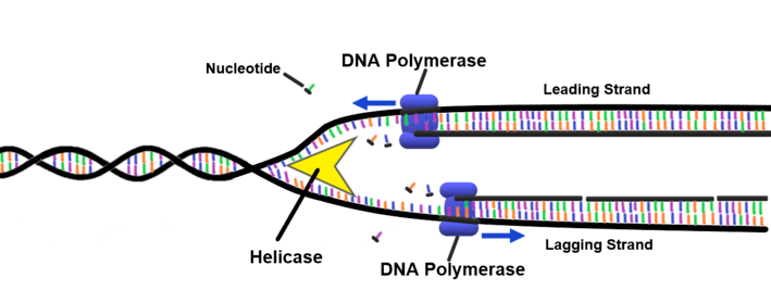 DNA polymerases use deoxyribonucleotides and synthesize the DNA molecules by assembling the nucleotides. It matches the correct nucleotides and then joins the adjacent nucleotides to each other. DNA polymerase can only add nucleotides to the free 3′-OH end of a growing strand, so every new strand is built in the 5′ to 3′ direction. It is also involved in proofreading and error correction. DNA polymerase has a single active site from which it catalyzes the addition of any of the four deoxynucleoside triphosphates. DNA polymerase can add as many as 1000 nucleotides per second to the primer strand.
Prokaryotic DNA polymerase
The prokaryotic DNA polymerases are DNA polymerases I, II, III, IV, and V.
DNA polymerase I
DNA polymerase I or Pol I is encoded by the polA gene. In a single bacteria, about 400 Pol I molecules are present. Before being dissociated from the template strand, DNA polymerase I only make an average of 20 phospho-diester bonds. After the formation of the Okazaki fragment, RNA primers are removed from the lagging strand. Then DNA polymerase I adds the DNA nucleotides.
Pol I performs the three enzymatic activities.
- It performs the 5’-3′ DNA-Dependent DNA polymerase activity. It requires the 3′ primer site and a template strand.
- It performs the 3’-5′ exonuclease activity. It means the ability to remove the nucleotides from the 3′ end of the chain. It helps in the proofreading activity.
- It performs the 5’-3′ exonuclease activity. It removes the nucleotides from the 5′ end of DNA or from an RNA primer. It helps in DNA replication and repair.
DNA polymerase II
The pol B gene encodes DNA polymerase II or Pol II. Polymerase II does not have the 5’-3′ exonuclease activity.
DNA polymerase III
It has proofreading activity and corrects DNA replication errors using the exonuclease activity working 3′ to 5′ direction. DNA polymerase III binds the RNA primer and keeps on elongating the DNA chain by adding the deoxynucleotides. It can add the nucleotides only in the 3′ hydroxyl end of the growing chain, due to which the synthesis of the daughter molecule occurs in the 5′ to 3′ direction.
The core of DNA polymerase III consists of three subunits: α, ε, and θ. (The full holoenzyme also includes a sliding clamp and a clamp-loader complex that hold it on the DNA.)
These subunits perform their functions. The ɑ is encoded by the dnaE gene, and it has polymerase activity. The ε subunit is encoded by the dnaQ gene and has the3’-5′ exonuclease activity. The holE gene encodes the θ subunit. It stimulates the ε subunit’s proofreading activity.
DNA polymerase IV
DNA polymerase IV is encoded by the dinB gene. It is used in DNA repair.
DNA polymerase V
DNA polymerase V is encoded by the umuC and umuD genes during the SOS repair of DNA. SOS repair is cells’ last resort repair mechanism when exposed to a high level of mutagen or radiation.
| Name of enzyme | Function |
|---|---|
| Helicase | Unwinds the double strand |
| Gyrase (topoisomerase II) | Relieves supercoiling ahead of the fork |
| Primase | Synthesizes the RNA primer |
| DNA polymerase III | Adds nucleotides (main synthesis) and proofreads |
| DNA polymerase I | Removes RNA primer and fills the gap with DNA |
| DNA topoisomerase I | Cuts and reseals a single strand |
| DNA ligase | Joins the fragments together |
Eukaryotic DNA polymerases
DNA replication in eukaryotes occurs in two different places, i.e., the nucleus and mitochondria. The eukaryotic DNA polymerases are α, β, γ, 𝛅 and ε
DNA polymerase alpha (ɑ)
Polymerase alpha initiates the synthesis of the strand on both the leading and lagging strand. DNA polymerase ɑ has primase activity, but the exonuclease activity is absent.
DNA polymerase beta (β)
It plays a crucial role in base excision repair (BER), essential for maintaining the DNA and in replication, recombination, etc.
DNA polymerase gamma (γ)
It is crucial for mitochondrial DNA replication and repair.
DNA polymerase delta (δ)
It is involved in DNA replication and repair in eukaryotes.
DNA polymerase epsilon (ε)
It helps synthesize the leading strand of DNA and base excision repair.
Leading strand vs lagging strand: where students get stuck
DNA polymerase can only build in the 5′ to 3′ direction. But the two template strands run in opposite directions. This one fact creates the whole leading/lagging problem.
On the leading strand, the template runs the "right way," so DNA polymerase III follows the helicase and builds one continuous strand. One primer, then non-stop synthesis.
On the lagging strand, the template runs the "wrong way." The polymerase cannot follow the fork continuously, so it works backward in short pieces called Okazaki fragments. Each fragment needs its own primer. DNA polymerase I then removes each RNA primer and fills the gap with DNA, and DNA ligase seals the nicks between fragments.
That is why the lagging strand needs so many more enzymes doing so much more work for the same length of DNA.
DNA ligase
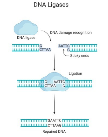 In the lagging strands, different Okazaki fragments are formed. DNA ligase helps join the strands together by forming the phosphodiester bond. A phosphodiester bond is formed between the 3′ hydroxyl end and 5′ phosphate end of the two DNA strands. Making the phosphodiester bond requires a free OH group at the 3′ end and phosphate group at the 5′ end of the other DNA strand. This reaction needs energy. Bacterial DNA ligase uses NAD+ as its energy source, while many other ligases use ATP. DNA ligase is used in DNA replication, repair, and recombination.
Why each enzyme is essential: what would fail without it
The clearest way to understand these enzymes is to ask what would go wrong if each one were missing. Replication is a chain, and removing any link stops the whole process.
Without topoisomerase (gyrase): as helicase unwinds the helix, the DNA ahead of the fork winds tighter and tighter, like twisting a rope. Without gyrase to relieve this strain, the DNA would become too tightly coiled for the fork to move, and replication would stall. This is exactly how fluoroquinolone antibiotics kill bacteria: they block gyrase, and the fork jams.
Without helicase: the two strands would never separate, so there would be no single-stranded template to copy. Nothing downstream could begin.
Without primase: DNA polymerase cannot start a strand from bare template. It can only extend an existing end. With no primer to provide that starting point, synthesis could never begin, on either strand.
Without DNA polymerase III: there would be no main synthesis. The new strands would not be built. This is the workhorse, and its proofreading is also a major reason replication is so accurate.
Without DNA polymerase I: the RNA primers would stay in the new DNA, and the gaps left after primer removal would not be filled. The lagging strand especially would remain a patchwork of RNA and DNA rather than clean DNA.
Without DNA ligase: the Okazaki fragments on the lagging strand would never be joined. The new strand would remain in pieces instead of one continuous molecule.
Read in order, this is the logic of the fork: relieve the strain, unwind, start, build, clean up, seal. Each enzyme solves a problem created by the step before it, which is why they act in a fixed sequence rather than all at once.
How to Remember
The order at the fork. Say it as a sentence: "Gyrase gives room, helicase opens, primase primes, Pol III writes, Pol I fixes, ligase seals." Six verbs, six enzymes, in the order they act.
Which polymerase is which (Pol I vs Pol III). The higher number does the bigger job. Pol III is the main workhorse that lays down most of the new DNA. Pol I is the cleanup crew that removes primers and fills the gaps. A memory check: if the question is about primer removal, the answer is the lower number, Pol I.
The two exonuclease directions. Proofreading removes a wrong nucleotide just added at the 3′ end, so proofreading is 3′ to 5′ exonuclease. Primer removal happens at the 5′ end of the primer, so it is 5′ to 3′ exonuclease. Self-check: "proofreading reads backward" (3′→5′).
Gyrase. It works ahead of the fork, like a person untwisting a garden hose before you can pull more of it. It is also the fluoroquinolone target, so "gyrase = quinolone" is worth one repetition.
Where Students Get Confused
Helicase vs topoisomerase. Helicase separates the two strands (breaks the base pairs). Topoisomerase/gyrase relieves the twisting strain that unwinding creates further along the molecule. Different problems, different enzymes.
Primase makes RNA, not DNA. The primer is a short stretch of RNA, not DNA. This is why it later has to be removed and replaced with DNA by Pol I.
Pol III proofreads; Pol I removes primers. Both are polymerases and both have exonuclease activity, so they get swapped. Pol III is main synthesis plus 3′→5′ proofreading. Pol I removes the RNA primer (5′→3′ exonuclease) and fills in.
"Only one primer" is only true for the leading strand. The lagging strand needs a new primer for every Okazaki fragment.
Prokaryotic vs eukaryotic names do not match one-to-one. Bacterial Pol III is the main replicase; in eukaryotes that job is split between Pol δ and Pol ε, and the primer is made by Pol α, which carries primase activity. Do not map bacterial and eukaryotic polymerases by number.
Key exam facts in one table
| Enzyme | Job at the fork | Direction / energy | Exam hook |
|---|---|---|---|
| Helicase (DnaB in bacteria) | Unwinds the double helix | Uses ATP | "Helix opener" |
| DNA gyrase (topoisomerase II) | Relieves supercoils ahead of the fork | Uses ATP | Fluoroquinolone target (ciprofloxacin, levofloxacin) |
| Topoisomerase I | Relaxes supercoils, single-strand cut | No ATP | Single-strand vs gyrase double-strand |
| Primase | Makes the RNA primer | Makes RNA, not DNA | Primer is RNA, later removed |
| DNA polymerase III | Main new-strand synthesis + proofreading | Builds 5′→3′; proofreads 3′→5′ | The workhorse |
| DNA polymerase I | Removes RNA primer, fills gap | 5′→3′ exonuclease removes primer | Cleanup crew |
| DNA ligase | Seals nicks between fragments | Uses ATP (or NAD+ in bacteria) | Joins Okazaki fragments |
Reference
- Madigan MT, Bender KS, Buckley DH, Sattley WM, Stahl DA. Brock Biology of Microorganisms. 16th ed. Pearson; 2021.
- Watson JD, Baker TA, Bell SP, Gann A, Levine M, Losick R. Molecular Biology of the Gene. 7th ed. Pearson; 2013.
- Timson DJ, Singleton MR, Wigley DB. DNA ligases in the repair and replication of DNA. Mutat Res. 2000;460(3-4):301-318. https://doi.org/10.1016/S0921-8777(00)00033-1
Frequently Asked Questions
What are the main enzymes involved in DNA replication?
What are the main enzymes involved in DNA replication?
Helicase unwinds the double helix, gyrase (a topoisomerase) relieves the strain ahead of the fork, primase makes RNA primers, DNA polymerase III carries out the main synthesis, DNA polymerase I removes the primers and fills the gaps, and DNA ligase seals the pieces together.
What is the difference between DNA polymerase I and III in bacteria?
What is the difference between DNA polymerase I and III in bacteria?
DNA polymerase III is the main enzyme that builds most of the new DNA strand and proofreads as it goes. DNA polymerase I is the cleanup enzyme: it removes the RNA primers and fills those gaps with DNA.
Why is the lagging strand made in fragments?
Why is the lagging strand made in fragments?
DNA polymerase can only build in the 5′ to 3′ direction. Because the two template strands run in opposite directions, the polymerase cannot follow the fork continuously on the lagging strand. It works in short pieces called Okazaki fragments, each needing its own primer.
What does DNA gyrase do, and why does it matter clinically?
What does DNA gyrase do, and why does it matter clinically?
DNA gyrase is a bacterial type II topoisomerase that removes the positive supercoils building up ahead of the replication fork. It is the target of fluoroquinolone antibiotics such as ciprofloxacin. Blocking gyrase stops replication.
Is the primer made of DNA or RNA?
Is the primer made of DNA or RNA?
The primer is a short piece of RNA, made by primase. It is later removed by DNA polymerase I and replaced with DNA.
What is the difference between the two exonuclease activities of DNA polymerase I?
What is the difference between the two exonuclease activities of DNA polymerase I?
The 3′ to 5′ exonuclease activity proofreads by removing a wrong nucleotide just added. The 5′ to 3′ exonuclease activity removes the RNA primer from ahead of the growing strand.
Which enzyme adds nucleotides during DNA replication?
Which enzyme adds nucleotides during DNA replication?
DNA polymerase adds nucleotides. In bacteria, the main synthesizing enzyme is DNA polymerase III, which reads the template strand and positions each matching nucleotide onto the growing new strand, building it in the 5′ to 3′ direction.
Are the enzymes the same in prokaryotes and eukaryotes?
Are the enzymes the same in prokaryotes and eukaryotes?
The jobs are the same, but the enzyme names differ and do not match one-to-one. Bacterial DNA polymerase III is the main replicase, while in eukaryotes that role is split between DNA polymerases δ and ε, and the primer is made by DNA polymerase α. Do not map the two sets by number.

Tankeshwar Acharya, MSc (Medical Microbiology)
Tankeshwar Acharya is an Assistant Professor in the Department of Microbiology at Patan Academy of Health Sciences (PAHS), Nepal, where he has been teaching and practicing clinical microbiology for over 14 years. He is the founder of Microbe Online, one of the leading free microbiology education resources on the web, covering bacteriology, mycology, parasitology, immunology, and clinical laboratory diagnostics written from direct experience in both the classroom and the diagnostic laboratory.
Comments
No comments yet. Be the first to share your thoughts.
Leave a comment
All comments are reviewed before they appear.