RNA Translation: Major Steps of Protein Synthesis
RNA Translation: Major Steps of Protein Synthesis
The DNA (deoxyribonucleic acid) has all the genetic instructions necessary for the proper functioning of any cell. The process of expressing genetic instructions in DNA into functional products (proteins) is called gene expression/protein synthesis. The gene expression occurs by the two processes viz; transcription and translation.
Then, what is translation? The translation is the process of translating the genetic information present in the copies of mRNA (complementary to DNA sequences) to form amino acids (foundation of polypeptides or proteins).
Proteins are vital components for both structural and functional roles in cells. Therefore, synthesizing protein (translation) is an essential task the cell performs. The ribosome, mRNA, and tRNA (transfer RNA) plays vital role in protein synthesis. As mentioned earlier, the DNA has all the required information that the mRNA decodes and then converts the decoded message to amino acids.
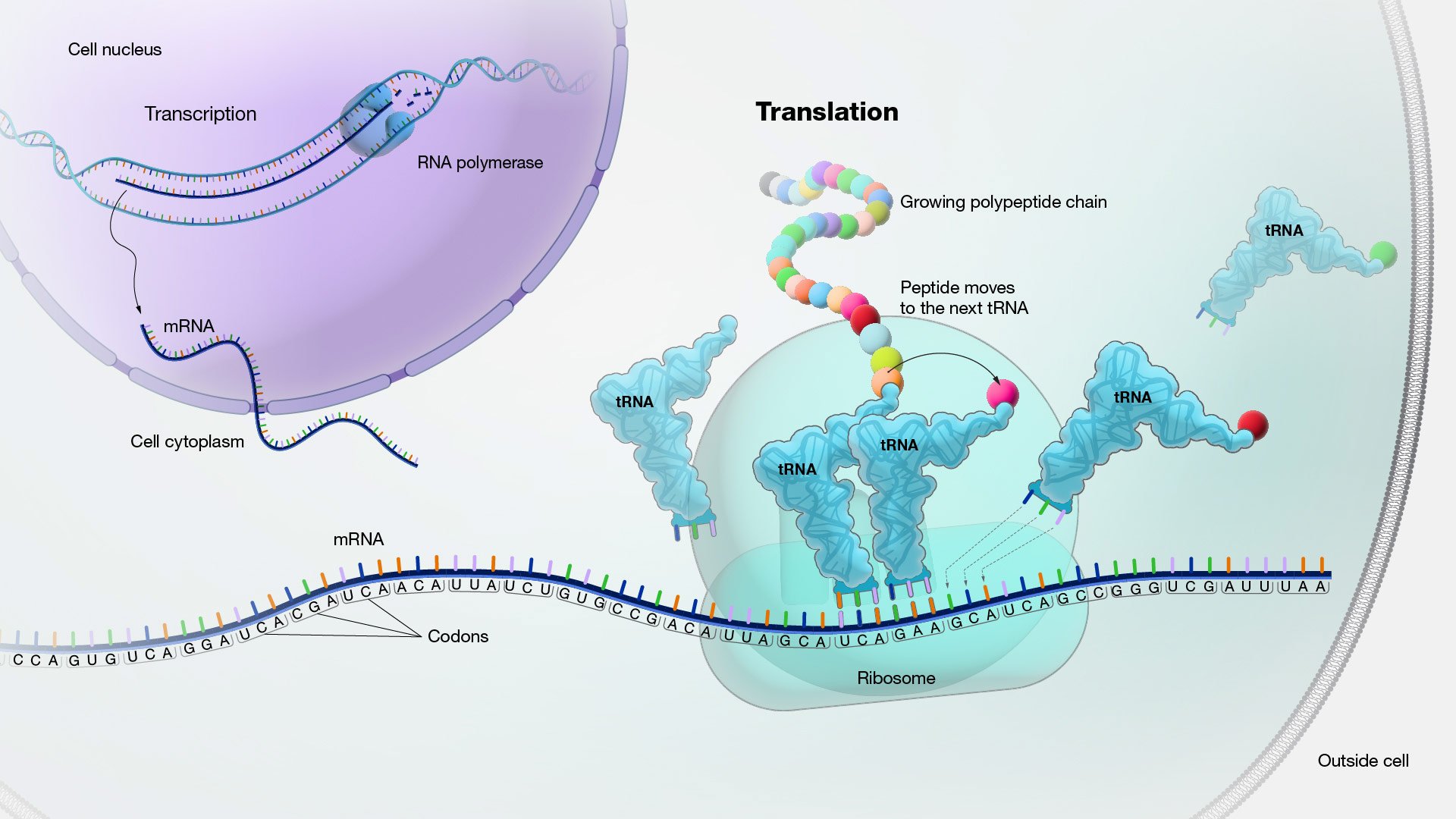 Figure: Source:https://www.genome.gov/genetics-glossary/Translation
Where does it occur?
The entire translation process occurs inside the cell organelle-ribosomes (made up of proteins and RNA). The ribosomes have two subunits; 70s (30s and 50s) in prokaryotes and 80s (40s and 60s) in eukaryotes. The subunits are made up of rRNA (ribosomal RNA) and tRNA. The two subunits have a small opening called a cleft and stay separated in normal conditions inside the cytoplasm. The mRNA passes through the cleft during protein synthesis. The tRNA acts like an adapter molecule; one end reads the codons in the mRNA, and the other binds to the specific amino acid. The aminoacyl-tRNA and mRNA are held closely together for complementary base pairing. The rRNA adds the newly synthesized amino acid to the growing chain of a polypeptide during translation.
aminoacyl tRNA- tRNA that is bound with amino acid.
Steps or Stages of Translation/Protein Synthesis
The protein synthesis or translation process occurs in four steps/stages; activation of amino acids, initiation of polypeptide synthesis, elongation of the polypeptide chains, and termination of polypeptide synthesis.
Activation of amino acids
Before synthesizing protein, amino acids (the essential compound for protein) must be activated. The activation of amino acids occurs with the help of tRNA, which translates the nucleic acid language into the language of proteins. The carboxyl group of amino acids takes part in activation. The enzyme aminoacyl tRNA synthetase catalyzes the reactions. The activation occurs in two steps; the formation of aminoacyl adenylate and the formation of aminoacyl tRNA.
**Step 1: Formation of aminoacyl adenylate-**The carboxyl group of amino acids binds with ɑ-phosphate of ATP by forming a high-energy acyl bond and aminoacyl adenylate (aa-AMP) as the end product. The 𝛽 and 𝛄 phosphates are released as PPi. The enzyme aminoacyl tRNA synthetase catalyzes the reaction in the presence of magnesium ions (Mg++).
Amino acid + ATP → Amino acyl adenylate (aa-AMP) + PPi; in the presence of aminoacyl tRNA synthetase and Mg++.
Step 2: Formation of aminoacyl tRNA- Thus formed aminoacyl adenylate reacts with tRNA in the presence of the enzyme aminoacyl tRNA synthetase and Mg++ forming aminoacyl tRNA and AMP as products. A high-energy ester bond is formed between the carboxyl group of amino acids and the 3′ hydroxyl group of terminal adenosine of tRNA.
Aminoacyl adenylate + tRNA → Aminoacyl tRNA + AMP; in the presence of aminoacyl tRNA synthetase and Mg++
Overall reaction:
Amino acid+ ATP + tRNA → Aminoacyl tRNA + AMP + PPi; enzyme- aminoacyl tRNA and Mg++
Initiation of polypeptide synthesis
For initiation of polypeptide synthesis/translation, a few components are required. They are; ribosomes, mRNA with the codons, and tRNA with methionine (first amino acids coded by AUG).
The first step is the activation of methionine
The step in eukaryotes is similar to activating any amino acids. Whereas, in prokaryotes, the activated methionine is treated with N10-formal tetrahydrofolate and forms fmet-tRNA.
In eukaryotes,
Methionine + ATP +tRNA → met-tRNA + AMP + PPi; in the presence of enzyme methionyl tRNA synthetase and Mg++.
In prokaryotes,
Transformylase enzyme transfers a formyl group from the N10-formal tetrahydrofolate to the amino group of met-tRNA. The transfer of the N formyl group prevents fmet from entering the polypeptide chain’s interior position.
- Methionine + ATP +tRNA → met-tRNA + AMP + PPi; in the presence of enzyme methionyl tRNA synthetase and Mg++
- N10-formal tetrahydrofolate + met-tRNA → fmet-tRNA +tetrahydrofolate; in presence of transformylase.
The second step is slightly different in eukaryotes and prokaryotes
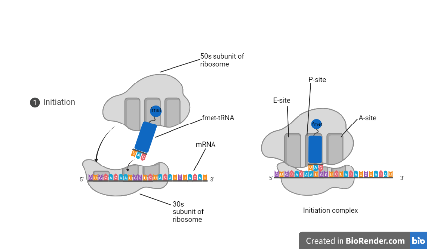 Figure: Initiation of translation in prokaryotes
In prokaryotes, three initiation factors, IF1, IF2, and IF3, are essential after the activation of methionine.
- The 30s ribosomal subunit binds to the IF1 and IF3; here, the IF3 prevents prematurely combining the larger (50s) and the smaller subunits (30s).
- Then the mRNA binds to the smaller subunit; the Shine-Dalgarno sequence points to the initiating 5′ AUG in the mRNA.
- The interaction of mRNA and 16s rRNA determines the precise position of the 5′ AUG.
- Three sites forms during the interaction of mRNA and the 30s subunit of the ribosome. A-site: Aminoacyl site where the IF2 binds, P-site: Peptidyl site (AUG binds), and E-site is the exit site. The 5′ AUG is confined in the P-site and attaches to the fmet-tRNA with the help of GTP and IF2.
- The GTP hydrolyzes and releases all the initiation factors. The newly formed complex (30s subunit + mRNA + fmet-tRNA) combines with the 50s subunit and forms the initiation complex. The initiation complex has 70s ribosome, mRNA, and fmet-tRNA
In eukaryotes, the multiple initiation factor eIF has similar functions to the initiation factor of the prokaryotes. For example, eIF3 and eIF1A and analogs to IF3 and IF1, respectively, eIF2 and eIF2B are two GTP binding proteins, etc. The activated met-tRNA attaches to the small subunit of ribosome (the 40s). The combination (met-tRNA+ small subunit of the ribosome) binds at the 5′ end of mRNA at the GTP cap. And then, the combination moves along the mRNA in the 3′ end. It stops when it finds the start codon. The larger subunit then joins with the newly formed combination of mRNA, 40s subunit, and met-tRNA, giving rise to the initiation complex.
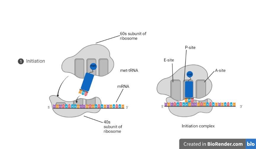 Figure: Initiation step in eukaryotes
Elongation of the polypeptide chain
The elongation of the polypeptide chain is the stage in translation where the chain of amino acids gets longer. The addition of amino acids occurs after the movement of the initiation complex in the 3′ direction of mRNA. The stage requires three elongation factors;EFTU, EFTS, and EFq. In eukaryotes, eEF1ɑ acts as an analog to EFTU, eEF2𝛽yis analog to EFTS, and eEF2is an analog of EFq. The stage can be understood in three steps; recognition, peptidyl transfer or transpeptidation, and translocation.
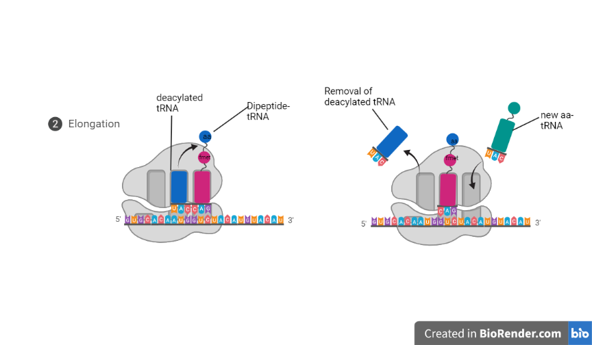 Figure: Elongation of polypeptide chains
Recognition
The P-site in the initiation complex is occupied by the met-tRNA (in eukaryotes) and fmet-tRNA (in prokaryotes). So, the recognition step occurs in the A-site of the initiation complex. The recognition has the following steps:
- A molecule of aminoacyl tRNA attaches to the A-site, which has a sequence of three bases complementary to the anticodon on tRNA.
- Two elongation factors (EFTU and EFTS) and GTP have roles in this step.
- EFTU firstly binds to the GTP. The EFTU-GTP then binds to aminoacyl tRNA. After that, EFTU-GTP-aminoacyl tRNA binds to the ribosome.
- The GTP hydrolysis and the GDP-EFTU complex release. The EFTS later dissociates the complex. Then, hydrolysis facilitates the attachment of aminoacyl tRNA into the A-site of the ribosome.
Peptidyl transfer or transpeptidation
In this step, the peptide bond forms between the terminal carboxyl group of the peptide on the P-site and the alpha-amino group of amino acids at the A-site. The enzyme peptidyl transferase catalyzes the reaction. The step does not require GTP and ATP. The tRNA in the P-site becomes uncharged and deacylated, and the dipeptide tRNA is bound to the A-site.
Translocation
In this step, the ribosomes move a codon towards the 3′ end of mRNA. The movement shifts the deacylated tRNA from the P-site to E-site. From the E-site, it releases into the cytosol. Then the anticodon of the dipeptidyl tRNA translocate from A-site to P-site. The translocation/movement of the ribosome requires EFG and a molecule of GTP. Now, the new codon in the A-site again recognizes the specific aminoacyl tRNA. The elongation step repeats and forms a long chain of polypeptides.
Termination of polypeptide synthesis
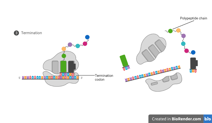 Figure: Termination of polypeptide synthesis
The presence of one of the three codons, UAA, UAG, and UGA, signals the termination of polypeptide synthesis. In prokaryotes, the three factors, RF1, RF2, and RF3 recognizes the termination signals. The RF1 recognizes UAG and UAA codons, and RF2 recognizes UGA and UAA codons. The RF3, along with GTP, releases the RF1 and RF2 and dissociates the combined ribosome into its subunits.
Whereas the eRF factor recognizes the stop codons in eukaryotes, and unchanged tRNA expels directly from the P-site.
Post Translational Modification
The nascent polypeptide chain undergoes numerous chemical, physical, and biological changes to change into the active protein, termed post-translational modification. The modifications are as follows:
Amino-terminal and carboxy-terminal modification: The removal of N-formyl methionine in bacteria may occur enzymatically to form the final functional protein. In 50% eukaryotic protein, the amino group in the amino-terminal residue is N-acetylated after translocation.
Loss of signal sequences: The loss of 15 to 30 residues at the amino-terminal directs the protein to its ultimate destination in the cell. Specific peptides ultimately remove such signal sequences.
Modification of individual amino acid: ATP enzymatically phosphorylates some proteins’ OH-group of ser, threonine, and tyrosine residues. Casein has many phospho-serine groups that bind to Ca++. So, casein provide Ca, PO4, and amino acid.
Attachment of carbohydrate: The glucose or carbohydrate group’s attachment to the polypeptide forms the glycoproteins.
Addition of the isoprenyl group: thioester bond adds the isoprenyl or isoprene group to cysteine residue. The isoprene group helps to anchor the protein in a membrane.
Addition of a prosthetic group: Prosthetic groups like the heme group are added to form hemoglobin, and the biotin molecule is the added molecule of acetyl CoA carboxylase.
Proteolytic processing: Sometimes larger inactive proteins trims to form smaller active proteins.
Formation of disulfide cross-links: The sulfide cross-linking occurs between cysteine residue in the eukaryotic cell, which protects the native confrontation of a protein molecule from denaturation in the extracellular environment. Phosphoserine is an example of this modification.
References
- Clancy, S. & Brown, W. (2008) Translation: DNA to mRNA to Protein. Nature Education 1(1):101
- Ganguly, P. (2022). Translation. Retrieved 8 August 2022, from https://www.genome.gov/genetics-glossary/Translation
- genetic code | Learn Science at Scitable. (2014). Retrieved 8 August 2022, from https://www.nature.com/scitable/definition/genetic-code-13/
- Snustad, D., & Simmons, M. (2012). Genetics (6th ed., pp. 286-315). Hoboken, NJ: Wiley.
- Watson, J., Baker, T., Bell, S., Gann, A., Levine, M., & Losick, R. (2003). Molecular biology of the gene (5th ed., pp. 411-478). Menlo Park, CA: Cold Spring Harbor Laboratory Press.

Tankeshwar Acharya, MSc (Medical Microbiology)
Tankeshwar Acharya is an Assistant Professor in the Department of Microbiology at Patan Academy of Health Sciences (PAHS), Nepal, where he has been teaching and practicing clinical microbiology for over 14 years. He is the founder of Microbe Online, one of the leading free microbiology education resources on the web, covering bacteriology, mycology, parasitology, immunology, and clinical laboratory diagnostics written from direct experience in both the classroom and the diagnostic laboratory.