
Cytoplasmic Granules in Bacteria: Types, Staining, and Diagnostic Significance

On this page
The stain that buys time before the culture comes back
A child arrives with a sore throat and a grayish membrane spreading across the tonsils, one that bleeds when a clinician tries to scrape it away. Diphtheria is on the differential, and it's a genuine emergency: the toxin can cause fatal myocarditis or airway obstruction, and antitoxin only works if given early. But confirming Corynebacterium diphtheriae by culture takes a day or two, and confirming that the strain actually produces toxin takes even longer. Waiting for either result before treating isn't a real option.
What clinicians actually have available within the hour is a direct smear from the membrane, stained with Albert's or Neisser's stain. Under the microscope, club-shaped bacilli arranged in angular clusters (often described as "Chinese letters") appear studded with dark-staining granules clustered at their poles, granules that stain a strikingly different color from the dye used to apply them. That visual signature alone is often distinctive enough to support starting antitoxin immediately, without waiting for the culture to catch up.
Those granules are called metachromatic granules, and this article is about what they actually are, chemically, why they stain the way they do, and the stains actually used to see them.
Cytoplasmic granules, also called inclusion bodies, are concentrated deposits of certain substances located in the cytoplasm of certain bacteria. They serve as storage reserves, most often for nutrients or energy, and their number and composition vary depending on the bacterial species and its nutritional environment.
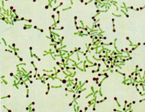 Figure: Metachromatic granules of Corynebacterium spp.
Types of Cytoplasmic Granules
- Polysaccharide granules (glycogen): a storage form of glucose.
- Polyphosphate granules (volutin/metachromatic granules): a storage form for inorganic phosphate, and the high-energy reserve responsible for the clinical stain pattern in the hook above.
- Poly-β-hydroxybutyrate (PHB) granules: a reserve carbon and energy source. Beyond storage, PHB granules have been shown to physically protect non-halophilic bacterial cells from the damaging effects of hypertonic (high-salt) environments, one reason PHB-accumulating organisms are of interest both microbiologically and industrially, since PHB is also a biodegradable bioplastic precursor.
- Elemental sulfur globules: accumulate in certain bacteria growing in hydrogen sulfide-rich environments.
- Magnetosomes: membrane-bound organelles containing magnetic crystals (usually magnetite or greigite) that let certain bacteria orient along magnetic field lines to navigate aquatic environments. Unlike the storage granules above, magnetosomes are true membrane-bound structures rather than simple deposited reserves.
Why Metachromatic Granules Stain the Way They Do
Volutin granules are called metachromatic because they stain a different color than the dye used to apply them, an intense reddish-purple with methylene blue, instead of the blue the dye itself actually is. Metachromatic granules are the characteristic feature of Corynebacterium diphtheriae.
Staining Methods Used to Visualize Cytoplasmic Granules
Albert's stain and Neisser's stain are the two stains actually used in clinical practice to demonstrate metachromatic granules, both considerably more specific and higher-contrast than plain methylene blue alone:
- Albert's stain uses toluidine blue with a malachite green mordant. Metachromatic granules stain bluish-black to greenish-black against a green-stained cytoplasm.
- Neisser's stain uses methylene blue and crystal violet as the primary stain, with a chrysoidine (vesuvin) counterstain. Granules stain blue-black against a yellow-brown cytoplasm.
Nile Red and Sudan Black B are lipophilic dyes used specifically for PHB granules; PHB granules fluoresce under Nile Red and appear black or dark blue with Sudan Black B.
Iodine stains glycogen granules brown.
Clinical Significance
A positive Albert's or Neisser's stain, combined with the classic club-shaped, angularly-arranged bacillary morphology, provides strong presumptive evidence for C. diphtheriae quickly enough to justify starting antitoxin treatment before culture results return. It is presumptive, not confirmatory: metachromatic granules aren't completely exclusive to C. diphtheriae, other Corynebacterium species can show similar granules to varying degrees, so culture and toxigenicity testing (such as the Elek test or PCR for the tox gene) remain necessary to confirm the diagnosis.
How to Remember
Anchor for the term "metachromatic": picture a guest showing up to a black-tie event in bright red instead of black, refusing to match the color they were "supposed" to wear. That's exactly what a metachromatic granule does to methylene blue: instead of taking on the dye's own blue color, it stains a completely different reddish-purple.
Anchor for the hook's clinical logic: a positive Albert's or Neisser's stain buys time, not a diagnosis. It's fast enough to justify starting antitoxin immediately, but culture and toxin testing still have to confirm what the stain only suggested.
Key exam facts in one table
| Fact | Detail |
|---|---|
| Definition | Concentrated cytoplasmic deposits (inclusion bodies) serving as nutrient/energy storage reserves |
| Volutin (metachromatic) granules | Composed of polyphosphate; characteristic of Corynebacterium diphtheriae |
| Why "metachromatic" | Stains a different color than the dye applied (reddish-purple with methylene blue, not blue) |
| Diagnostic stains for metachromatic granules | Albert's stain (bluish-black granules, green cytoplasm) and Neisser's stain (blue-black granules, yellow-brown cytoplasm) |
| PHB granules | Carbon/energy reserve; also physically protect cells against hypertonic stress; stain with Nile Red or Sudan Black B |
| Glycogen granules | Glucose storage; stains brown with iodine |
| Magnetosomes | Membrane-bound organelles (not simple storage deposits) containing magnetite/greigite for magnetic navigation |
| Clinical use | Rapid presumptive diagnosis of C. diphtheriae from a direct smear, supporting early antitoxin treatment before culture confirmation |
| Confirmatory testing still required | Culture plus toxigenicity testing (Elek test or tox gene PCR); granules are suggestive, not exclusive to C. diphtheriae |
Where Students Get Confused
- Treating a positive metachromatic stain as a confirmed diagnosis. It's a rapid presumptive finding, not a substitute for culture and toxigenicity testing, since other Corynebacterium species can show similar granules.
- Confusing "metachromatic" with simply "stains intensely." The term specifically means the granule takes on a different color than the dye itself, not just a darker version of the same color.
- Assuming plain methylene blue is equivalent to Albert's or Neisser's stain. The mordant in Albert's stain and the counterstain in Neisser's stain are what produce the sharp, diagnostically reliable contrast; plain methylene blue alone is far less specific.
- Lumping magnetosomes in with storage granules. Magnetosomes are true membrane-bound organelles used for navigation, structurally different from the simple, non-membrane-bound storage deposits (volutin, PHB, glycogen, sulfur) that make up the rest of this topic.
References
- Obruca, S., Sedlacek, P., Mravec, F., Krzyzanek, V., Nebesarova, J., Samek, O., Kucera, D., Benesova, P., Hrubanova, K., Milerova, M., & Marova, I. (2017). The presence of PHB granules in cytoplasm protects non-halophilic bacterial cells against the harmful impact of hypertonic environments. New Biotechnology, 39(Pt A), 68–80. https://doi.org/10.1016/j.nbt.2017.07.008
- Procop, G. W., & Koneman, E. W. (2016). Koneman's Color Atlas and Textbook of Diagnostic Microbiology (7th ed.). Lippincott Williams and Wilkins.
Frequently Asked Questions
What are cytoplasmic granules in bacteria?
What are metachromatic granules?
What stains are used to see metachromatic granules?
Can a positive metachromatic granule stain confirm a diphtheria diagnosis on its own?
What are PHB granules used for?
Are magnetosomes the same as other cytoplasmic granules?

Tankeshwar Acharya, MSc (Medical Microbiology)
Tankeshwar Acharya is an Assistant Professor in the Department of Microbiology at Patan Academy of Health Sciences (PAHS), Nepal, where he has been teaching and practicing clinical microbiology for over 14 years. He is the founder of Microbe Online, one of the leading free microbiology education resources on the web, covering bacteriology, mycology, parasitology, immunology, and clinical laboratory diagnostics written from direct experience in both the classroom and the diagnostic laboratory.
Comments
No comments yet. Be the first to share your thoughts.
Leave a comment
All comments are reviewed before they appear.