Albert Stain: Principle, Composition, Procedure, and Results
Albert stain detects metachromatic (volutin) granules in Corynebacterium diphtheriae — a key presumptive identification test for diphtheria. Learn the two-reagent principle, step-by-step procedure, and how to interpret results.
A child presents with a grey pseudomembrane covering the tonsils and pharynx, a "bull-neck" appearance from cervical lymphadenopathy, and a hoarse voice. The clinician suspects diphtheria. A throat swab is taken and plated on Löffler's serum medium. After overnight incubation, small grey colonies are visible. A smear is prepared and stained with Albert stain.
Under oil immersion: blue-green bacilli arranged in V and L formations — Chinese letter pattern. At the poles of the cells: dark bluish-black granules, sharply demarcated from the green cytoplasm. These are metachromatic (volutin) granules — the hallmark of Corynebacterium diphtheriae on Albert stain.
Albert stain is the standard presumptive identification test for C. diphtheriae in the clinical laboratory. A positive result — the right morphology with the right granules — is sufficient to start diphtheria antitoxin treatment immediately while awaiting confirmatory toxigenicity testing.
Albert stain is a type of differential stain used for staining high-molecular-weight polymers of polyphosphate known as metachromatic granules or volutin granules found in Corynebacterium diphtheriae. Metachromatic granules are also found in Yersinia pestis, and Mycobacterium species.
While metachromatic granules are also found in Yersinia pestis and some Mycobacterium species, Albert staining is almost exclusively used for C. diphtheriae in clinical practice. Y. pestis is identified by its characteristic bipolar (safety pin) staining on Wayson's stain rather than Albert stain, and Mycobacterium species are identified by Ziehl-Neelsen acid-fast staining. Albert stain should be specifically requested when diphtheria is clinically suspected — it is not a routine stain for these other organisms.
It is named metachromatic because of its property of changing color i.e when stained with blue stain they appear red in color. When grown in Loffler’s slopes, C. diphtheriae produces a large number of granules.
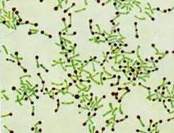 Figure: Fig: Albert Staining –Corynebacterium diphtheriae
Principle of Albert Staining
Albert stain is basically made up of two stains that are toluidine blue’ O’ and malachite green both of which are basic dyes with high affinity for acidic tissue components like cytoplasm. The pH of Albert stain is adjusted to 2.8 by using acetic acid which becomes basic for volutin granules as the pH of volutin granule is highly acidic.
Therefore on applying Albert’s stain to the smear, toluidine blue’ O’ stains volutin granules i. e the most acidic part of cell and malachite green stains the cytoplasm blue-green. On adding Albert’s iodine due to effect of iodine, the metachromatic property is not observed and granules appear blue in color.
Composition of Albert Stain
Albert stain is composed of two reagents:
Albert’s A solution consist of
- Toludine blue 0.15 gm
- Malachite green 0.20 gm
- Glacial acetic acid 1 ml
- Alcohol (95% ethanol) 2ml
- Dissolve the dyes in alcohol and add to the distilled water and acetic acid.
- Allow the stain to stand for one day and then filter.
- Add Distilled water to make the final volume 100ml
Albert’s B solution consist of
- Iodine 2gm
- Potassium iodide (KI) 3 gm
Dissolve KI in water and then add iodine. Dissolve iodine in potassium iodide solution
Requirements: Smear on a glass slide, staining rack, Albert’s A solution, Albert’s B solution, blotting paper, immersion oil, microscope
Procedure of Albert Staining
- Prepare a smear on clean grease free slide.
- Air dry and heat fix the smear.
- Treat the smear with Albert’s stain and allow it to react for about 7 mins.
- Drain of the excess stain do not water wash the slide with water.
Why no water wash between Albert's A and Albert's B: Water washing at this stage would remove the malachite green from the cytoplasm before Albert's iodine (Albert's B) has fixed it, resulting in pale or absent cytoplasmic staining. The iodine step must follow directly after draining the excess stain. Water washing is only performed after Albert's iodine has been applied and has acted for the full 2 minutes. - Flood the smear with Albert’s iodine for 2 minutes.
- Wash the slide with water, air dry and observe under oil immersion lens.
Result
If Corynebacterium diphtheriae is present in the sample it appears green colored rod-shaped bacteria arranged at an angle to each other, resembling English letter ‘L’, ‘V’, or Chinese letter pattern along with bluish-black metachromatic granules at the poles.
Uses of Albert Stain
Primary use — presumptive identification of Corynebacterium diphtheriae: Albert stain is the standard method for demonstrating metachromatic granules in throat swab cultures when diphtheria is clinically suspected. A positive result — blue-green bacilli in Chinese letter arrangement with polar bluish-black granules — provides strong presumptive evidence of C. diphtheriae and justifies immediate clinical action.
Important limitation: Albert stain is a presumptive test only. Two further steps are required for definitive diagnosis:
- Species confirmation: Biochemical tests distinguish C. diphtheriae from other Corynebacterium species and diphtheroids, which are normal commensal organisms that may produce similar morphology
- Toxigenicity testing: Only toxin-producing strains cause diphtheria. The Elek test (immunodiffusion) or PCR for the tox gene confirms whether the isolate carries the diphtheria toxin gene — the essential virulence factor
Distinguishing pathogenic from non-pathogenic corynebacteria: Non-pathogenic diphtheroids (e.g. C. xerosis, C. pseudodiphtheriticum) are normal throat commensals that may grow on Löffler's medium. They generally produce fewer or no metachromatic granules compared to C. diphtheriae, but this distinction is not absolute — toxigenicity testing is mandatory before a pathogenic diagnosis is made.
For the complete clinical picture of diphtheria — pathogenesis, clinical manifestations, toxin mechanism, and full laboratory diagnosis — see: Corynebacterium diphtheriae: Disease, Properties, and Laboratory Diagnosis
How to Remember: Albert Stain
"Albert's A stains the cell; Albert's B fixes the granules": Albert's A (toluidine blue O + malachite green) stains the cytoplasm blue-green and the granules dark. Albert's B (iodine solution) fixes and intensifies the granule color to bluish-black while leaving the cytoplasm green. Two reagents, two jobs.
The color result — "Green cells, Black granules":
- Cytoplasm → blue-green (malachite green)
- Metachromatic granules → bluish-black (toluidine blue + iodine intensification)
- Arrangement → Chinese letters (V, L, Y shapes from snapping cell division)
Why the granules are dark and not blue: Toluidine blue stains the highly acidic volutin granules metachromatically — changing from blue to dark bluish-black. The addition of iodine (Albert's B) intensifies this color change. This is the same metachromatic principle as toluidine blue alone, but with higher contrast.
The clinical sequence to remember: Pseudomembrane + bull-neck → Throat swab → Löffler's medium → Albert stain (granules = presumptive) → Elek test (toxin = confirmatory)
Key Exam Facts in One Table
| Feature | Detail |
|---|---|
| Primary organism | Corynebacterium diphtheriae |
| Structure demonstrated | Metachromatic (volutin) granules — polymerised polyphosphate |
| Albert's A components | Toluidine blue O + malachite green + glacial acetic acid + ethanol |
| Albert's B components | Iodine + potassium iodide (Lugol's-type iodine) |
| Cytoplasm color | Blue-green |
| Metachromatic granule color | Bluish-black (dark) |
| Cell arrangement | Chinese letter / V / L / Y pattern (snapping division) |
| Why no water wash between steps | Prevents removal of malachite green before iodine fixation |
| Presumptive vs confirmatory | Presumptive only — Elek test or PCR for tox gene needed for toxin confirmation |
| Other organisms with metachromatic granules | Y. pestis (identified by Wayson's stain), some Mycobacterium spp |
| Culture medium for C. diphtheriae | Löffler's serum medium (stimulates granule production) |
| Toluidine blue alone vs Albert stain | Both demonstrate granules; Albert stain has higher contrast and is standard |
References
- Forbes BA, Sahm DF, Weissfeld AS. Bailey & Scott's Diagnostic Microbiology. 14th ed. Elsevier; 2023.
- Koneman EW, Allen SD, Janda WM, Schreckenberger PC, Winn WC. Koneman's Color Atlas and Textbook of Diagnostic Microbiology. 6th ed. Lippincott Williams & Wilkins; 2006.
- Murray PR, Rosenthal KS, Pfaller MA. Medical Microbiology. 9th ed. Elsevier; 2020.
- Levinson WE. Review of Medical Microbiology and Immunology. 17th ed. McGraw-Hill; 2022.
Frequently Asked Questions
What does Albert stain demonstrate and why is it used for Corynebacterium diphtheriae?
What are the two reagents in Albert stain and what does each do?
What is the arrangement of Corynebacterium diphtheriae on Albert stain?

Tankeshwar Acharya, MSc (Medical Microbiology)
Tankeshwar Acharya is an Assistant Professor in the Department of Microbiology at Patan Academy of Health Sciences (PAHS), Nepal, where he has been teaching and practicing clinical microbiology for over 14 years. He is the founder of Microbe Online, one of the leading free microbiology education resources on the web, covering bacteriology, mycology, parasitology, immunology, and clinical laboratory diagnostics written from direct experience in both the classroom and the diagnostic laboratory.