Aflatoxin Testing: Materials and Methods
Aflatoxin Testing: Materials and Methods
Mycotoxins, the secondary metabolites of fungi, are toxic compounds to animals and humans (mycotoxicosis). 25% of the agricultural products worldwide get contaminated with mycotoxins. The ingestion of mycotoxins causes various acute and chronic biological effects, including carcinomas, mutations, genetic variation, and immunological effects.
Aflatoxins are a group of 16 structurally related mycotoxins that are carcinogenic, teratogenic, and hepatocarcinogenic. The methods generally used for detecting and quantifying aflatoxins are analytical, chromatographic, immunochemical, immunosensors, and spectroscopic techniques.
Aflatoxin (AF)
Sources of aflatoxins: Molds like Aspergillus flavus, A. fumigatus, A. parasiticus, Penicillium puberculum, P. citrinin, and P. expansum produces these toxins in foods like corn, peanuts, etc.
Factors favoring the production: Temperature and grain moisture.
Types of aflatoxins: B1, B2, G1, G2, M1, and M2 are the family of aflatoxins in food. B1 toxin is the most potent naturally occurring carcinogen. B-type has a characteristic cyclopentane E-ring, and G-type has a xanthone ring instead of cyclopentane. AFM1 and AFM2 are the metabolites of AFB1 and AFB2. If a dairy cow consumes B1 contaminated feed, the excretion of M1 occurs in milk.
Mechanism of action: Aflatoxins disrupt the breakdown of fats in the liver. Fatty liver or steatosis is a short-term or long-term condition of aflatoxins poisoning. The aflatoxin also inhibits mRNA synthesis by binding to guanine in DNA, leading to cancer.
Toxicity: AF is more toxic in children than adults. Its susceptibility increases during nutrition deficiency.
Clinical sign: It decreases the growth rate.The toxin reduces feed efficiency.Mild anemia. Increased susceptibility to infectious disease.
Treatment: Detoxification using sodium calcium aluminosilicate (HSCAS).Vitamin E and selenium are provided as supplements.
Prevention:Inhibition of mold growth.Treatment of grain with anhydrous ammonia for 10-14 days.
Materials for Aflatoxin Testing
Aflatoxin in food after consumption poses a significant health risk due to its severe toxicity. So, aflatoxin testing is very crucial in foods. Due to its low concentration, analytical methods like chromatographic, spectroscopic, electrochemical sensors, and immunochemical techniques are preferred. The sample is suspected food material.
The materials required for aflatoxin testing depend on the methods used for extraction, detection, and quantification. The materials needed are as follows:
For extraction and purification
- An organic solvent like methanol, acetone, or acetonitrile mixed or not mixed with water.
- Solid matrix and method/water and acetonitrile/water mixture
- Flask, centrifuge tube, or vials and ultrasonic bath
- Supercritical fluid (CO2)
- Immunoaffinity column for purification
Chromatographic method
- Frits
- Columns
- Flow cells
- Pumps
- Detectors
- Collectors
- Cylinder with gas (gas chromatography)
- Silica gel, aluminum foil, solvent (thin layer chromatography)
- Solvent (high-performance liquid chromatography)
Spectroscopic analysis
Immunological detection
- Pipettes
- For ELISA (enzyme-linked immunosorbent assay): Coated plate with 96-wells, reader, conjugate, conjugates, and stop solution.
- For RIA (radioimmunoassay):
- Lateral flow kit
Immunosensor detection
- QCM (quartz crystal microbalance) sensor
- SPR (surface plasma resonance) sensor
- Electrochemical immunosensor
Methods of Aflatoxin Testing
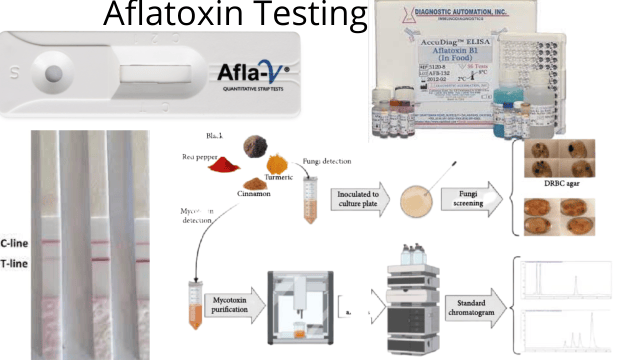 Figure: Different methods of aflatoxin testing
The first step of testing aflatoxin is extraction. Then it is followed by purification and concentration. The final step is detection and quantification.
Extraction and Purification (Clean-up)
Extraction is key to better detection and quantification of aflatoxin in food samples. The extraction and purification of aflatoxin can occur in the following ways.
- Liquid-liquid extraction (LLE): Aflatoxin is highly soluble in polar-protic solutions like acetone, chloroform, methanol, and acetonitrile. These can either be mixed with water or used in their pure state. It does not produce clean analytes.
- Liquid-solid extraction (LSE): A fixed amount of solid food particles is mixed using centrifugal force (mixer or vortex) with an extraction reagent, either acetonitrile/water or methanol/water, in varying ratios. It is the most common method, followed by the purification method like IAC (immunoaffinity column).
- Ultrasound extraction: The mixed sample from LSE containing flask, centrifuge tube, or vial) is dipped into the ultrasonic bath with water. The ultrasound helps quick transfer the toxin from the sample to the extraction reagent.
- Supercritical fluid extraction (SFE): The use of supercritical fluid CO2 to extract apolar aflatoxin has shown promising results. However, it is not ideal for polar aflatoxins because it has a low recovery of the toxin and a higher concentration of co-extracts.
- Solid phase extraction (SPE): It is a popular method of clean-up or purification. It uses a C-18 (octadecylsilane) column. Its application is in the immunoaffinity clean-up column (IAC) and SPME (solid-phase microextraction). SPME is compatible with chromatographic techniques, and IAC has columns with antibodies specific to the toxins. SPME has high specificity in foods like nuts, spices, dried fruits, and cereals. In contrast, IAC is useful in pasteurized milk. During IAC, the crude extracted sample is applied to the column. The aflatoxin binds to a specific antibody immobilized in the column. A washing step is required after IAC.
Detection and Quantification
Various methods have been applied to detect and quantify extracted and purified aflatoxins. They are chromatographic techniques (TLC, HPLC, and GC), Spectroscopic techniques (fluorescence and infrared spectroscopy), immunological techniques (RIA, ELISA, and later flow immunoassay), and immunosensors (electrochemical, optical, and QMC).
Chromatographic Techniques
Chromatographic techniques involve interaction between a mobile and a stationary phase. The mobile phase is usually a liquid, gas, or supercritical fluid that moves around the stationary phase (solid or liquid). The mixture with desired components gets distributed between the two phases (mobile and stationary). The general practice is dissolving the mixture into the mobile phase and applying it as a spot in the stationary phase. The mobile phase carries the mixture along the stationary phase, and the speed of different components of the mixture varies, which is the basis of separation.
The chromatographic techniques used commonly are thin-layer chromatography (TLC), gas chromatography (GC), and high-performance liquid chromatography (HPLC).
Thin layer chromatography (TLC)
TLC has been widely used in detecting aflatoxins in food and has found aflatoxins as low as 1-20 ppb (parts per billion). Its stationary phase comprises silica, alumina, or cellulose immobilized in glass or plastic. The mobile phase used for separating aflatoxins is a mixture of the sample with acetonitrile, methanol, and water. The difference in solubility of aflatoxins determines the interaction time between the mobile and stationary phases. Depending on the interaction time and molecular weight of the toxin, it either adheres to the stationary or mobile phase. The longer it adheres to the stationary phase, the later it separates from the mixture and vice versa. TLC helps to detect different types of toxins in a single test. Although it is sensitive, it requires highly trained professionals and lacks precision due to applying samples, plate development, and interpretation errors.
HPTLC (high-performance thin layer chromatography) has been developed in order to improve the limitation of TLC. The HPTLC is an automated process where sample application, plate development, and interpretation are automatic. HPTLC is currently the most precise and efficient technique for determining mycotoxins.
High-performance liquid chromatography (HPLC)
HPLC is the most common technique used for the separation of organic components. It has an absorbent inside a glass or plastic tube (column) as a stationary phase and an aqueous/organic solvent as a mobile phase. The sample is injected into the stationary phase and flows through it with the help of the mobile phase. A pump provides high pressure for fast movement of the mobile phase through the column (stationary phase). The separation occurs due to the different affinities of the components toward the stationary and mobile phases. The higher the affinity, the longer analyte reacts with the particular phase. If an analyte interacts longer with the mobile phase, it separates faster.
The time taken for the elution (separation followed by removal from the setup) of analytes (mixture) is called retention time. The retention time is detected using UV, fluorescent, or diode array detectors. HPLC is used widely in the determination of aflatoxins. However, it requires rigorous sample purification using immunoaffinity columns and tiresome pre and post-column derivatization for detecting AFB1 and AFG1 due to the use of UV for detection. Hence to overcome the derivatization, HPLC is combined with mass spectroscopy limiting the sample size for generating structural information. But HPLC-MS/MS is bulky and suitable only in laboratory environments.
Gas chromatography (GC)
In GC, the mobile phase is carrier gas, and the stationary phase is liquid coated on inert solid particles confined to a long stainless steel or glass tube (column). The stationary phase needs to maintain an appropriate temperature for separation. The sample is vaporized into its gaseous state and carried by the carrier gas along the stationary phase. Like all chromatographic techniques, the basis of the separation of components is their distribution among the two phases. The sample’s movement depends on the components’ affinity with the stationary phase. The higher the affinity, the slower the movement and the longer it takes to separate and vice versa. Once the separation completes, the separated particles are detected using FID (flame ionization detector), or electron capture detector (ECD), and a mass spectrometer. Since aflatoxins are non-volatile, these may need derivation for detection and are less commonly used.
Spectroscopic Techniques
Fluorescence spectroscopy
Aflatoxins are fluorescing compounds that emit light of various wavelengths after absorption. The fluorometric method help quantify aflatoxins in the range of 5-5000 ppb. However, derivatization is required for better analysis, and the limit for detection is slightly higher than the limit of 4 μg/Kg set by European standards.
Infrared spectroscopy
Infrared spectroscopy depends on the difference in molecular vibrations after irradiation with infrared. Atomic size, bond length, and strength vary from one molecule to another. The absorption rate of infrared radiation by a particular bond depends on the bond type and mode of vibrations. So, different frequency ranges of infrared radiation are passed through the sample during the analysis of compounds using an infrared spectrometer. The absorbed radiant energy by every bond of the sample molecule is then measured. The measurement helps construct the spectrum graph, and no two organic molecules have the same spectrum, helping identify the organic compound. It has been used in determining aflatoxins in peanuts, peanut cakes, and corn kernels.
Immunological Techniques
Immunological techniques involve the specific binding of antibodies and antigens. Various immunological methods have been developed based on the specificity and high-affinity interaction between antibodies and antigens and are expanded to receptor-ligand bonding. These interactions can also be quantified using the absorbance of photons of light energy by spectrophotometer. The other ways of detection and quantification are labeling with radioisotopes, fluorophores, and enzymes. The immunochemical methods used commonly are radioimmunoassay (RIA), enzyme-linked immunosorbent assay (ELISA), immunosensors, and ICA (immunocolumn assay).
Radioimmunoassay (RIA)
RIA relies on the competitive binding of the radiolabeled antigens and non-radiolabeled antigens with available antibodies. The unlabeled antigen competes with the radioactive antigen for binding with a fixed number of antibodies. The amount of radiolabeled antigen and antibody is known, whereas the quantity of non-labeled antigens is unknown, but the amount is inversely proportional to the amount of radiolabeled antigens. RIA is used in the qualitative and quantitative determination of aflatoxins. Although the RIA can analyze multiple samples at once, these require antigens in the pure state, and the radioactive isotope is hazardous and hard to dispose of and store.
ELISA (enzyme-linked immunosorbent assay)
Another immunochemical method that overcomes the hazardous limitation of RIA is ELISA, where an antigen or antibody is bound with enzymes. The principle of ELISA is similar to that of other immunochemical methods that rely on specific antigen-antibody binding and increased sensitivity due to antigen or antibody linked with an enzyme that combines with a particular substrate.
ELISA is available in a kit and can easily detect aflatoxins in agricultural products. Although it requires many washing steps, it is cost-effective, can analyze 96 samples simultaneously, and carries no health hazards.
Lateral flow devices (kit)
Lateral flow devices are immunochromatographic assays. The principle is based on the antigen-antibody interaction. These consist of a porous membrane for flowing samples, an absorbent pad for increasing the volume of flowing liquid, and a sample pad to ensure contact between the liquid sample and the membrane. The devices use colloidal gold or gold-coated antibodies to detect the red-colored binding zones. The sample moves through the sample pad due to capillary action. When the sample with aflatoxins reaches the gold particles, these bind with the particle giving the red colored line. A device made by Delmulle et al., can detect 5 μg/Kg AFB1 in pig feed within 10 minutes. These devices are easy to use, cost less, and provide rapid on-site detection.
Immunosensor Techniques for Detection
Immunosenors are biosensors that use antigens or antibodies as recognition components combined with signal transducers like graphite, gold, or carbon. These help to detect binding between complementary species. Based on the signal transduction used, immunosensors are of three types; piezoelectric, optical, and electrochemical.
Electrochemical immunosensors
These devices use antibodies added in a biorecognition layer for producing electroactive signals that are detectable by transducers resulting in the generation of measurable signals. Aflatoxin analysis can be done using various types of electrochemical immunosensors. The most common type consists of antibodies immobilized to the surface of the glassy carbon electrode, and an enzyme is a biological component used. However, there has also been the construction of non-enzymatic electrochemical immunosensors using chitosan, gold particles, anti-aflatoxin B1 and Fe3O4 as electrodes.
Optical immunosensors
Surface plasmon resonance (SPR) is the optical immunosensors used for detecting aflatoxins. It depends on measuring the refractive index produced after binding of the analyte with its biospecific partner (specific antibodies) that is immobilized in the sensor surface. It is used in detected AFB1 using monoclonal or polyclonal antibodies of AFB1. OWLS (optical waveguide light-mode spectroscopy) is another label-free biosensor that relies on fluorescence excitation for measuring the binding events at the surface of the waveguide. OWLS detects aflatoxins and ochratoxins; the detection range is 0.5-10 ng/ml in wheat flour and barley.
Piezoelectric QCMs (quartz crystal microbalances)
These are label-free devices used in detecting antigens. T depends on the change in mass of the electrode when antigens react with the immobilized antibody in the quartz crystal surface. The difference in mass is proportional to the concentration of the antibody-antigen complex. QCM sensor is coated with gold for detecting aflatoxin B1 in the range of 0.5-10 ppb.
References and further reading
- Abbas, M. (2021). Chromatographic Techniques for Estimation of Aflatoxins in Food Commodities. In (Ed.), Aflatoxins – Occurrence, Detoxification, Determination and Health Risks. IntechOpen. https://doi.org/10.5772/intechopen.98508
- Wacoo, A., Wendiro, D., Vuzi, P., & Hawumba, J. (2014). Methods for Detection of Aflatoxins in Agricultural Food Crops. Journal Of Applied Chemistry, 2014, 1-15. https://doi.org/10.1155/2014/706291
- Miklós, G., Angeli, C., Ambrus, Á., Nagy, A., Kardos, V., & Zentai, A. et al. (2020). Detection of Aflatoxins in Different Matrices and Food-Chain Positions. Frontiers In Microbiology, 11. https://doi.org/10.3389/fmicb.2020.01916
- Eurofins Aflatoxin Testing. Aflatoxin Testing. Retrieved 1 August 2022, from https://www.eurofins.com/food-and-feed-testing/food-testing-services/mycotoxin-testing/aflatoxin-testing/.
- Leszczyńska, J., MasŁowska, J., Owczarek, A., & Kucharska, U. (2013). Determination of aflatoxins in food products by the ELISA method. Czech Journal Of Food Sciences, 19(No. 1), 8-12. https://doi.org/10.17221/6567-cjfs
- Espinosa-Calderon, A., Miguel, L., Francisco, R., Roberto, J., Guevara Gonzalez, R., & Torres-Pacheco, I. (2011). Methods for Detection and Quantification of Aflatoxins. Aflatoxins – Detection, Measurement And Control. https://doi.org/10.5772/28439

Tankeshwar Acharya, MSc (Medical Microbiology)
Tankeshwar Acharya is an Assistant Professor in the Department of Microbiology at Patan Academy of Health Sciences (PAHS), Nepal, where he has been teaching and practicing clinical microbiology for over 14 years. He is the founder of Microbe Online, one of the leading free microbiology education resources on the web, covering bacteriology, mycology, parasitology, immunology, and clinical laboratory diagnostics written from direct experience in both the classroom and the diagnostic laboratory.