
Lyme Disease (Borrelia burgdorferi): Stages, Diagnosis, and How to Treat It
How Borrelia burgdorferi causes Lyme disease, the three stages from the erythema migrans rash to Lyme arthritis, why early Lyme is treated on the rash alone, and how two-tier serology is interpreted.

On this page
A 45-year-old hiker returns from a camping trip in rural Connecticut. Three weeks later he develops a circular expanding rash on his thigh: red at the edge, clearing in the center, now 12 cm across. He has had mild fever, fatigue, and joint aches for the past week. He did not notice a tick bite.
This is Lyme disease until proven otherwise. The rash, called erythema migrans, is the clinical hallmark, present in roughly 70 to 80% of cases. The 20 to 30% who never develop it are the diagnostic challenge.
The most important thing to understand about Lyme disease is that its diagnosis is often clinical, not serological. In an endemic area, the erythema migrans rash is enough to diagnose and treat Lyme disease on the spot, without waiting for a blood test, because antibodies take weeks to appear and the test is often negative this early. Getting this right, treating early on the rash, and knowing when serology helps and when it misleads, is what this page is about.
Lyme disease (Lyme borreliosis) is caused by Borrelia burgdorferi sensu lato, a complex of at least three genomospecies: B. burgdorferi sensu stricto (predominant in North America), B. afzelii and B. garinii (predominant in Europe and Asia). It is the most common vector-borne disease in North America and Europe, with over 30,000 cases reported annually in the USA and an estimated 65,000 in Europe: though both figures are considered significant underestimates of true incidence.
- High-risk regions (Northeastern & Midwestern USA, Central Europe).
- Seasonal variation (peak in spring/summer due to tick activity).
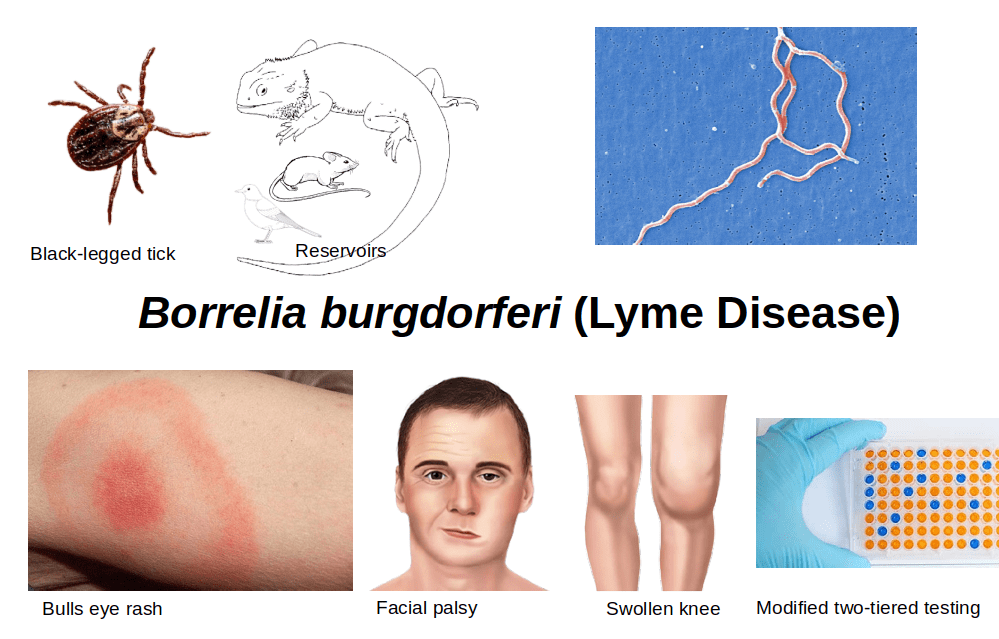 Figure: Main features of Borrelia burgdorferi & Lyme Disease
Characteristics of Borrelia burgdorferi
Borrelia burgdorferi is a helical-shaped spirochete that is poorly gram-stained (other spirochetes do not take up Gram stain). It stains weakly Gram-negative (safranin, the counterstain, is the last dye applied), but in practice Borrelia is not classified as Gram-positive or Gram-negative and is not identified by Gram stain. It is better seen with Giemsa or Wright stain, dark-field microscopy, or silver staining.
Borrelia can be stained with Giemsa stain or other blood stains and can be seen in the standard light microscope. It is better viewed under a dark ground microscope or by silver impregnation staining. The ultrastructure of Borrelia is similar to Treponema and Leptospira, with minor differences.
- Size: Larger, 10-30 μm in length (longer than a human red blood cell, which is 7 μm in diameter) and 0.2-0.5 μm in width.
- Spirals: fewer in number (about 3 to 10 per organism), with wider, looser coils than Treponema.
- Endoflagella: More in number (7-11), attached subterminally at the pole.
Why Borrelia is hard to detect: Unlike most bacteria, B. burgdorferi is present in blood only transiently and at very low density: which is why blood culture is insensitive and direct microscopy of blood rarely useful in Lyme disease. The organism rapidly disseminates to joints, nervous system, and cardiac tissue, where it persists. This tissue tropism explains the clinical stages of disease and the diagnostic challenge, you are rarely chasing the organism in blood.
Transmission
Rodents and deer are the main reservoirs of Lyme disease. It is widespread in the USA and reported in other parts of the world.
Lyme disease is transmitted by the bite of black-legged tick (Ixodes scapularis or Ixodes pacificus). All three stages of ticks (i.e. larval, nymphal, and adult stages) can transmit the infection, but it is thought that nymphs infect more humans than adult ticks because they are so hard to see (<2 mm).
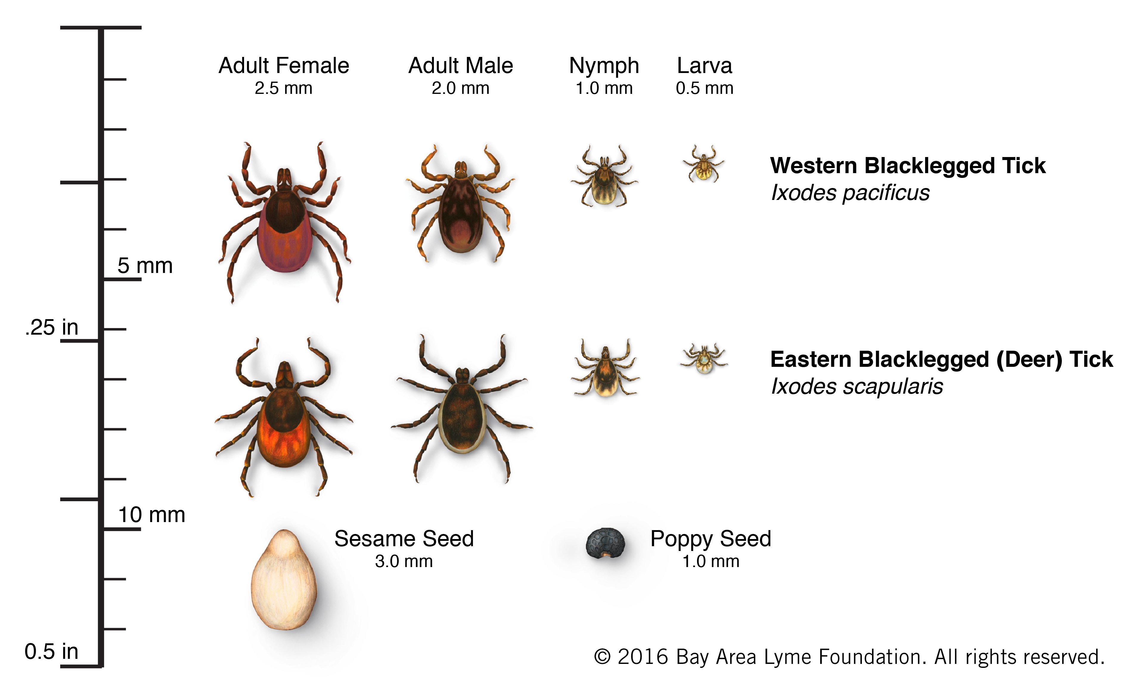 Figure: Life stages of tick
Borrelia burgdorferi expresses outer-surface protein A (OspA) in the midgut of the tick which is required for its survival in tick. When the bacterium reaches the salivary gland of the tick, it expresses protein OspC that binds to a tick salivary gland protein (Salp15). This attachment is crucial for transmission.
There is no evidence of person-person transmission of Lyme disease. There is no credible evidence that Lyme disease can be transmitted through air, food, water, or from the bites of mosquitoes, flies, fleas, or lice.
The tick must attach at least 24 hours for transmission. Removing a tick quickly (within 24 hours) can greatly reduce the chance of getting Lyme disease.
The 24-Hour Rule and Its Nuances
The tick must be attached for at least 24–36 hours to transmit B. burgdorferi, the organism must migrate from the tick's midgut (where it expresses OspA for survival) to the salivary glands (where it switches to OspC for transmission). This migration takes time, which is why prompt tick removal is protective.
Practical implications:
- A tick found engorged (fed for >36 hours) represents a meaningful transmission risk
- A tick found unengorged (flat, clearly recently attached) represents low but non-zero risk
- The decision to give prophylactic doxycycline after a tick bite depends on the tick species, the estimated attachment duration, and local endemicity, not on the presence or absence of symptoms. The dose and timing are a clinical decision.
Tick identification matters: Only Ixodes scapularis (black-legged tick, "deer tick") and Ixodes pacificus (western black-legged tick) transmit Lyme disease in North America. Dermacentor species (dog ticks, wood ticks) and Amblyomma americanum (lone star tick) do NOT transmit B. burgdorferi, a common source of patient and clinician confusion.
Clinical Manifestations: Three Stages
Lyme disease progresses through three stages if untreated. Not every patient passes through all three, some present in Stage 2 or 3 without a recognized prior rash.
Stage 1: Early Localized Infection (Days 3–30 After Bite)
Erythema migrans (EM) is the defining feature, an expanding annular (ring-shaped) skin lesion at the site of the tick bite. It:
- Appears 3–30 days after the bite (median 7 days)
- Expands gradually, must reach ≥5 cm in diameter for clinical case definition
- Is present in approximately 70–80% of cases, 20–30% of patients never develop it
- Does NOT always show the classic "bull's-eye" (target) pattern, many cases are uniformly red without central clearing
- Is not painful or itchy in most cases, thus patients may not notice it
Associated symptoms: Flu-like illness: fever, chills, myalgia, arthralgia, headache, fatigue. These are non-specific and easily attributed to viral illness, especially without the rash.
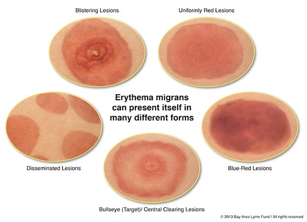 Figure: Different types of rash are seen in Lyme disease. Not everyone will have a “bullseye” rash.
Clinical pearl: In an endemic area, erythema migrans alone is sufficient to diagnose and treat Lyme disease, serological testing is not required and may be negative at this early stage (antibodies take 2–6 weeks to develop). Treating before seroconversion gives the best outcomes and prevents progression.
Stage 2: Early Disseminated Infection (Weeks to Months After Bite)
If untreated, B. burgdorferi disseminates haematogenously. Manifestations include:
Neurological (Lyme neuroborreliosis) ~15% of untreated cases:
- Facial nerve palsy (Bell's palsy): bilateral facial palsy should always raise suspicion for Lyme disease in an endemic area
- Meningitis: typically lymphocytic, with CSF pleocytosis
- Radiculopathy: shooting pains, numbness, weakness
- Bannwarth syndrome (Europe): the classic European presentation: painful radiculopathy + CSF pleocytosis + cranial nerve palsy, particularly facial palsy
Cardiac ~8% of untreated cases:
- Atrioventricular (AV) block: ranging from first-degree to complete heart block
- Lyme carditis is usually self-limiting but complete heart block requires temporary pacing
- Young patient + new AV block in endemic area = Lyme carditis until proven otherwise
Secondary skin lesions:
- Multiple secondary erythema migrans lesions at sites distant from the original bite
Stage 3: Late Disseminated Infection (Months to Years After Bite)
Lyme arthritis, the dominant manifestation:
- Intermittent or persistent arthritis affecting one or a few large joints, most commonly the knee
- Episodes of joint swelling lasting weeks to months
- Synovial fluid shows inflammatory cells (typically 10,000–25,000 WBC/mm³)
- Distinguished from septic arthritis by the much lower WBC count and clinical context
Late Lyme arthritis is partly immune-mediated: in some patients the joint inflammation persists even after the organism is cleared by antibiotics (sometimes called antibiotic-refractory Lyme arthritis), because the immune response continues. This is why a minority of arthritis does not resolve immediately with treatment, and it connects to the post-treatment syndrome below.
Late neurological Lyme (uncommon):
- Encephalopathy: cognitive difficulties, memory problems
- Peripheral neuropathy
Post-treatment Lyme disease syndrome (PTLDS):
- Some patients have persistent fatigue, musculoskeletal pain, and cognitive difficulties after completing antibiotic treatment
- Cause is debated, not evidence of ongoing active infection; additional antibiotics do not help
- Important to distinguish from active Lyme disease requiring further treatment
| Stage | Timing | Key Manifestations | Diagnostic Approach |
|---|---|---|---|
| Early localized | Days 3–30 | Erythema migrans (70–80%), flu-like illness | Clinical diagnosis; serology often negative, do not wait for it |
| Early disseminated | Weeks–months | Bell's palsy, meningitis, radiculopathy, AV block, multiple EM | Serology (ELISA + Western blot); CSF if neurological; ECG if cardiac |
| Late disseminated | Months–years | Lyme arthritis (knee), encephalopathy, neuropathy | Serology; synovial fluid PCR; joint fluid analysis |
Laboratory Diagnosis
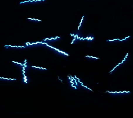 Figure: Borrelia in dark-field microscopy
Specimen Selection
Specimen depends on clinical stage and presentation:
- Early localized: Clinical diagnosis is sufficient if EM is present — serology not needed
- Early disseminated (neurological): Serum + CSF (cell count, protein, glucose, intrathecal antibody index)
- Early disseminated (cardiac): Serum serology + ECG
- Late disseminated: Serum serology + synovial fluid (PCR, cell count)
Serology: The Primary Diagnostic Tool
The CDC recommends a two-tier testing algorithm:
Tier 1: ELISA (enzyme immunoassay):
- Detects IgM and IgG antibodies against B. burgdorferi
- High sensitivity but lower specificity: false positives occur with other spirochetal infections (syphilis, leptospirosis), EBV, autoimmune diseases
- If negative → stop. Lyme disease is unlikely (unless testing in first 2 weeks of illness)
- If positive or equivocal → proceed to Tier 2
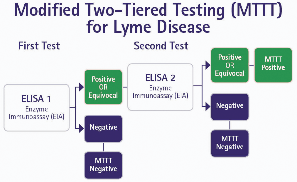 Figure: Modified two tiered testing for Lyme disease
Tier 2: Western Blot (immunoblot):
- More specific: detects antibodies against specific B. burgdorferi proteins
- IgM Western blot (interpret only in first 4 weeks of illness): positive if ≥2 of 3 bands (OspC/24 kDa, 39 kDa, 41 kDa)
- IgG Western blot (use after 4 weeks): positive if ≥5 of 10 bands
Critical point: IgM Western blot should NOT be used after 4 weeks of illness, the false-positive rate is unacceptably high and leads to overdiagnosis. After 4 weeks, only IgG Western blot is interpretable.
Modified two-tier testing (MTTT): A newer approach replacing the second-tier Western blot with a second ELISA using a different antigen preparation is now endorsed by CDC as an acceptable alternative to the classic two-tier algorithm.
Culture
B. burgdorferi can be cultured in Barbour-Stoenner-Kelly (BSK) medium: a specialised liquid medium. Sensitivity is low (<50% in early disease, much lower in late disease). Culture is a research tool, not a clinical diagnostic tool. Incubation requires weeks.
PCR
- Synovial fluid PCR is the most useful PCR application: sensitivity ~70% in Lyme arthritis, much better than culture
- Blood PCR: low sensitivity, not recommended for routine diagnosis
- CSF PCR: low sensitivity for CNS Lyme; intrathecal antibody index is preferred
Microscopy
Direct visualization of B. burgdorferi in clinical specimens:
- Dark-field microscopy: can demonstrate spirochetes in blood during early disease but sensitivity is poor; not used routinely
- Giemsa or Wright stain: spirochetes visible in blood smear; again low sensitivity
- Silver impregnation staining (Warthin-Starry or Dieterle stain): used in tissue sections from skin biopsy of EM lesion
Treatment
Treatment depends on clinical stage and manifestations:
- Early localized disease: oral doxycycline (amoxicillin or cefuroxime as alternatives).
- Neurological disease or severe carditis: IV ceftriaxone.
- Lyme arthritis: oral doxycycline, longer course.
- Children under 8 and pregnant women: doxycycline avoided; amoxicillin or cefuroxime used.
- Post-exposure prophylaxis after a high-risk Ixodes bite exists but is a clinical decision.
Borrelia Species: Lyme Disease vs. Relapsing Fever
| Feature | Lyme Disease | Relapsing Fever |
|---|---|---|
| Main species | B. burgdorferi, B. afzelii, B. garinii | B. recurrentis (epidemic), B. hermsii, B. duttoni (endemic) |
| Vector | Ixodes tick | Body louse (B. recurrentis); Ornithodoros tick (others) |
| Reservoir | Rodents, deer | Rodents (endemic); humans (epidemic) |
| Key mechanism | Multi-stage tissue dissemination | Antigenic variation → relapsing fever |
| Clinical hallmark | Erythema migrans, arthritis, neurological | Recurrent febrile episodes |
| Diagnosis | Two-tier serology (ELISA + Western blot) | Blood smear (Giemsa/Wright) during febrile episode |
| Treatment | Doxycycline (oral); ceftriaxone (severe) | Doxycycline |
See also: Relapsing fever
How to Remember
The name story: Named after Old Lyme, Connecticut, where a 1975 cluster of childhood arthritis cases triggered investigation. The organism is named after Willy Burgdorfer, who identified the spirochete in 1982.
Three stages: three systems:
- Stage 1 = Skin (erythema migrans)
- Stage 2 = Heart and Nerves (AV block, Bell's palsy, meningitis)
- Stage 3 = Joints (knee arthritis)
Memory aid: SKiN, then Heart/Nerves, then JoiNts. The disease moves inward from the skin.
The proportions worth knowing: 20 to 30% never develop EM, about 15% of untreated cases develop neurological disease, and about 8% develop cardiac disease.
Two-tier testing logic: ELISA first (sensitive screen) → Western blot only if ELISA positive (specific confirmation). Never order Western blot as a first test. Never interpret IgM Western blot after 4 weeks.
Tick attachment time: 24–36 hours minimum for transmission. A flat, unengorged tick just removed = very low risk. A swollen, engorged tick = consider prophylactic doxycycline (a clinical decision).
Key Exam Facts in One Table
| Feature | Detail |
|---|---|
| Causative organism | Borrelia burgdorferi sensu lato (spirochete) |
| Vector | Ixodes scapularis / I. pacificus (North America); I. ricinus (Europe) |
| Reservoir | White-footed mouse (Peromyscus leucopus); deer amplify tick population |
| Minimum attachment for transmission | 24–36 hours |
| Staining | Giemsa, Wright, silver impregnation; poorly Gram-stained |
| Stage 1 manifestation | Erythema migrans (≥5 cm); present in 70–80% |
| Stage 2 manifestations | Bell's palsy, meningitis, AV block, radiculopathy |
| Stage 3 manifestation | Lyme arthritis (knee); encephalopathy |
| Diagnostic algorithm | Two-tier: ELISA → Western blot (IgG after 4 weeks) |
| Best specimen for arthritis | Synovial fluid PCR |
| Culture medium | Barbour-Stoenner-Kelly (BSK) medium |
| Key European syndrome | Bannwarth syndrome (radiculopathy + facial palsy + CSF pleocytosis) |
Where Students Get Confused
Erythema migrans does not always look like a bull's-eye. Many EM rashes are uniformly red with no central clearing. Waiting for a "classic target" rash before diagnosing means missing cases. An expanding red rash ≥5 cm after a plausible tick exposure is enough.
Do not wait for serology in early Lyme. Antibodies take weeks to develop, so serology is often negative when the EM rash is present. In an endemic area, the rash alone justifies treatment. A negative early test does not rule Lyme out.
IgM Western blot is only valid in the first 4 weeks. After 4 weeks, a positive IgM blot is more likely a false positive than real, and using it leads to overdiagnosis. After 4 weeks, only the IgG blot counts.
Not every tick transmits Lyme. Only Ixodes (black-legged/deer) ticks transmit B. burgdorferi. Dog ticks (Dermacentor) and the lone star tick (Amblyomma) do not. A bite from the wrong tick is not a Lyme risk.
Lyme arthritis is not septic arthritis. The synovial fluid WBC in Lyme arthritis (roughly 10,000-25,000) is far lower than in bacterial septic arthritis. The clinical context (endemic area, prior stages) and the lower count separate them.
Post-treatment symptoms are not ongoing infection. Persistent fatigue and aches after treatment (PTLDS) are not evidence of live organisms, and more antibiotics do not help. This is distinct from genuinely inadequately treated disease.
References
- Steere AC, Strle F, Wormser GP, et al. Lyme borreliosis. Nat Rev Dis Primers. 2016;2:16090. https://doi.org/10.1038/nrdp.2016.90
- Wormser GP, Dattwyler RJ, Shapiro ED, et al. The clinical assessment, treatment, and prevention of Lyme disease, human granulocytic anaplasmosis, and babesiosis: clinical practice guidelines by the Infectious Diseases Society of America. Clin Infect Dis. 2006;43(9):1089–1134. https://doi.org/10.1086/508667
- Centers for Disease Control and Prevention. Lyme Disease: Two-step Laboratory Testing Process. https://www.cdc.gov/lyme/diagnosistesting/labtest/twostepp/index.html
- Murray PR, Rosenthal KS, Pfaller MA. Medical Microbiology. 9th ed. Elsevier; 2020.
- Shapiro ED. Lyme disease. N Engl J Med. 2014;370(18):1724–1731. https://doi.org/10.1056/NEJMcp1314325
Frequently Asked Questions
What is the rash of Lyme disease?
What is the rash of Lyme disease?
Erythema migrans: an expanding red rash at the site of the tick bite, appearing 3 to 30 days later. It reaches at least 5 cm and may or may not have the classic bull's-eye appearance. It is present in about 70 to 80% of cases.
Do you need a blood test to diagnose Lyme disease?
Do you need a blood test to diagnose Lyme disease?
Not in early disease with a typical rash. In an endemic area, the erythema migrans rash alone is enough to diagnose and treat Lyme disease. Antibodies take weeks to appear, so blood tests are often negative early and should not delay treatment.
How long must a tick be attached to transmit Lyme disease?
How long must a tick be attached to transmit Lyme disease?
Usually 24 to 36 hours. The organism has to move from the tick's gut to its salivary glands before it can be transmitted, which takes time. Removing a tick promptly greatly reduces the risk.
What is two-tier testing for Lyme disease?
What is two-tier testing for Lyme disease?
A two-step blood test: first an ELISA (a sensitive screen), and only if that is positive or equivocal, a confirmatory Western blot (more specific). A newer version replaces the second step with a second ELISA. The IgM Western blot is only reliable in the first 4 weeks of illness.
Which ticks transmit Lyme disease?
Which ticks transmit Lyme disease?
Only Ixodes ticks (the black-legged or deer tick, Ixodes scapularis and I. pacificus in North America, I. ricinus in Europe). Dog ticks and the lone star tick do not transmit Borrelia burgdorferi.
What are the stages of Lyme disease?
What are the stages of Lyme disease?
Three: early localized (the erythema migrans rash and flu-like illness), early disseminated (facial palsy, meningitis, heart block, multiple rashes), and late (mainly Lyme arthritis of the knee). Not everyone passes through all three.

Tankeshwar Acharya, MSc (Medical Microbiology)
Tankeshwar Acharya is an Assistant Professor in the Department of Microbiology at Patan Academy of Health Sciences (PAHS), Nepal, where he has been teaching and practicing clinical microbiology for over 14 years. He is the founder of Microbe Online, one of the leading free microbiology education resources on the web, covering bacteriology, mycology, parasitology, immunology, and clinical laboratory diagnostics written from direct experience in both the classroom and the diagnostic laboratory.
Comments
No comments yet. Be the first to share your thoughts.
Leave a comment
All comments are reviewed before they appear.