
Biochemical Tests Used to Identify Bacteria in the Microbiology Laboratory

On this page
Bacteria cannot be identified by appearance alone. Two organisms may look identical under the microscope (for example, both gram-positive cocci), yet belong to entirely different species with different clinical significance, antibiotic susceptibility profiles, and treatment implications. Biochemical tests solve this problem by exploiting differences in bacterial metabolism.
A biochemical test works by exposing a bacterial isolate to a specific substrate, nutrient, or chemical and observing the metabolic response whether the organism produces a particular enzyme, ferments a particular sugar, or generates a particular end product. Since metabolic pathways are genetically determined and species-specific, the pattern of positive and negative biochemical reactions creates a metabolic "fingerprint" that identifies the organism.
In practice, no single biochemical test identifies a bacterium conclusively. Identification requires a battery of tests (a combination of reactions interpreted together). This is the basis of both traditional manual biochemical testing and commercial systems like the API 20E strip, which runs 20 biochemical tests simultaneously.
Biochemical tests are broadly organized into the following categories:
- Enzyme detection tests: detect specific enzymes produced by the organism
- Carbohydrate fermentation and utilization tests: detect acid or gas production from specific sugars
- Amino acid degradation tests: detect decarboxylation or deamination of amino acids
- Miscellaneous metabolic tests: detect hydrolysis of substrates, gas production, or other metabolic reactions
- Commercial identification systems: miniaturized panels of multiple biochemical tests
Enzyme Detection Tests
These tests detect the presence or absence of specific enzymes produced by the bacterial isolate.
Catalase Test
Principle: Detects the enzyme catalase, which breaks down hydrogen peroxide (H₂O₂) into water and oxygen gas, producing visible bubbling. Positive result: Immediate vigorous bubbling within 5–10 seconds Primary use: Differentiates Staphylococcus spp. (positive) from Streptococcus and Enterococcus spp. (negative)See more about Catalase Test
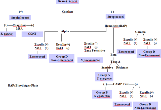 Flow chart of gram-positive cocci identification
Oxidase Test
Principle: Detects the presence of cytochrome c oxidase, a terminal electron carrier in the bacterial respiratory chain. Positive result: Development of purple/blue color within 10–60 seconds on oxidase reagent strip or disk Primary use: Differentiates oxidase-positive organisms (Pseudomonas, Neisseria, Vibrio, Campylobacter, Pasteurella) from oxidase-negative Enterobacteriaceae (E. coli, Klebsiella, Salmonella). Read more: Oxidase Test
Coagulase Test
Principle: Detects coagulase enzyme (bound and/or free) that converts fibrinogen to fibrin, clotting plasma. Positive result: Clumping (slide test) or clot formation (tube test) Primary use: Identifies Staphylococcus aureus (positive) and differentiates it from coagulase-negative staphylococci (CoNS) such as S. epidermidis and S. saprophyticus. Read in details about Coagulase Test
DNase Test
Principle: Detects the enzyme deoxyribonuclease (DNase), which hydrolyzes DNA in the medium. Positive result: Clear halo around colonies after flooding with HCl (DNA precipitation method) or zone of depolymerization on toluidine blue DNase agar Primary use: Differentiates Staphylococcus aureus (positive) from other staphylococci; also used to identify Serratia marcescens and Moraxella catarrhalis. Read more about DNase Test
Urease Test
Principle: Detects the enzyme urease, which hydrolyzes urea into ammonia and carbon dioxide, raising the pH and turning phenol red indicator pink/magenta.
Positive result: Pink/magenta to deep pink color change in Christensen's urea agar Primary use: Identifies Proteus spp. (rapid, strong positive), Helicobacter pylori, Morganella morganii, Yersinia enterocolitica; helps differentiate Klebsiella (positive) from E. coli (negative). Read more: Urease Test
Phenylalanine Deaminase Test
Principle: Detects the enzyme phenylalanine deaminase, which deaminates phenylalanine to phenylpyruvic acid. The acid reacts with ferric chloride to produce a green color. Positive result: Green color development after adding 10% FeCl₃ Primary use: Differentiates Proteus, Morganella, and Providencia spp. (positive) from other members of Enterobacteriaceae (negative). See more: Phenylalanine Deaminase Test
Lipase Test (Lipid Hydrolysis Test)
Principle: Detects bacterial lipase enzymes that hydrolyze tributyrin or other lipid substrates into fatty acids and glycerol. Positive result: Clear halo around colonies on tributyrin agar Primary use: Identifies lipolytic organisms including Pseudomonas spp., Clostridium perfringens, Staphylococcus aureus, and some Bacillus spp. Read more: Lipid Hydrolysis Test
Starch Hydrolysis Test
Principle: Detects amylase enzymes that hydrolyze starch. After incubation, flooding with iodine solution produces a blue-black color in areas of intact starch; clear halos indicate hydrolysis.
Positive result: Clear halo around colonies after iodine flooding
Primary use: Identifies amylase-producing organisms including Bacillus spp., Clostridium spp., and some streptococci. See more: Starch Hydrolysis Test
Gelatin Hydrolysis Test
Principle: Detects gelatinase enzymes that liquefy gelatin. After incubation at low temperature, liquefied gelatin indicates gelatinase production.
Positive result: Gelatin remains liquid after refrigeration (negative: solidifies) Primary use: Identifies Pseudomonas aeruginosa, Serratia marcescens, Proteus spp., and some Clostridium spp.; helps differentiate within Enterobacteriaceae. See more: Gelatin Hydrolysis Test
Beta-Glucuronidase Test (MUG Test)
Principle: Detects β-D-glucuronidase enzyme using the fluorogenic substrate 4-methylumbelliferyl-β-D-glucuronide (MUG). Positive organisms produce a fluorescent compound visible under UV light.
Positive result: Blue fluorescence under long-wave UV (365 nm)
Primary use: Rapid identification of Escherichia coli (positive) from other Enterobacteriaceae. Read more: MUG Test
CAMP Test
Principle: Detects the CAMP factor, a diffusible protein produced by certain organisms that acts synergistically with S. aureus beta-lysin to produce enhanced hemolysis on blood agar.
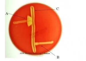 Positive result: Arrowhead-shaped zone of enhanced hemolysis at the junction of the CAMP factor and S. aureus beta-lysin streaks
Primary use: Identifies Streptococcus agalactiae (Group B strep) positive from from S. pyogenes (Group A), negative. Also used to identify Listeria monocytogenes and Clostridium perfringens (reverse CAMP test). Read more: CAMP Test
Carbohydrate Fermentation and Utilization Tests
These tests detect the ability of an organism to ferment or utilize specific carbohydrates, producing acid, gas, or both.
Carbohydrate (Sugar) Fermentation Test
Principle: Detects the fermentation of specific sugars (glucose, lactose, sucrose, mannitol, etc.) with acid and/or gas production. pH indicator (phenol red or Andrade's) changes color when acid is produced; a Durham tube traps gas. Positive result: Color change from red to yellow (acid); bubble in Durham tube (gas) Primary use: Differentiates members of Enterobacteriaceae; distinguishes fermenters from non-fermenters; part of the IMViC battery → Full article: Carbohydrate Fermentation Test
Oxidative-Fermentative (O-F) Test
Principle: Determines whether an organism metabolizes glucose oxidatively (aerobically) or fermentatively (anaerobically), or not at all (non-reactive). Positive result: Acid production in open tube only (oxidative); acid in both open and sealed tubes (fermentative); no change in either (non-reactive/alkaline) Primary use: Essential for distinguishing glucose-fermenting Enterobacteriaceae from non-fermenting gram-negative rods such as Pseudomonas, Acinetobacter, and Stenotrophomonas → Full article: Oxidative-Fermentative Test
Citrate Utilization Test
Principle: Determines whether an organism can use sodium citrate as its sole carbon source. Growth on Simmons citrate agar produces ammonia, raising pH and turning bromothymol blue indicator from green to blue. Positive result: Brilliant blue color and visible growth on Simmons citrate agar Primary use: Part of the IMViC battery; differentiates Klebsiella and Enterobacter (positive) from E. coli (negative); also differentiates Salmonella (positive) from Shigella (negative) → Full article: Citrate Utilization Test
ONPG Test (Beta-Galactosidase Test)
Principle: Detects β-galactosidase enzyme using the substrate o-nitrophenyl-β-D-galactopyranoside (ONPG). β-galactosidase cleaves ONPG, releasing yellow o-nitrophenol. Positive result: Yellow color development Primary use: Identifies late or slow lactose fermenters that possess β-galactosidase but lack the lactose permease needed for rapid fermentation; used to differentiate Salmonella (negative) from Citrobacter (positive); identifies Vibrio cholerae → Full article: ONPG Test
Litmus Milk Test
Principle: Detects multiple metabolic activities in litmus milk medium: acid/alkaline production, clot formation, peptonization, and litmus reduction. Each reaction produces a characteristic color or physical change. Positive result: Variable: acid (pink), alkaline (blue), clot (firm curd), peptonization (clear), reduction (white) Primary use: Identifies Enterococcus spp. (litmus reduction); characterizes Clostridium spp.; differentiates lactic acid bacteria. Read more: Litmus Milk Test
IMViC Battery (Indole, Methyl Red, Voges-Proskauer, Citrate)
The IMViC tests are a classic battery of four biochemical tests used specifically to differentiate members of the Enterobacteriaceae family, particularly E. coli from Klebsiella/Enterobacter.
| Organism | Indole (I) | Methyl Red (M) | Voges-Proskauer (V) | Citrate (C) |
|---|---|---|---|---|
| Escherichia coli | + | + | − | − |
| Klebsiella pneumoniae | − | − | + | + |
| Klebsiella aerogenes (formerly Enterobacter aerogenes) | − | − | + | + |
| Citrobacter freundii | − | + | − | + |
Read more about: IMViC Tests
Indole Test
Principle: Detects the ability of an organism to split tryptophan into indole, pyruvic acid, and ammonia using tryptophanase. Kovács or Ehrlich's reagent reacts with indole to produce a red/pink color. Positive result: Red/pink ring at the surface of the medium after adding Kovács reagent Primary use: Differentiates E. coli (positive) from most other Enterobacteriaceae; part of IMViC; identifies Proteus vulgaris (+) from P. mirabilis (−). See more : Indole Test
Methyl Red (MR) Test
Principle: Determines whether an organism performs mixed acid fermentation, producing sufficient stable acid end products to maintain a low pH. Methyl red indicator turns red at pH ≤4.4. Positive result: Stable red color after adding methyl red indicator Primary use: Part of IMViC; differentiates E. coli (positive) from Klebsiella and Enterobacter (negative). Read more: Methyl Red Test
Voges-Proskauer (VP) Test
Principle: Detects acetoin (acetylmethylcarbinol), an intermediate produced during butylene glycol fermentation. Acetoin reacts with KOH and alpha-naphthol to produce a red color. Positive result: Red color development within 15–60 minutes after adding VP reagents Primary use: Part of IMViC; differentiates Klebsiella and Enterobacter (positive) from E. coli (negative); identifies Staphylococcus aureus and Bacillus cereus (positive). See more: Voges-Proskauer Test
Amino Acid Degradation Tests
Lysine Decarboxylase Test
Principle: Detects lysine decarboxylase enzyme, which decarboxylates lysine to produce cadaverine. The alkaline cadaverine reverses the initial acid pH, turning bromocresol purple indicator back to purple. Positive result: Purple color in sealed tube (after initial yellow acid phase)
Primary use: Differentiates Salmonella (positive) from Shigella (negative); differentiates Klebsiella (positive) from Enterobacter on Lysine Iron Agar. (Note: Shigella are lysine decarboxylase negative as a group. S. sonnei is distinctive for being ornithine decarboxylase positive, which is a separate test, not LDC.). See more: Lysine Iron Agar (LIA)
Mixed Acid Fermentation
Principle: Certain bacteria ferment glucose through the mixed acid pathway, producing acetic, lactic, succinic, and formic acids, plus CO₂ and H₂ gas. The specific pattern of acids and gases produced is species-characteristic. Primary use: Differentiates enterics and fermentative gram-negative rods based on their specific acid and gas production patterns. Read more: Mixed Acid Fermentation
Multi-Test Media
These media detect multiple biochemical reactions simultaneously in a single tube.
Triple Sugar Iron (TSI) Agar
Principle: Detects fermentation of glucose, lactose, and sucrose, plus H₂S production and gas production, all in one tube. The slant reflects aerobic metabolism (lactose/sucrose); the butt reflects anaerobic glucose fermentation; black precipitate indicates H₂S; cracks/displacement indicate gas. Reading key: Alkaline slant/acid butt (K/A) = glucose only; acid slant/acid butt (A/A) = glucose + lactose/sucrose; alkaline slant/alkaline butt (K/K) = non-fermenter Primary use: First-line differentiation of Enterobacteriaceae from non-fermenters; preliminary identification of Salmonella (K/A + H₂S) and Shigella (K/A, no gas, no H₂S). Read more: Triple Sugar Iron Agar
SIM Medium (Sulfide-Indole-Motility)
Principle: Detects three characteristics simultaneously: H₂S production (sulfide), indole production, and motility, in a single semisolid tube. Reading: Black precipitate = H₂S positive; red ring with Kovács reagent = indole positive; diffuse turbid growth away from stab line = motility positive Primary use: Part of the standard Enterobacteriaceae identification battery alongside TSI; particularly useful for differentiating Salmonella (H₂S+, indole−, motile) from Shigella (all negative/non-motile). Read more: SIM Medium
Antibiotic and Chemical Susceptibility Tests (Used as Biochemical Identifiers)
Some antibiotic susceptibility patterns are so species-specific that they serve as identification tests rather than treatment guides.
Bacitracin Sensitivity Test
Principle: Streptococcus pyogenes (Group A strep) is uniquely sensitive to very low concentrations of bacitracin (0.04 unit disk). Other beta-hemolytic streptococci are resistant. Positive result: Any zone of inhibition around the bacitracin disk Primary use: Presumptive identification of S. pyogenes from other beta-hemolytic streptococci. Read more: Bacitracin Test
Optochin (Ethylhydrocupreine) Sensitivity Test
Principle: Streptococcus pneumoniae is uniquely sensitive to optochin. A zone of inhibition ≥14 mm (6 mm disk) indicates susceptibility. Positive result: Zone of inhibition ≥14 mm around optochin disk Primary use: Differentiates S. pneumoniae (sensitive) from viridans streptococci (resistant). Read more: Optochin Test
Novobiocin Susceptibility Test
Principle: Staphylococcus saprophyticus is novobiocin-resistant, while S. epidermidis is susceptible. A zone ≤16 mm indicates resistance. Positive result (resistance): Zone of inhibition ≤16 mm Primary use: Differentiates S. saprophyticus (resistant) from S. epidermidis (susceptible). Red more: Novobiocin Susceptibility Test
Miscellaneous Biochemical Tests
Bile Solubility Test
Principle: Sodium deoxycholate (bile salt) activates autolytic enzymes in S. pneumoniae, causing rapid lysis of colonies. Other alpha-hemolytic streptococci are resistant. Positive result: Lysis (disappearance) of colonies after bile salt application Primary use: Differentiates Streptococcus pneumoniae (positive) from viridans streptococci (negative). Read more: Bile Solubility Test
Bile Esculin Test
Principle: Detects the ability to grow in the presence of 40% bile and hydrolyze esculin. Esculin hydrolysis products react with ferric ions to produce a dark brown/black precipitate. Positive result: Blackening of bile esculin agar within 48 hours Primary use: Differentiates Group D streptococci and Enterococcus spp. (positive) from non-Group D streptococci (negative). Read more: Bile Esculin Test
Hippurate Hydrolysis Test
Principle: Detects the enzyme hippuricase, which hydrolyzes sodium hippurate to benzoic acid and glycine. Glycine is detected by ninhydrin reagent, producing a deep purple color. Positive result: Deep purple color after ninhydrin treatment Primary use: Identifies Streptococcus agalactiae (Group B) and Campylobacter jejuni (positive); differentiates C. jejuni from C. coli (negative). Read more: Hippurate Hydrolysis Test
PYR Test (Pyrrolidonyl Arylamidase Test)
Principle: Detects pyrrolidonyl arylamidase enzyme, which hydrolyzes the substrate L-pyrrolidonyl-β-naphthylamide (PYR). The product reacts with a cinnamaldehyde reagent to produce a red/pink color. Positive result: Bright red/pink color within 5 minutes Primary use: Identifies Streptococcus pyogenes (positive) and Enterococcus spp. (positive); differentiates S. lugdunensis from other CoNS; helps confirm S. pyogenes alongside bacitracin sensitivity. See more: PYR Test
Nitrate Reduction Test
Principle: Detects the ability of an organism to reduce nitrate (NO₃⁻) to nitrite (NO₂⁻) or further to nitrogen gas. Nitrite reacts with sulfanilic acid and alpha-naphthylamine to produce a red color. Positive result: Red color after adding reagents (nitrite present); or no color + gas bubble/zinc reduction (nitrate reduced all the way to N₂) Primary use: Differentiates members of Enterobacteriaceae; identifies Pseudomonas aeruginosa (positive, denitrifies to N₂); used in Mycobacterium identification. See more: Nitrate Reduction Test
Modified Oxidase Test (Microdase Test)
Principle: A modified oxidase disk test that detects cytochrome oxidase in staphylococci and micrococci. The standard oxidase test is unreliable for distinguishing these gram-positive cocci, so the microdase (modified) disk is used instead. Positive result: Blue/purple color within 2 minutes on microdase disk Primary use: Differentiates Micrococcus spp. (positive) from Staphylococcus spp. (negative). Read more: Modified Oxidase Test
Commercial Identification Systems
API 20E and API System (bioMérieux)
The API (Analytical Profile Index) system is a commercial miniaturized biochemical identification system. The API 20E strip contains 20 micro-tubes with dehydrated biochemical substrates. After inoculation with a bacterial suspension and incubation at 37°C for 18–24 hours, color changes in each well are read and converted into a 7-digit numerical profile that is looked up in the API database for species identification.
Available systems include:
- API 20E: Enterobacteriaceae and other gram-negative rods
- API 20 NE: Non-Enterobacteriaceae gram-negative rods (Pseudomonas, Acinetobacter, Burkholderia)
- API Staph: Staphylococci
- API 20 Strep: Streptococci and enterococci
- API 20A: Anaerobes
- API Coryne: Corynebacterium and related organisms
→ Full article: API 20E Test System → Full article: API and RAPID ID Systems
Quick Reference: Tests by Organism Group
In practice, tests are run in sequence, not all at once. The Gram stain and colony morphology come first and decide which column below you are in.
- For gram-positive cocci, the usual order is catalase (splits staphylococci from streptococci and enterococci), then coagulase for the catalase-positive cocci and hemolysis-guided tests (bacitracin, optochin, bile solubility, CAMP) for the catalase-negative ones.
- For gram-negative rods, oxidase comes first (it separates the non-fermenters and organisms like Vibrio and Pseudomonas from the Enterobacteriaceae), followed by TSI and the IMViC or SIM battery to place the organism within the family. Each test narrows the field for the next, which is why the panel, not any single result, makes the identification.
For gram-positive cocci
| Test | Purpose |
|---|---|
| Catalase | Staphylococcus (+) vs Streptococcus/Enterococcus (−) |
| Coagulase | S. aureus (+) vs CoNS (−) |
| DNase | S. aureus (+) vs other staphylococci |
| PYR test | S. pyogenes (+) and Enterococcus (+) |
| Bacitracin | S. pyogenes (sensitive) vs other beta-hemolytic strep |
| Optochin | S. pneumoniae (sensitive) vs viridans strep |
| Bile solubility | S. pneumoniae (lyses) vs viridans strep |
| CAMP test | S. agalactiae (positive arrowhead) |
| Hippurate hydrolysis | S. agalactiae (+) |
| Bile esculin | Enterococcus and Group D strep (+) |
| Novobiocin | S. saprophyticus (resistant) vs S. epidermidis (sensitive) |
| Modified oxidase | Micrococcus (+) vs Staphylococcus (−) |
For gram-negative rods (Enterobacteriaceae)
| Test | Purpose |
|---|---|
| IMViC battery | Differentiates E. coli, Klebsiella, Enterobacter, Citrobacter |
| TSI agar | Fermentation pattern; H₂S; gas; preliminary Salmonella/Shigella ID |
| SIM medium | H₂S, indole, motility simultaneously |
| Urease | Proteus (rapid +), H. pylori, Klebsiella (+) vs E. coli (−) |
| Lysine decarboxylase | Salmonella (+) vs Shigella (−) |
| Phenylalanine deaminase | Proteus, Morganella, Providencia (+) |
| ONPG | Late lactose fermenters; Citrobacter (+) vs Salmonella (−) |
| Nitrate reduction | Denitrifiers; Pseudomonas distinction |
| Oxidative-fermentative | Fermenters vs non-fermenters |
For non-fermenting gram-negative rods
| Test | Purpose |
|---|---|
| Oxidase | Pseudomonas (+) vs Acinetobacter (−) |
| O-F test | Non-fermentative (alkaline/no change) vs fermentative |
| Motility | Pseudomonas (motile) vs Acinetobacter (non-motile) |
| Nitrate | P. aeruginosa (denitrifies to N₂) |
Frequently Asked Questions
What is the purpose of biochemical tests in microbiology?
Biochemical tests detect specific metabolic activities: enzyme production, sugar fermentation, gas production. Since metabolic pathways are genetically determined, the pattern of positive and negative reactions creates a unique fingerprint identifying a bacterial species. A battery of tests is always interpreted together for definitive identification.
What is the IMViC battery and which organisms does it differentiate?
IMViC = Indole, Methyl Red, Voges-Proskauer, Citrate. E. coli: + + - -. Klebsiella pneumoniae and Klebsiella aerogenes: - - + +. Differentiates members of Enterobacteriaceae.
Why is the catalase test performed before the coagulase test?
Catalase immediately separates Staphylococcus (positive) from Streptococcus/Enterococcus (negative). Only after confirming catalase positivity does the coagulase test make sense to separate S. aureus from coagulase-negative staphylococci.
What is the difference between the oxidase and catalase tests?
Catalase: detects H2O2 breakdown, differentiates Staphylococcus from Streptococcus (gram-positive cocci). Oxidase: detects cytochrome c oxidase, differentiates Pseudomonas from Enterobacteriaceae (gram-negative rods).
What is the significance of urease production?
Rapid strong urease (2-4 hours): Proteus spp. and H. pylori. Moderate urease: differentiates Klebsiella (positive) from E. coli (negative). Also used in Mycobacterium species differentiation.
What is the API 20E system?
Why are multiple biochemical tests needed?
No single test is specific enough: most have exceptions and cross-reactions. A battery of 5-10 tests interpreted together provides the specificity needed for definitive species-level identification.
What is the difference between fermentation and oxidation in the O-F test?
Fermentative: acid in both open and sealed tubes. Oxidative: acid only in open tube. Non-reactive: no acid in either tube. Distinguishes Enterobacteriaceae fermenters from non-fermenters like Pseudomonas and Acinetobacter.
References and Further Reading
- Tille, P. M. (2022). Bailey and Scott's Diagnostic Microbiology (15th ed.). St. Louis: Elsevier.
- Mahon, C. R., Lehman, D. C., & Manuselis, G. (2019). Textbook of Diagnostic Microbiology (6th ed.). Saunders.
- Garcia, L. S. (Ed.). (2016). Clinical Microbiology Procedures Handbook (4th ed.). ASM Press.
- Procop, G. W., Church, D. L., Hall, G. S., Janda, W. M., Koneman, E. W., Schreckenberger, P. C., & Woods, G. L. (2017). Koneman's Color Atlas and Textbook of Diagnostic Microbiology (7th ed.). Philadelphia: Wolters Kluwer.
- Madigan, M. T., Bender, K. S., Buckley, D. H., Sattley, W. M., & Stahl, D. A. (2021). Brock Biology of Microorganisms (16th ed.). Pearson.

Tankeshwar Acharya, MSc (Medical Microbiology)
Tankeshwar Acharya is an Assistant Professor in the Department of Microbiology at Patan Academy of Health Sciences (PAHS), Nepal, where he has been teaching and practicing clinical microbiology for over 14 years. He is the founder of Microbe Online, one of the leading free microbiology education resources on the web, covering bacteriology, mycology, parasitology, immunology, and clinical laboratory diagnostics written from direct experience in both the classroom and the diagnostic laboratory.
Comments
No comments yet. Be the first to share your thoughts.
Leave a comment
All comments are reviewed before they appear.