Pyrogen and Bacterial Endotoxin Testing Methods
Pyrogen and Bacterial Endotoxin Testing Methods
Bacterial endotoxin is the lipopolysaccharide (LPS) present in the cell wall of Gram-negative bacteria. Endotoxin is responsible for inducing fever when introduced in the blood or CSF (cerebrospinal fluid), hence also called, pyrogen.
Testing bacterial endotoxin is essential before releasing sterile products, especially in pharmaceutical industries. Bacterial endotoxin or pyrogen testing are in-vitro testings of endotoxin before the release of medical effects like medicines and medical devices associated with cardiovascular, lymphatic systems, or cerebrospinal fluid.
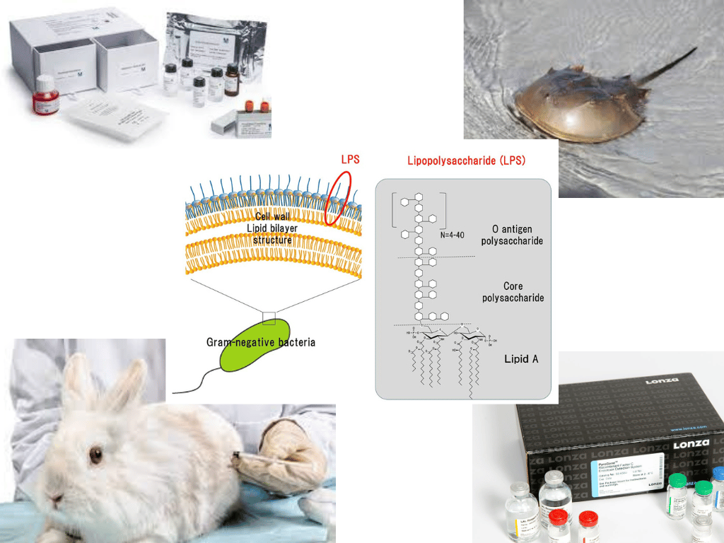 Figure: Pyrogen and Bacterial Endotoxin testing
Importance of Bacterial Endotoxin Testing
Gram-negative bacteria are ubiquitous, and endotoxin produced by the bacteria can induce fever as an inflammatory response. The endotoxin can contaminate the medical devices, injectable drugs, water, or any object in contact with the patient’s blood. This response can sometimes be fatal, especially in immunocompromised people, children, old population.
Pyrogen or bacterial endotoxin testing are FDA-approved method of determining pyrogenic elements like endotoxin. These tests are essential for decreasing the risk of cross-infection in patients.
Pyrogen testing differs from sterility testing, and both tests should be carried out simultaneously. Sterility testing usually detects viable organisms or spores produced by microorganisms. The sterility testing cannot detect endotoxin, so conducting pyrogen testing is necessary.
Methods of Bacterial Endotoxin and Pyrogen Testing
The methods of detecting bacterial endotoxin and pyrogen are simple, easy to perform, and feasible. Different methods for testing the bacterial endotoxin are bacterial endotoxin test (BET) or Limulus amebocyte lysate (LAL) assay, recombinant Factor C (rFC) assay, monocyte activation test (MAT), and rabbit pyrogen test (RPT).
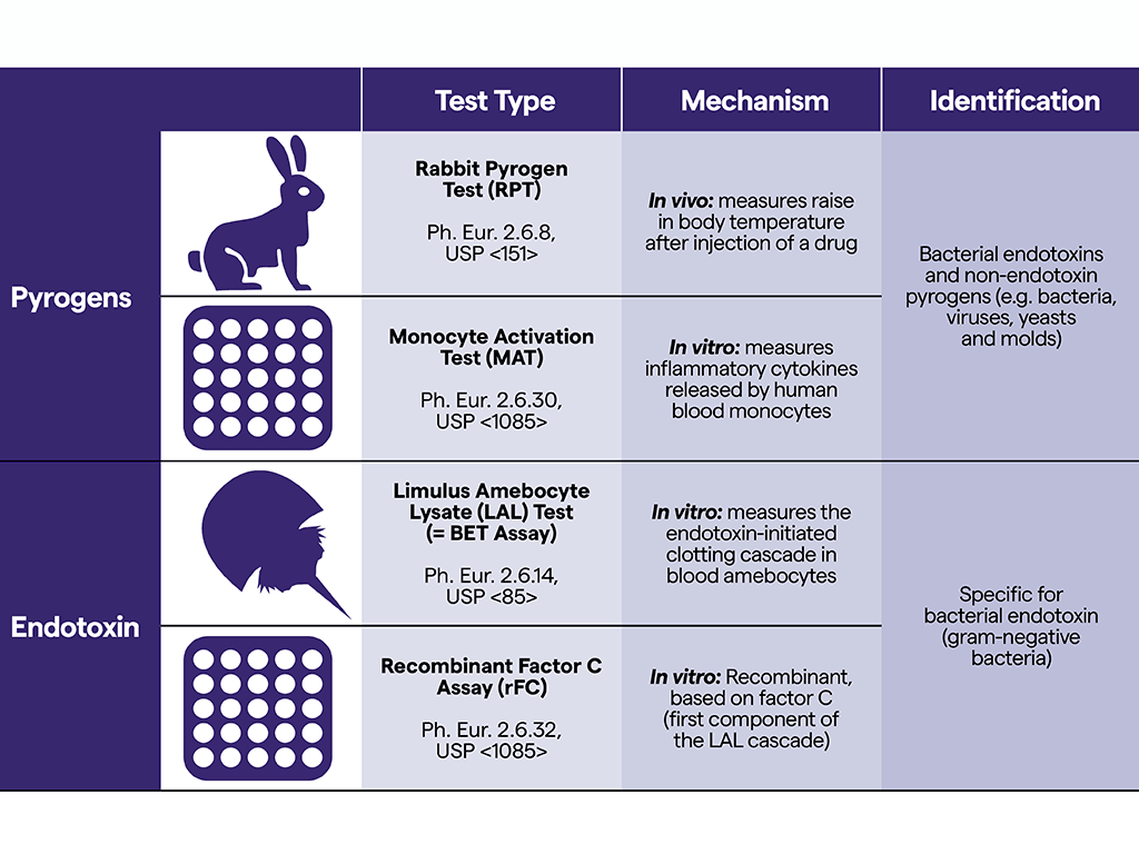 Figure: Suitability of different pyrogenic methods
Rabbit Pyrogen Test
The rabbit pyrogen test is a more invasive method of detecting the presence of pyrogen in the sample that involves injecting the sample into a number of rabbits. The presence of pyrogenic substances leads to the rabbit generating fever after 3-6 hours of injection. It is also termed the Sham test.
Since this method involved animal testing, it is widely replaced by monocyte activation test (MAT) and Limulus amoebocyte lysate (LAL) assay.
Principle of RPT
The basis of the rabbit pyrogen test (RPT) is measuring the rise in the temperature of rabbits after injecting a sterile solution of samples to be tested intravenously. If the sample consists of pyrogens, the temperature of rabbits will increase by 0.6℃.
Procedure of RPT
The procedure for conducting a rabbit pyrogen test (RPT) is broadly divided into two steps; preliminary and main test.
Preliminary test
- Select three rabbits that are not used for at least two weeks. Condition them for 1-3 weeks.
- Stop the supply of food for 2 hours of the test. Record their temperature. It should be around 38.5℃.
- Inject with pyrogen-free saline water through IV after 90 minutes.
- Check the temperature after 30 minutes of injection and continue checking the temperature for another 3 hours.
- If the temperature rise by 0.6℃, reject the animal and select another. Repeat the preliminary test in the new animal.
Main test
- First, mix the sample with pyrogen-free water and heat it to 38℃ before injection.
- Withhold food to the rabbits selected by preliminary test before 2 hours of the experiment.
- Record initial temperatures (the mean of two temperatures at 30 minutes intervals). Rabbits showing a difference of >0.2℃ in the two readings cannot be used in the experiment.
- Inject, not less than 0.5 ml/Kg and not more than 10 ml/kg, prepared sample in the marginal vein of the ear of the three rabbits.
- Record the temperature of animals for 3 hours 30 minutes intervals.
Interpretation of Results
Case I: No rabbits show an individual rise of 0.6℃ in the temperature, i.e., the sum of the increase in temperature in the three rabbits does not exceed 1.4℃. It means the absence of pyrogen.
Case II: If two or three rabbits show a rise in temperature of ≥0.6℃ or the sum of temperature rise exceeds 1.4℃. It indicated the presence of pyrogen in the sample.
Application of RPT
The rabbit pyrogen test is the FDA-approved method of detecting pyrogens, not only endotoxin but non-endotoxin. It is a simple method of detecting pyrogens and is commonly used by pharmaceutical companies before releasing their products.
Bacterial Endotoxin Test or Limulus amebocyte Lysate Assay
The bacterial endotoxin test is an in vitro method of determining endotoxin using Limulus amoebocyte lysate. The LAL test is the most widely used and recommended test for determining endotoxin in all international pharmacopeias.
Principle of BET or LAL Assay
The main reagent, Limulus amoebocyte lysate (LAL), is an aqueous extract of blood cells (amoebocytes) obtained from the horseshoe crab (Limulus polyphemus).The proenzyme in the LAL reagent reacts with lipopolysaccharide (LPS) , a Gram-negative bacterial endotoxin, to produce the activated enzyme coagulase. This activation of the coagulase enzyme depends mainly on the concentration of the endotoxin present in the sample. Now, the activated enzyme (coagulase) hydrolyses certain bonds inside the coagulogen (clotting protein), which is also present in the LAL reagent. Once the hydrolysis completes, the resultant coagulin binds and forms a clot. There are four FDA-approved methods for determining the clot formed: thegel clot, the colorimeter, the spectrophotometric, and the chromogenic assay.
The overall reaction of the LAL assay
Proenzyme →Coagulase; catalyzed by Gram-negative bacterial endotoxin
Coagulogen →Coagulin; catalyzed by activated coagulase
Procedure of LAL Test
Reagents provided in the LAL test kit
- Lyophilized Limulus amebocyte lysate (LAL): The lysate is prepared from the circulating amoebocytes of the horseshoe crab, Limulus polyphemus. It is standardized as per FDA reference standard endotoxin. It also consists of buffered mono and divalent cations. This reagent is then lyophilized and sealed under a vacuum. It needs to be reconstituted with LAL reagent water. The storage of the reagent needs to be done at 2-8°C. Exposure to bright light and temperature above 37°C for a prolonged period may result in insoluble and yellow coloration in the reagent. The reagent is then not usable.
- Lyophilized E. coli Endotoxin O55:B5 (Test control organism): Purified endotoxin E. coli strain O55:B5 is lyophilized. When prepared by the instructions, each vial acts as the standard control endotoxin (CSE). Its potency is under the current FDA reference standard endotoxin (RSE). The appropriate RSE/CSE ratio and resultant CSE potency are provided in the certificate of analysis. The vials are stored at 2-8°C before reconstitution. Potency is calculated as below:
Potency (EU/ml)=RSE/CSE (in EU/ng) ✖ _ng/vial ÷ 5.0 ml/vial.
Reagent preparation
LAL preparation
- Reconstitute the lyophilized lysate by adding 1.8 ml LAL reagent water, the 16-vial or 5.2 ml, to the 50-test vial. Mix by swirling thoroughly for at least 30 seconds. Avoid shaking, as the contents will foam.
- Storage of reconstituted lysate is done for up to 24 hours at 2-8°C without loss of sensitivity. Similarly, the reconstituted lysate can be stored below -10°C for up to four weeks in convenient volumes.
Control standard E. coli preparation
- Reconstitute the vial of endotoxin with 5 ml LAL reagent water.
- Mix by vortexing the vial of endotoxin for 15 minutes.
- Dilute the endotoxin with LAL reagent water to 1 EU/ml concentration. The dilution is done by diluting the reconstituted endotoxin to 1/X; here, X= CSE potency in EU/ml as specified on the certificate of analysis.
- Mix by vortexing for 60 seconds before proceeding.
- Using the 1 EU/ml endotoxin solution, prepare a serial two-fold dilution series. Vortex each dilution before proceeding to the successive dilution.
Steps of the LAL test
- Each assay needs serial two-fold dilutions of the CSE with labeled lysate sensitivity, diluted test samples, and LAL reagent. Water acts as the negative control.
- Transfer 0.1 ml of standard, sample, or water to the 10 x 75 mm reaction tube.
- Add 0.1 ml of the reconstituted lysate to each tube, beginning with the blank and moving from the lowest to the highest endotoxin concentration.
- Immediately mix the contents thoroughly and place the tube in 37±1°C non-circulating hot water or a dry heat bath. This process should be done for each dilution of endotoxin.
- Similarly, run the unknown test sample in parallel with CSE.
- The incubation time starts with placing each tube in the 37± 1°C bath. The tubes are not disturbed for 60±2 minutes.
- After completion of incubation, carefully remove the tube and invert 180°.
Result Interpretation of LAL Test
The result interpretation is based on gel formation (gel clot LAL assay), colorimetric analysis, spectrophotometric analysis, or the appearance of color in the chromogenic assay. The chromogenic LAL assay uses a synthetic chromogenic peptide substrate which can be cleaved into clotting enzyme, resulting in a product that exhibits yellow color.
The spectrophotometric analysis uses the native substrate coagulogen cleaved to coagulin. The coagulin then begins to self-associate increasing turbidity. The densities of turbidity are correlated with endotoxin concentration.
The gel clot method is the most common method of performing the LAL test.
The following is the result of the gel clot assay:
- Positive reaction= Formation of firm gel which remains intact momentarily when the tube is inverted.
- Negative reaction= absence of solid clot after inversion. But the lysate may show increased turbidity or viscosity.
Things to consider
- Storage of lyophilized LAL reagent the E. coli O55:B5 at an appropriate temperature of 2-8°C is a must.
- Do not reconstitute the reagent of the endotoxin until immediately before use.
- Store reconstituted endotoxin at 2-8°C for four weeks.
- Prepare 1.0 EU/ml dilution in quantities as needed and do not store or use diluted endotoxins for more than a day.
- Freezing of reconstituted lysate reagent at -10°C for four weeks is possible. Thaw the frozen liquid lysate immediately before use. Freeze and thaw only one.
Application of LAL Test
- The LAL test is widely used in the pharmaceutical industries before the release of pharmaceutical products. As endotoxins are the most common pyrogens in the pharmaceutical industry, the LAL test is an easy and quick way to detect endotoxin and a suitable replacement for the pyrogen tests on rabbits.
- It is also helpful in industries that produce medical devices.
- LAL test is helpful in areas that prefer avoiding animal testing because, unlike other tests, the reagent for this test is extracted from crabs that are returned to live in their natural habitat.
Recombinant Factor C (rFC) Assay
When Limulus polyphemus is infected by a gram-negative bacteria, it results in fatal intravascular coagulation. At the genetic level, it has been known that the endotoxin activates a serine protease catalytic coagulation cascade which results in the gelato of Limulus blood. This cascade is used during the LAL assay. Factor C is the first component of the cascade activated by endotoxin binding. Factor C activates Factor B. Another alternative pathway is where factor G is activated by glucan binding. Both Factor C and G change the proclotting enzyme to the clotting enzyme. Factor C can selectively recognize endotoxin and trigger the protease cascade. Factor C has been purified and cloned to create an endotoxin-specific assay. The activated recombinant Factor C acts upon the fluorogenic substance in the assay mixture and produces a fluorescent signal directly proportional to the endotoxin concentration in the sample.
Principle of rFC Assay
The endotoxin binding activates recombinant Factor C. This active binding cleaves a synthetic substrate resulting in the generation of a fluorogenic compound. The assay is carried out on a 96-well plate. The fluorescence is measured at time zero and after an hour of incubation at 37°C ± 1 °C in microplate reading using an excitation wavelength of 380/440 nm. The log net fluorescence (difference between one hour and the time zero reading= ∆RFU) is proportional to the log endotoxin concentration and linearly in the 0.005-5.0 EU/ml range. The calculation of endotoxin in a sample is relative to a standard curve.
Procedure of rFC Assay
- Bring all the test reagents and sample to room temperature.
- Add 100 µl of the blank, endotoxin standards, and samples to the desired wells of the microplate.
- Spike the samples with endotoxin standards using concentration relative to the possible background endotoxin in the dilution.
- Pre-incubate the plate at 37°C ± 1°C in the reader for 10 minutes.
- Prepare a working reagent with a fluorogenic substrate, assay buffer, and rFC enzyme solution in the ratio of 5:4:1.
- Dispense 100 µl of the working agent to each well carefully.
- Now, read the fluorescence. It is the fluorescence at time zero.
- Incubate the plate for an hour and read the plate. It is a one-hour reading.
- Correct the difference between the one-hour reading with time zero reading with the blanks.
- Plot the log net ∆RFU against the log endotoxin concentration in a linear regression curve and then calculate the concentration of endotoxin using the standard curve.
Application of rFC Assay
- It is applicable for determining bacterial endotoxin in medical devices, parental animal and human drugs, and biological products.
- It is a good alternative for LAL in determining bacterial endotoxins.
Monocyte Activation Test
The monocyte activation test (MAT) helps detect and quantify substances that activate human monocytes for releasing mediators responsible for fever response. MAT is another suitable replacement for the rabbit pyrogen test (RPT). This test explores human fever response, providing better information on pyrogenic activity than RPT. This test not only determines the endotoxin pyrogens but also helps determine non-endotoxin pyrogens.
Principle of MAT
The MAT test detects cytokines, prostaglandins like IL (interleukin) 1-3, and tumor necrosis factor ɑ (TNF-ɑ) produced by monocytes-activated pyrogenic substances. These interleukins are then detected using ELISA (enzyme-linked immunosorbent assay).
The general principle of MAT
Pyrogens +Human White blood cells →Interleukins→ Detection using ELISA and quantification in IU/ml.
Procedure of MAT
The general procedure for MAT includes three basic steps; activation of monocyte, incubation for generating IL-6, and analysis using software for quantification.
Requirements for MAT
Following are the materials and instruments required for performing MAT:
- Biological safety cabinet
- Humidified incubator
- Water bath
- Vortex mixer
- Single/multi-channel adjustable volume pipettes suitable for 5 to 1,000 μl (electronic recommended)
- IL-6 ELISA kit
- Microtiter plate reader
- Software
- Pyrogen-free pipette tips and 50 ml flacons
- Eppendorf Microcentrifuge 2 ml tubes or 24-well flat bottom plates
- 96-well microtiter plates
- Polystyrene disposable pipettes
- Flasks, beakers, and liquid containers necessary for the preparation of reagents
- Isopropyl alcohol (70%)
**Steps of monocyte activation test (MAT)**
- Firstly thaw medium A (to reconstitute LPS standard) and medium B (culture media) in a water bath.
- In order to prepare 100 EU/ml LPS standard, add 950 μl medium to the available thawed LPS standard vial. Vortex for 60 seconds. Then dilute 100 EU/ml LPS standard in medium B but do not vortex mix gently using pipettes to 0.004-0.5 EU/ml. Now, add at least a duplicate to the culture plate.
- In this step, prepare the required dilution of the sample in medium B and add the same dilutions to the culture plate.
- This step prepares peripheral blood monocytes (PBMC) by thawing the cryopreserved PBMC using a water bath. Mix 1 ml thawed PBMC in 9 ml medium B slowly. Then add 100 μl cell suspension to the plate containing 100 μl of sample dilutions and the standard LPS titration.
- Then, incubate the plate in a humified incubator at 37°C in the presence of 5% CO2 for 19-20 hours. This helps activate human monocytes to release cytokine in the presence of pyrogenic substances.
- After incubation, separate the supernatant from cells by transferring 170 μl of supernatant to a 96-well culture plate.
- Pre-dilute the supernatant for IL-6 measurement. At least test two dilutions prepared from the MAT supernatant; one for ELISA and another for concluding all the high- and low- concentrations of IL-6 in the supernatant.
- Quantify the IL-6 concentration using the software.
Uses of MAT
- MAT is a suitable alternative for rabbit pyrogen testing in the pharmaceutical industry. Because it is less time-consuming, more accurate, cost-effective, and can detect endotoxin as well as non-endotoxin pyrogens.
- It helps detect pyrogens from medical devices and water sources.
References
- Bacterial Endotoxin Testing | Charles River. Criver.com. Retrieved 16 October 2022, from https://www.criver.com/products-services/qc-microbial-solutions/endotoxin-testing?region=3681.
- BACTERIAL ENDOTOXINS TEST. Houshiji.com. (2012). Retrieved 16 October 2022, from https://www.houshiji.com/Uploads/Download/5a27396519552.pdf.
- Bacterial Endotoxins/Pyrogens. U.S. Food and Drug Administration. (2014). Retrieved 16 October 2022, from https://www.fda.gov/inspections-compliance-enforcement-and-criminal-investigations/inspection-technical-guides/bacterial-endotoxinspyrogens.
- Limulus Amebocyte Lysate (LAL) PYROGENTTMPlus. Lonzabio.jp. Retrieved 16 October 2022, from http://www.lonzabio.jp/products/endotoxin/pdf/00190168_en.pdf.
- Monocyte Activation Test Kit Manual. Mstechno.co.jp. Retrieved 16 October 2022, from https://www.mstechno.co.jp/html/protocolFile/ms622eec285d459/MAT-MANUAL-US-2022.pdf.
- Monocyte Activation Test (MAT). Sigmaaldrich.com. Retrieved 16 October 2022, from https://www.sigmaaldrich.com/deepweb/assets/sigmaaldrich/marketing/global/documents/109/402/pyromat-white-paper-wp1071en-mk.pdf.
- Monocyte Activation Test. Solvias.com. Retrieved 16 October 2022, from https://www.solvias.com/docs/download/en/000_Brochures_amp_Flyers/Monocyte_Activation_Test.pdf.
- PyroGene™ Recombinant Factor C. Bionordika.no. Retrieved 16 October 2022, from https://bionordika.no/application/files/9416/0285/6144/PyroGene_instructions.pdf.

Tankeshwar Acharya, MSc (Medical Microbiology)
Tankeshwar Acharya is an Assistant Professor in the Department of Microbiology at Patan Academy of Health Sciences (PAHS), Nepal, where he has been teaching and practicing clinical microbiology for over 14 years. He is the founder of Microbe Online, one of the leading free microbiology education resources on the web, covering bacteriology, mycology, parasitology, immunology, and clinical laboratory diagnostics written from direct experience in both the classroom and the diagnostic laboratory.