
Pocket Microscope: Parts, Working Principle, and Uses
A pocket microscope is a small optical microscope you look through with your eye, magnifying about 20x to 250x. Here is how it works, its parts, its uses, and how it differs from a handheld digital microscope.

On this page
A pocket microscope is a small, portable optical microscope that you hold in your hand and look through with your eye. It is the size of a pen or a small torch, and it magnifies from about 20x to 250x. A small handheld microscope you look through is called a pocket microscope (or a field microscope); if it has no eyepiece and shows the image on a phone or computer screen instead, that is a handheld digital microscope, a different instrument.
Optically, a pocket microscope is a high-power magnifier. A precision lens system produces an enlarged image of an object held close beneath it, and an LED lights the object so you can see fine detail in the field, away from a bench. Its whole value is being simple, cheap, and portable: something you can carry in a pocket and use anywhere.
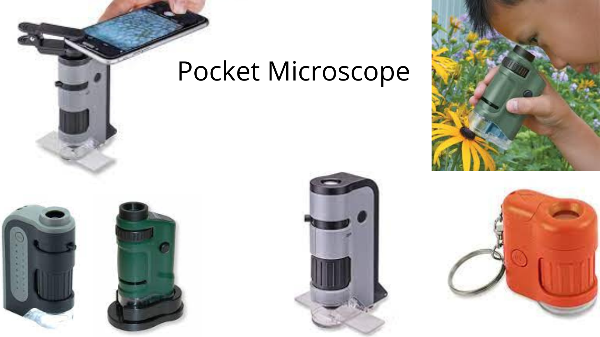 Figure: Different types of pocket microscope
Working Principle of Pocket Microscope
A pocket microscope works as a high-power magnifier. You hold it directly over the object, with the lens end close to the surface, and look through the eyepiece at the other end. The lens system produces an enlarged image that your eye views directly.
The LED lights the object in one of two ways. For a solid, opaque object such as a coin, a leaf, or a circuit board, the light reflects off the surface (reflected, or incident, light). This is the usual field setting. Some pocket microscopes have a clear base that also lets light through from below, which helps when viewing a thin or partly transparent specimen on a slide (transmitted light).
Unlike a compound microscope, a pocket microscope does not use a two-stage objective-and-eyepiece relay to reach high magnification. It is closer to a strong magnifying glass with its own light. This is why it is simple and cheap, and also why its magnification and resolving power are far lower than a compound microscope's.
Parts of Pocket Microscope
A pocket microscope fits perfectly in the hand clutch but has parts quite similar to the parts of the general light microscope. The parts are as follows:
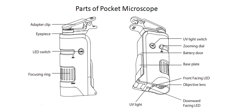 Figure: Parts of pocket microscope
- Light source: most pocket microscopes use an LED. Some offer both white LED and UV light. There are often two LEDs, one directed at the surface and one downward through a clear base. The light switch usually sits near the eyepiece.
- Stage: The stage is the place that holds the sample. It is in the bottom part of the microscope.
- Eyepiece: It is in the topmost part of the microscope. The eyepiece is the microscope’s part that helps view the magnified image.
- Battery: Most of these microscopes use batteries as the power source.
- Battery Chamber/Holder: A battery chamber/holder is the part that holds the battery used by the microscope.
- Zooming Dial: Moving the dials help to change the magnification of the microscope. It is also called a fine adjustment knob.
- Focus rings: The focus rings help to focus the image at low power. It is also called a coarse adjustment knob.
- Objective lens: It is present in the bottom part of the microscope.
- Additional parts: some models add an adapter clip that holds a smartphone over the eyepiece so you can photograph the magnified image. Some add a foldable base plate that clips a slide in place during viewing.
Procedure for using Pocket Microscope
The following steps are followed for using it:
- Firstly, select the object you want to view under the microscope.
- If you are using a slide, pick up the sample with a piece of clear tape and secure it on the slide. Some models take a standard slide with a coverslip.
- Then place the microscope at a 90° angle above the slide holder.
- But if you are viewing objects like flowers, fruits, etc., place the microscope directly above the object at a 90° angle.
- Now, turn on the light switch and start viewing from the eyepiece.
- Then, move the coarse knob or focus ring (present at the top) to fix the focus (moving it closer or farther from the objective).
- Now, move the fine adjustment or zooming dial to focus on the object up close and in higher magnification.
- If your model has a phone adapter, clip it over the eyepiece and attach your smartphone to photograph or record the magnified image.
Uses of Pocket Microscope
A pocket microscope is used wherever fine surface detail has to be checked away from a lab bench:
- Fieldwork and nature study: examining flowers, leaves, fruits, insects, minerals, and rocks on site.
- Precision trades: watchmaking, jewelry inspection and finishing, and dentistry, where small parts and fine detail must be seen clearly.
- Inspection and engineering: checking circuit boards, glassware, metal objects, and structural defects in machinery.
- Hobbies and education: coin and stamp collecting, and simple science exploration for students and children.
Advantages of Pocket Microscope
The advantages of these kind of microscopes are as follows:
- It is perfect for traveling with for fulfilling outdoor curiosity.
- It is cost-efficient and perfect for children with hobbies of exploring the miniature world.
- These microscopes are easy to use, so amateur or budding scientists can benefit from them.
Limitation of Pocket Microscope
The only limitation of the pocket microscope is its magnification range (20x-250x), which is not suitable for viewing bacterial cells.
Choosing a Magnification
Pocket microscopes are sold in a few magnification tiers, and higher is not always better. Lower magnification gives a wider field and a steadier image, which is easier to use in the field:
- About 20x to 40x: a wide, bright, easy-to-hold view. Good for coins, stamps, insects, plants, and general inspection.
- About 60x to 120x: more detail on small features, with a narrower field and a shakier image that needs a steady rest.
- About 100x to 250x: the top of the range. It shows fine surface detail but has a very small field, a short working distance, and is hard to hold steady by hand.
Even at 250x, a pocket microscope cannot resolve bacteria. Its magnification and resolving power are far below a compound microscope's.
How to Remember
- Pocket vs handheld digital, in one look: if you put your eye to it, it is a pocket microscope (optical). If the image goes to a screen, it is a handheld digital microscope. Both fit in a pocket; the eyepiece is the difference.
- Why higher magnification is not always better: as magnification goes up, the field of view shrinks and every hand tremor is magnified too. For handheld field use, a steady 40x often beats a shaky 250x.
Key exam facts
| Feature | Pocket microscope |
|---|---|
| Type | Optical; a high-power simple magnifier |
| Viewing | Through an eyepiece, with the eye |
| Magnification | About 20x to 250x |
| Light source | LED; reflected light on opaque objects, transmitted light on clear-base models |
| Size and use | Pen or torch sized; portable, for field and bench-side inspection |
| Also called | Field microscope |
| Key limit | Cannot resolve bacteria; low magnification and resolving power |
| Defining trait | Optical, with an eyepiece you look through |
Where Students Get Confused
- Calling a pocket microscope a small compound microscope. It is not. A compound microscope uses an objective and an eyepiece in a two-stage relay to reach high magnification. A pocket microscope is a high-power magnifier: a single lens system that produces one enlarged image for the eye. This is why its magnification and resolving power are much lower.
- Confusing it with a handheld digital microscope. Both are small and portable, but a pocket microscope is optical, with an eyepiece you look through. A handheld digital microscope has no eyepiece and shows the image on a screen.
- Assuming higher magnification is always better. Above roughly 100x, the field of view is tiny and the image is hard to hold steady by hand. For most field tasks, a lower magnification is more useful.
- Expecting it to show bacteria. Even at 250x it cannot. Bacteria need a compound microscope at about 1000x with oil immersion.
References
- Introduction to Lenses and Geometrical Optics. Nikon MicroscopyU. https://www.microscopyu.com/microscopy-basics/introduction-to-lenses-and-geometrical-optics (accessed August 2026).
- Simple Magnification and the Simple Magnifier. Molecular Expressions, Florida State University. https://micro.magnet.fsu.edu/primer/anatomy/magnification.html (accessed August 2026).
- Reflected (Episcopic) Light Microscopy. Nikon MicroscopyU. https://www.microscopyu.com/techniques/reflected-light (accessed August 2026, for the reflected-vs-transmitted illumination point).
Frequently Asked Questions
What is a small handheld microscope called?
What is a small handheld microscope called?
A small handheld microscope that you look through with your eye is called a pocket microscope, or a field microscope. If it has no eyepiece and shows the image on a phone or computer screen instead, it is a handheld digital microscope, which is a different instrument.
How does a pocket microscope work?
How does a pocket microscope work?
It works as a high-power magnifier. You hold it over the object with the lens end close to the surface and look through the eyepiece. A lens system produces an enlarged image that the eye views directly, and an LED lights the object, usually by reflected light off the surface.
What is the difference between a pocket microscope and a handheld digital microscope?
What is the difference between a pocket microscope and a handheld digital microscope?
A pocket microscope is optical: you look through an eyepiece. A handheld digital microscope uses an electronic image sensor instead of an eyepiece and shows the image on a screen. Both are small and portable, but the detector is different.
Can a pocket microscope see bacteria?
Can a pocket microscope see bacteria?
No. Its magnification is about 20x to 250x, far below the roughly 1000x with oil immersion needed to see bacteria. A pocket microscope is made for surface detail on coins, insects, plants, and circuit boards, not for stained bacterial slides.
What magnification pocket microscope should I choose?
What magnification pocket microscope should I choose?
For general field and inspection use, about 20x to 40x is easiest, giving a wide, steady view. Higher magnifications (up to 250x) show finer detail but have a very small field and are hard to hold steady by hand.

Tankeshwar Acharya, MSc (Medical Microbiology)
Tankeshwar Acharya is an Assistant Professor in the Department of Microbiology at Patan Academy of Health Sciences (PAHS), Nepal, where he has been teaching and practicing clinical microbiology for over 14 years. He is the founder of Microbe Online, one of the leading free microbiology education resources on the web, covering bacteriology, mycology, parasitology, immunology, and clinical laboratory diagnostics written from direct experience in both the classroom and the diagnostic laboratory.
Comments
No comments yet. Be the first to share your thoughts.
Leave a comment
All comments are reviewed before they appear.