Peroxisomes are the membrane-bound cell organelles of the eukaryotic cells. In 1954, J. Rhodin first described the peroxisomes. Later in 1967, Christian de Duve identified it as the cell organelle. The term peroxisomes comes due to their crucial role in the metabolism of hydrogen peroxide.
Peroxisomes are small vesicles and consist of digestive and oxidative enzymes. Peroxisomes convert hydrogen peroxide (H202) into water and oxygen with the action of the catalase enzyme. The peroxisome also palys some role in the biosynthesis of lipids and oxidation of fatty acids. Peroxisome doesn’t have their own DNA, but like the mitochondria and chloroplast, replication occurs by division.
Proliferation occurs in peroxisome when needed in large amounts. Peroxisome has the unique property of having a single unit membrane, whereas other cellular organelles, like mitochondria, consist of a double membrane.
Table of Contents
Location of Peroxisomes
Peroxisomes are present in most eukaryotic cells except in the mature erythrocyte. Peroxisomes are present in animal cells and the leaf of higher plants. In the plant cell, peroxisome is in association with the endoplasmic reticulum, chloroplasts, and the mitochondria and helps in photorespiration. Since peroxisome also assist in toxification, they are in large amounts in the liver and kidney cells. In the fibroblasts, peroxisome occurs as the individual organelle, whereas in the liver cells, they exist in the form of interconnected tubules forming the peroxisome reticulum.
Structure of Peroxisomes

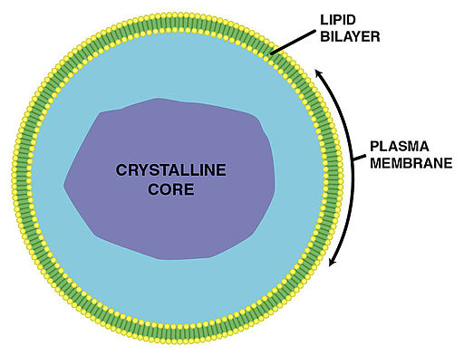
Peroxisomes comprises of the phospholipid bilayer and many membrane-bound proteins. The phospholipid bilayer is a thin covering, made up of two layers of lipid or fat molecules. Membrane proteins help to move the contents in and out of the cell. Peroxisomes are of different shapes and sizes, but in the cross-section, they are seen in a circular shape.
The diameter of peroxisomes ranges from 0.2 to 1.5 µm. In most mammalian tissues, its diameter is 0.15 to 0.25 µm. In the rat liver cells, its diameter is 0.5 μm. The diameter of the peroxisomes varies as per the energy requirement of the cell. Peroxisomes are smaller in the carbohydrate-rich cells, and in the lipid-rich cells, they are large in size and number. The number of peroxisomes per cell varies based on the type of tissue and cell. In the fibroblast, a few peroxisomes are present. Within the peroxisome, the granular matrix is present, which is covered by the single-unit membrane.
This membrane is made up of lipids and protein molecules. The matrix of the peroxisomes consists of fibrils or a crystalloid structure in which about 60 enzymes are present. Examples of peroxisome enzymes are urate oxidase, D-amino acid oxidase, and catalase.
Peroxisomes can be present in any of the two forms. It can be present either as the individual microperoxisomes or as a peroxisome reticulum in which it exists as a network of interconnected tubules. When they are interconnected, they are seen as clusters within a cell. In the preputial gland, these peroxisomes are interconnected, forming the large, cup-shaped structure within the cell. In some cases, as in the festucoid grasses, the matrix of peroxisomes consists of the fibrils or numerous threads structures.
Functions of Peroxisomes
Peroxisomal respiration
The peroxisome is involved in the metabolism of hydrogen peroxide (H202). The enzymes present in the peroxisome produce and degrade the H202. Catalase enzyme converts hydrogen peroxide into water and oxygen. Hydrogen peroxide is a highly reactive molecule and, when present in a large quantity, can be toxic and react with many other molecules in the cell. Catalase enzyme thus neutralizes its toxicity.
Biosynthesis of the lipid
Cholesterol and dolichol are synthesized both in the endoplasmic reticulum and peroxisomes. Then the synthesized cholesterol is used to produce bile acid in the liver cells. Another vital role of the peroxisome is plasmogen formation. The enzymes present in the peroxisomes synthesize the plasmalogens, which form the myelin sheath in the nerve fibers. Plasmalogens are a family of phospholipids and are essential components of the heart and brain tissue. Phospholipids are present in abundance in the myelin. When the plasmalogens are deficient, it causes various abnormalities in the myelination of the nerve cell. So due to this reason, peroxisomal disorders affect the nervous system. In patients with Alzheimer’s, there is a reduced level of plasmalogens.
Fatty acid β-oxidation
The peroxisome is the only site of fatty acid oxidation in yeast and plant. Mitochondria don’t catalyze fatty acid oxidation, so for fat utilization, peroxisome is crucial in both the plants and yeast cells. In beta (β) oxidation, the long chain fatty acids are broken down in the acetyl-CoA, which occurs in both the peroxisome and mitochondria of animals. Then the adenosine triphosphate (ATP) is produced, which is the high-energy molecule
Photorespiration
Peroxisomes in the green leaves perform photorespiration. This process protects plants from photooxidative damage. Photorespiration is the respiratory process occurring in higher plants in the presence of light. In this process, oxygen is taken, and carbon dioxide is released from the cell. During the photorespiration in plant cells, peroxisome also recycles carbon from phosphoglycolate.
Seed germination
The peroxisome in the seed converts the stored fatty acids to carbohydrates, which is essential for seed germination and its growth and development.
Purine catabolism and Bioluminescence
The uric acid oxidase enzyme in the peroxisomes degrades the purines, polyamines, and amino acids. In the peroxisomes of the firefly, luciferase enzymes are present, due to which it emits its light.
References
- Verma, P. S., & Agrawal, V. K. (2006). Cell Biology, Genetics, Molecular Biology, Evolution & Ecology (First edition). S.Chand and Company Ltd.
- Alberts, B. (2004). Essential cell biology. New York, NY: Garland Science Pub.
- Okumoto, K., Tamura, S., Honsho, M., & Fujiki, Y. (2020). Peroxisome: Metabolic Functions and Biogenesis. Advances in experimental medicine and biology, 1299, 3–17. https://doi.org/10.1007/978-3-030-60204-8_1