
Northern Blotting: Principle, Procedure, and Gene Expression Uses
Northern blotting explained for students: step-by-step procedure, why RNA must be denatured, how band intensity shows gene expression level, and applications.

On this page
Two tissue samples sit on the bench: one from healthy liver, one from a tumor. Both carry the same genes. The question is not which genes are present, but which genes are switched on, and how strongly. A gene that is barely active in healthy tissue may be running at full volume in the tumor.
Northern blotting answers this by measuring RNA. Because a cell makes RNA from a gene only when that gene is active, the amount of a specific messenger RNA (mRNA) tells you how strongly that gene is being expressed.
Southern blotting asks whether a DNA sequence is present. Northern blotting asks a different question: how much of a gene's RNA is a cell actually making? This article walks through how it works, step by step, and how the band on the film becomes a readout of gene expression.
Objective of Northern Blotting
Northern blotting is used to:
- Measure how much of a specific mRNA a tissue is making, which reflects how strongly the matching gene is expressed.
- Compare gene expression between tissues or between conditions (for example, healthy versus diseased).
- Detect the size of a transcript, which can reveal abnormal or alternatively spliced RNA.
Principle of Northern Blotting
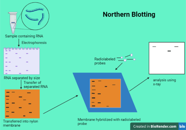
- The principle of the method is the separation of RNA according to size by denaturing agarose gel electrophoresis, then transferring the separated RNA onto a nylon membrane. The gel is denaturing (it contains formaldehyde) for a reason specific to RNA: unlike DNA, single-stranded RNA folds back on itself into secondary structures. If those folds are not removed, RNA molecules of the same length migrate differently and the size separation is wrong. Denaturing keeps every RNA molecule unfolded so it separates by true length.
- Consequently, the detection of the desired RNA is done by using the radiolabeled probe with all or a part of the base sequence of the targeted RNA hybridizing with the immobilized and separated RNA
 ## Materials and Equipment Required
- Agarose
- Formaldehyde
- 20X MAE
- Gel electrophoresis equipment
- 20X SSPE
- RNA loading buffer
- Loading dye (10X)
- Nylon hybridization membrane (3MM Whatman paper)
- Capillary system
- Ultraviolet light transilluminator
- NTPs: UTP, ATP, GTP, CTP
- Microwave
- Centrifuge
- Micropipette
- Micropipette tips
- Centrifuge tubes
- DNA or RNA template (for making the labeled probe)
Procedure of Northern Blotting
Northern blotting has six stages:
| Stage | What happens | Why it matters |
|---|---|---|
| 1. Denature and separate RNA | RNA is unfolded, then run on a denaturing gel | RNA folds on itself; denaturing lets it separate by true size |
| 2. Transfer (blotting) | Separated RNA is moved onto a nylon membrane | The membrane is workable and can be probed; the gel cannot |
| 3. Fix the RNA | RNA is cross-linked to the membrane by UV | Stops RNA washing off during later steps |
| 4. Label the probe | A probe matching the target is tagged with a label | The probe is what finds and marks the target RNA |
| 5. Hybridization | The probe binds its complementary target RNA | This is the specific step: the probe finds only the target mRNA |
| 6. Wash and detect | Unbound probe is washed off, the signal is read | The band and its intensity are the result you read |
Stage 1: RNA denaturation and separation
- Prepare a denaturing agarose gel (agarose plus formaldehyde) and pour it into a casting tray. The formaldehyde is what keeps the RNA unfolded.
- Let the gel solidify, then equilibrate it with running buffer for about 30 minutes.
- Mix the RNA sample with RNA loading buffer, then heat it to about 65°C for 12 to 15 minutes. This heating step fully denatures the RNA, unfolding its secondary structure so it will separate by true size. This is the single most important difference from a Southern blot, and it must not be skipped.
- Prepare the RNA size marker (ladder) with loading dye in a separate tube. The marker lets you read transcript sizes later.
- Load the denatured RNA samples and the marker into the wells using RNase-free tips.
- Run the gel at about 125 V for approximately 3 hours, until the dye has migrated far enough down the gel.
Integrity check: on a stained gel, intact total RNA shows two sharp bands, the 28S and 18S ribosomal RNA, with the 28S band about twice as bright as the 18S. This roughly 2:1 ratio is the quick check that the RNA is intact and not degraded. A low smear instead of sharp bands means the RNA has broken down and the blot will not be reliable.
Stage 2: Transfer of the separated RNA (blotting)
- Cut a nylon membrane and 3MM Whatman paper to the same size as the gel.
- Place the gel on a sponge for support inside a vacuum container holding transfer buffer (SSC).
- Position the gel so the separated RNA faces upward.
- Place the wet nylon membrane on top of the gel, then the 3MM Whatman paper on top of the membrane.
- Apply vacuum pressure to the container. The buffer pulls the RNA out of the gel and onto the membrane, keeping the band positions unchanged.
- Allow about 90 minutes for the transfer.
Stage 3: Fixing the RNA to the membrane
After transfer, cross-link the RNA to the membrane using short-wavelength UV light. This permanently attaches the RNA so it does not wash off during hybridization. (A nylon membrane is used rather than nitrocellulose because RNA binds it more firmly and it survives UV cross-linking.)
Stage 4: Labeling the probe
- Prepare a probe whose sequence is complementary to the target RNA. The probe can be DNA or RNA (a riboprobe).
- Label the probe, most commonly with a radioactive tag such as ³²P.
- The labeled probe is now ready to be added to the membrane. Because it is complementary to the target, it will base-pair only with the RNA you are looking for.
Stage 5: Hybridization
- Place the membrane in hybridization solution containing the labeled probe.
- Allow the probe to hybridize, binding wherever its complementary target RNA sits on the membrane.
Stage 6: Washing and detection
- Wash the membrane to remove unbound probe. Without thorough washing, leftover probe raises background and hides the real signal.
- Detect the bound probe. For a radioactive probe, expose the membrane to X-ray film (autoradiography); the film darkens wherever the probe bound.
- Read the result. A band appears at the size of the target transcript, and its intensity can be measured with software.
How to read a Northern blot result
A Northern blot result is a band, or a set of bands, on film. Three features carry the meaning.
Is there a band at all? A band means the target mRNA is present in that sample. No band means the gene is not expressed in that tissue, or is expressed below the detection limit. This alone answers "is this gene switched on here?"
Where is the band? The band's position, read against the RNA size marker, gives the size of the transcript. A band at the expected size is the normal mRNA. A band at an unexpected size can mean an alternatively spliced transcript, a degradation product, or an abnormal RNA. Northern blotting is one of the few methods that shows transcript size directly.
How intense is the band? This is what makes Northern blotting a measure of gene expression. A darker, thicker band means more of that mRNA is present, which means the gene is more strongly expressed. A faint band means low expression. Comparing band intensity across lanes lets you compare expression across tissues or conditions.
Always compare against a loading control. Before concluding that one lane has more mRNA than another, you must know that equal amounts of total RNA were loaded in each lane. The 28S and 18S rRNA bands (or a probe for a housekeeping gene) act as the loading control. If the rRNA bands are equal across lanes but your target band is darker in one lane, that difference is real. If the rRNA bands themselves differ, the lanes were unequally loaded and you cannot trust the comparison.
Worked example: comparing gene expression between two tissues
Suppose you want to know whether a particular gene is more active in tumor tissue than in healthy tissue. Both tissues carry the same gene in their DNA, so a Southern blot could not tell them apart. Northern blotting can, because it measures the RNA the gene produces.
Here is the walkthrough:
- Extract total RNA from both samples, healthy tissue in one tube and tumor tissue in another. Keep everything RNase-free, because RNA degrades easily.
- Denature and run both samples side by side in separate lanes on a denaturing gel, with equal amounts of total RNA loaded in each lane.
- Blot and fix the RNA onto the membrane.
- Probe with a labeled probe complementary to the mRNA of your gene of interest. The probe binds only that mRNA, in whichever lane it is present.
- Wash, expose, and read the film.
Now read the two lanes:
- A band in both lanes, darker in the tumor lane: the gene is expressed in both tissues but more strongly in the tumor. This is over-expression in the tumor.
- A band only in the tumor lane: the gene is switched on in the tumor and silent (or below detection) in healthy tissue.
- A band at a different size in the tumor lane: the tumor is making an abnormally sized transcript, which can point to alternative splicing or a rearrangement.
Check the loading control first. Before trusting any of these readings, look at the 28S and 18S rRNA bands. If they are equal in both lanes, the amount of total RNA loaded was equal, so a difference in your target band is a real difference in expression. If the rRNA bands are unequal, one lane simply had more RNA loaded, and the target-band difference may be an artifact, not real biology. This is why intensity is only meaningful relative to a loading control, never on its own.
This is the core skill: a Northern blot does not just say "the gene is there." It says how much RNA the gene is making, and lets you compare that between samples. That is what "measuring gene expression" means in practice.
Applications of Northern Blotting
Northern blotting detects and measures a specific RNA in a sample. Its main uses:
- Measuring gene expression. Its central use: showing whether a gene is expressed in a tissue and how strongly, by reading band intensity. (See the worked example above.)
- Comparing expression across conditions. Comparing the same gene between healthy and diseased tissue, between treated and untreated cells, or across developmental stages.
- Determining transcript size. The band position gives the size of the mRNA, which few other methods show directly.
- Detecting alternative splicing and abnormal transcripts. A transcript at an unexpected size can reveal splice variants or abnormal RNA.
- Studying RNA integrity and processing. It can show whether an RNA is full-length or degraded, and can be used to study RNA processing.
- Confirming results from other methods. It is used to validate gene-expression findings from techniques such as RT-PCR or microarrays, because it directly shows both size and amount.
Things to Remember
The precautionary measure necessary for performing this method are:
- Maintain RNase free environment to keep RNA intact.
- Follow personal safety measures.
- Cool the molten agarose slightly before adding formaldehyde, to limit fumes and premature setting.
- Handle and dispose of radioactive labeling probes according to the protocol.
Advantages of Northern Blotting
- Shows transcript size directly. Unlike RT-PCR, it tells you the actual size of the RNA, which reveals splice variants and abnormal transcripts.
- Detects small changes in RNA. It can distinguish transcripts that differ only slightly in size.
- Quantitative. Band intensity gives a real measure of how much mRNA is present, so expression levels can be compared across samples.
- High specificity. A well-designed probe binds only its complementary target, so the signal is specific to one RNA.
Disadvantages of Northern Blotting
- Less sensitive than newer methods. It needs relatively large amounts of high-quality RNA and detects low-abundance transcripts poorly, compared with RT-PCR or RNA sequencing.
- RNase-free conditions are demanding. RNA degrades easily, so every reagent and surface must be kept RNase-free. This is hard to maintain and is a common cause of failed blots.
- Time-consuming and skill-intensive. The full procedure takes a long time and needs careful technique.
- Low throughput. Analyzing many genes at once is tedious, which is why microarrays and RNA sequencing have largely replaced it for large-scale expression studies.
Southern, Northern, and Western blot: what is the difference?
All three separate molecules by size, transfer them to a membrane, and detect a specific target. They differ in what they detect.
| Blot | Detects | Probe or detector | Key extra step |
|---|---|---|---|
| Southern | DNA | Labeled DNA or RNA probe | Cut DNA with restriction enzymes |
| Northern | RNA | Labeled DNA or RNA probe | Denature RNA (no cutting) |
| Western | Protein | Antibody | Denature protein with SDS |
Note one difference specific to Northern: the RNA is not cut with enzymes the way DNA is in a Southern blot. RNA transcripts are already a workable size, so they are denatured and run whole.
A common memory aid: Southern = DNA, Northern = RNA, Western = Protein. Only Southern is named after a person (Edwin Southern); the others are wordplay on his name.
How to remember
Northern reads RNA, so it reads gene volume. DNA is the same in every cell; RNA is not. The amount of an mRNA is a volume dial for how loudly a gene is being expressed. Northern blotting reads that dial. Hold this and you know what the technique is for, not just what it detects.
Denature RNA or the sizes lie. RNA folds on itself. A folded RNA runs at the wrong size, so the whole result is wrong. The heat-and-formaldehyde denaturing step is the one thing that separates a Northern from a Southern, and it is the step people forget. "Unfold before you measure."
Darker band, louder gene, but check the loading control. Band intensity means expression level only if equal RNA was loaded in each lane. The 28S and 18S rRNA bands are your proof of equal loading. Intensity without a loading control is a guess, not a measurement.
28S over 18S, roughly two to one. Intact RNA shows two sharp ribosomal bands, the upper (28S) about twice as bright as the lower (18S). See that 2:1 pattern and your RNA is good. See a smear and it has degraded.
Key exam facts
| Point | Fact | Memory aid |
|---|---|---|
| What it detects | RNA, especially mRNA | Northern = RNA |
| Main purpose | Measuring gene expression | Reads how loud a gene is running |
| Defining step | Denaturing (formaldehyde) gel | Unfold RNA or the sizes lie |
| Why denature | RNA forms secondary structures | Folded RNA runs at the wrong size |
| No cutting step | RNA is run whole, not cut | Transcripts are already a workable size |
| Membrane | Nylon (not nitrocellulose) | RNA binds nylon and survives UV fixing |
| Probe | DNA or RNA, complementary to target, labeled (often ³²P) | Probe finds only its match |
| Integrity check | Sharp 28S and 18S rRNA bands, ~2:1 | Also serves as loading control |
| Band intensity | Reflects amount of mRNA = expression level | Darker band, louder gene |
| Transcript size | Read from band position vs marker | Shows splice variants and abnormal RNA |
| Main limitation | Low sensitivity and throughput vs RT-PCR/RNA-seq | Needs lots of clean RNA |
Where students get confused
"Why denature RNA when Southern blot cuts DNA instead?" Different problems. DNA is one huge molecule, so it must be cut into fragments to be sortable. RNA transcripts are already separate and a workable size, so no cutting is needed. But single-stranded RNA folds back on itself, and a folded molecule runs at the wrong apparent size. So RNA is denatured (unfolded) rather than cut. Cutting solves DNA's size problem; denaturing solves RNA's folding problem.
"Why nylon and not nitrocellulose?" RNA binds nylon more firmly, and nylon survives the UV cross-linking used to fix RNA in place. Nitrocellulose is more fragile and binds RNA less well. For RNA, nylon is the safer choice.
"Does band intensity really mean expression level?" Yes, but only relative to a loading control. A darker band means more mRNA only if the same amount of total RNA was loaded in every lane. That is what the 28S and 18S rRNA bands confirm. Without checking equal loading, a darker band might just mean more RNA was loaded in that lane, not that the gene is more active.
"Is the probe the same as the target RNA?" No, complementary, not identical. The probe is a labeled single strand whose sequence base-pairs with the target RNA. It carries the label; the target is the RNA on the membrane you are trying to find and measure. The probe seeks; the target is sought.
"Northern blot measures protein, right?" No. It is easy to mix up because the intro example involves proteins, but Northern blotting measures RNA. It tells you how much mRNA a cell is making, which predicts protein but does not measure it. Measuring protein directly is the Western blot.
Frequently Asked Questions
What is Northern blotting used for?
What is Northern blotting used for?
It detects and measures a specific RNA, usually mRNA, in a sample. Its main use is measuring gene expression: showing whether a gene is switched on in a tissue and how strongly, by reading the intensity of the band. It also shows transcript size, which can reveal splice variants and abnormal RNA.
What is the principle of Northern blotting?
What is the principle of Northern blotting?
RNA is separated by size on a denaturing agarose gel, transferred to a nylon membrane, and fixed in place. A labeled probe complementary to the target RNA then binds only its matching sequence, and that binding is detected as a band. Band intensity reflects how much of the RNA is present.
Why is the gel denaturing in Northern blotting?
Why is the gel denaturing in Northern blotting?
Single-stranded RNA folds back on itself into secondary structures. A folded RNA runs at the wrong apparent size, so the separation would be wrong. A denaturing gel (containing formaldehyde), together with heating the sample, keeps the RNA unfolded so it separates by true length. This is the main difference from a Southern blot.
How does Northern blotting measure gene expression?
How does Northern blotting measure gene expression?
A cell makes mRNA from a gene only when that gene is active, so the amount of a specific mRNA reflects how strongly the gene is expressed. On the blot, a darker or thicker band means more mRNA and therefore stronger expression. Comparing band intensity across lanes compares expression across tissues or conditions, as long as equal RNA was loaded (checked with the rRNA bands).
What is the difference between Southern and Northern blotting?
What is the difference between Southern and Northern blotting?
Southern blotting detects DNA; Northern blotting detects RNA. Southern blot cuts the DNA with restriction enzymes first; Northern blot does not cut the RNA but denatures it instead. Both use a labeled nucleic acid probe. The name Northern is a wordplay on Southern, which was named after Edwin Southern.
Why is RNase-free technique so important in Northern blotting?
Why is RNase-free technique so important in Northern blotting?
RNA is far less stable than DNA and is quickly destroyed by RNase enzymes, which are present on skin and surfaces everywhere. If RNA degrades before or during the blot, the result is unreliable or absent. Keeping every reagent, tip, and surface RNase-free is essential and is a common reason Northern blots fail.
Why is nylon membrane used instead of nitrocellulose?
Why is nylon membrane used instead of nitrocellulose?
RNA binds nylon more firmly than nitrocellulose, and nylon survives the UV cross-linking used to fix the RNA to the membrane. This makes nylon the more reliable choice for RNA.
What does an abnormally sized band mean on a Northern blot?
What does an abnormally sized band mean on a Northern blot?
The band position gives the transcript size. A band at an unexpected size can indicate an alternatively spliced transcript, an abnormal or rearranged RNA, or partial degradation. Showing transcript size directly is one of the strengths of Northern blotting.
References
- He S, Green R. Northern Blotting. Methods in Enzymology. 2013;530:75-87. doi:10.1016/B978-0-12-420037-1.00003-8
- Lovatt D, Eberwine J. Northern Blotting. In: Brenner's Encyclopedia of Genetics. 2nd ed. 2013:105-107. doi:10.1016/B978-0-12-374984-0.01065-2
- Trayhurn P. Northern blotting. Proceedings of the Nutrition Society. 1996;55(1B):583-589. doi:10.1079/pns19960051
- Green MR, Sambrook J. Molecular Cloning: A Laboratory Manual. 4th ed. Cold Spring Harbor, NY: Cold Spring Harbor Laboratory Press; 2012.
- Streit S, Michalski CW, Erkan M, Kleeff J, Friess H. Northern blot analysis for detection and quantification of RNA in pancreatic cancer cells and tissues. Nature Protocols. 2009;4(1):37-43. doi:10.1038/nprot.2008.216

Tankeshwar Acharya, MSc (Medical Microbiology)
Tankeshwar Acharya is an Assistant Professor in the Department of Microbiology at Patan Academy of Health Sciences (PAHS), Nepal, where he has been teaching and practicing clinical microbiology for over 14 years. He is the founder of Microbe Online, one of the leading free microbiology education resources on the web, covering bacteriology, mycology, parasitology, immunology, and clinical laboratory diagnostics written from direct experience in both the classroom and the diagnostic laboratory.
Comments
No comments yet. Be the first to share your thoughts.
Leave a comment
All comments are reviewed before they appear.