
Southern Blotting: Principle, Steps, DNA Fingerprinting, Applications
Southern blotting explained for students: full step-by-step procedure, how DNA fingerprinting works with a VNTR probe, result interpretation, and applications.

On this page
Two DNA samples are on the bench: one from a crime scene, one from a suspect. The question the lab must answer is whether they came from the same person. Southern blotting answers it by turning each sample into a pattern of bands, a set of lines at different heights that acts like a barcode for that person's DNA. Line up the two barcodes and you can see at a glance whether they match. This article walks through how the technique works, step by step, and how those bands become a DNA fingerprint.
Southern blotting is a molecular technique to find target DNA sequences in a sample. It is a multi-step process, that begins with the electrophoresis of DNA, transfer of DNA fragments onto a nitrocellulose or nylon membrane, and exposing those fragments to a DNA probe labeled with a radioactive or chemical tag.
The Southern blotting technique was named after Edwin Southern who introduced the technique in 1975.
Principle
The target DNA is broken into small fragments using restriction endonucleases and is separated by electrophoresis. Following separation, the double-stranded pieces of DNA are denatured into single strands within the gel and transferred from the gel onto a blotting membrane. The membrane is then treated with a small piece of DNA or RNA called a probe, which has a complementary sequence to the target DNA.
The probe also has a radioactive atom or a fluorescent dye label, that following hybridization, permits the DNA fragment of interest to be detected from different DNA fragments present on the membrane.
Requirements
- Agarose gel
- Cellulose nitrate or nitrocellulose membrane filter with uniform porosity.
- Ethidium bromide for staining the DNA
- Enzymes: restriction endonucleases (to cut the DNA).
- DNA loading buffer, TBE Buffer for electrophoresis
Steps involved in Southern blotting
Southern blotting has six stages:
| Stage | What happens | Why it matters |
|---|---|---|
| 1. Cut and separate DNA | DNA is cut with restriction enzymes and run on a gel | Fragments separate by size, spreading the genome into a ladder of bands |
| 2. Denature and transfer (blotting) | Double-stranded DNA is made single-stranded, then moved onto a membrane | Single strands can pair with the probe; the membrane is workable, the gel is not |
| 3. Bake or UV-fix | The DNA is fixed permanently to the membrane | Stops the DNA washing off during the later steps |
| 4. Pre-hybridization (blocking) | The membrane is coated with non-target DNA | Stops the probe sticking where it should not, lowering background |
| 5. Hybridization | A labeled probe is added and binds its complementary sequence | This is the specific step: the probe finds only the target DNA |
| 6. Visualization | The bound probe is detected, usually on X-ray film | This is the band pattern you actually read |
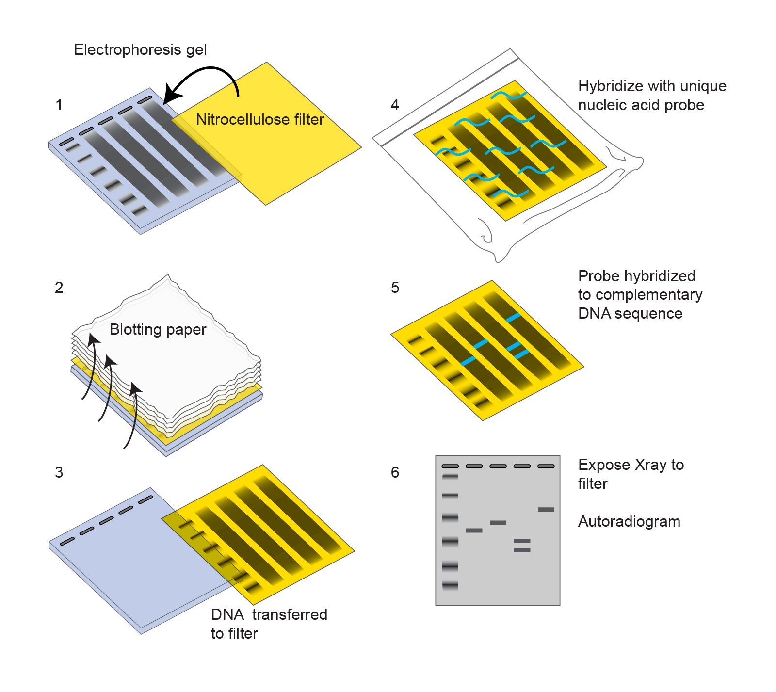 Figure: Southern blotting technique (Image source: National Human Genome Research Institute)
Stage 1: Extraction, fragmentation, and separation of target DNA
- Extract DNA from the target source.
- Cut the DNA into small fragments using a restriction endonuclease enzyme, which cuts at specific recognition sequences.
- Separate the fragments by gel electrophoresis on an agarose gel, which sorts them by molecular weight. Acrylamide gels can be used instead for better resolution of small DNA fragments (under 800 bp).
- Denature the DNA. The fragments are double-stranded, so dip the gel in an alkaline solution to separate them into single strands. Single strands are needed so the probe can pair with the target later.
Stage 2: Blotting (transfer to the membrane)
Blotting means transferring the separated DNA fragments onto a nitrocellulose or nylon membrane. This transfer step is what gives the technique its name, though "Southern blotting" is used for the whole procedure.
- Transfer is done by either capillary blotting or electroblotting.
- In capillary blotting, place a sheet of membrane on the gel (or below it, depending on transfer direction) and apply gentle, even pressure, either by suction or by stacking paper towels and a weight on top. Even contact between gel and membrane is essential for a clean transfer.
- The DNA fragments are pulled onto the membrane by capillary action, creating an imprint (blot) of the gel pattern. The bands keep the same positions they had in the gel.
- Gently remove the portion of membrane touching the gel using a blade.
Stage 3: Baking (fixing the DNA to the membrane)
Bake the membrane in a vacuum or regular oven at 80°C for 2 hours, or fix the DNA using UV crosslinking with short-wavelength UV light. Either method permanently attaches the transferred DNA so it does not wash off during hybridization.
Stage 4: Pre-hybridization and blocking
Pre-hybridization coats the membrane with non-target DNA so the probe cannot stick to empty spaces, which would raise background. Salmon or herring sperm DNA is commonly used as the blocking agent.
- Incubate the membrane in Denhardt's solution for 1 hour or more, depending on the reaction.
- After warming the pre-hybridization solution to 42°C, add heat-denatured (snap-chilled) salmon sperm DNA at 50 µg/mL.
Stage 5: Hybridization with the probe
Hybridization is where the labeled probe binds its complementary target sequence on the membrane.
- Carry out hybridization in a sealed bag containing the blot and the hybridization fluid with the labeled probe.
- Allow 1 to 16 hours, depending on the complexity and concentration of the probe.
The probe carries a label (a radioactive atom, or a fluorescent or chromogenic tag) so its binding site can be found in the next step.
Stage 6: Visualization of the result
- Wash the excess unbound probe off the membrane with a buffer.
- Detect the bound probe:
- a radioactive probe is detected on X-ray film by autoradiography,
- a fluorescent probe is detected by fluorescence imaging,
- a chromogenic probe is detected by color developing directly on the membrane.
The result is a pattern of bands showing exactly where the probe bound.
How to read a Southern blot result
A Southern blot result is a pattern of bands. Reading it means asking what each band tells you.
One band means one match. If the probe hybridizes to a single DNA fragment, you see one band. This says the sample contains exactly one region complementary to the probe, and that region sits at one fragment size.
Multiple bands mean multiple matches. If the probe binds several similar sequences, you get several bands. This happens when the target sequence is repeated across the genome, or when the probe recognizes a family of related sequences.
Band position tells you fragment size. Each band sits at the size of the DNA fragment it is on, read against a size marker. A shift in band position between two samples means the fragment carrying the target is a different length in each, which is the entire basis of DNA fingerprinting below.
No band means no match, or a failed run. Before calling a sample negative, confirm the run worked: a control sample with a known target should give its expected band. If the control is blank, the problem is the run, not the sample.
Stringency changes what counts as a match. Raising the hybridization temperature or lowering the salt concentration makes conditions more stringent, so the probe only stays bound to closely matching sequences. Lower stringency lets the probe bind sequences that are less similar. Adjusting stringency lets you choose between finding only exact matches and finding a wider family of related sequences.
Worked example: DNA fingerprinting with a radiolabeled VNTR probe
DNA fingerprinting is the classic use of Southern blotting, and it is the clearest way to see why the technique matters. It relies on one fact about the human genome: certain regions, called VNTRs (variable number of tandem repeats), are short DNA sequences repeated many times in a row, and the number of repeats differs from person to person. One person may have 8 repeats at a given spot; another may have 20. These are also called minisatellites.
Because the number of repeats varies, the length of the DNA fragment carrying that region varies too. More repeats means a longer fragment; fewer repeats means a shorter fragment. That difference in fragment length is called a restriction fragment length polymorphism (RFLP). Southern blotting turns these length differences into a band pattern.
Here is the walkthrough, step by step:
- Cut the DNA. Digest each person's DNA with a restriction enzyme that cuts on either side of the VNTR regions but not within the repeats. Each VNTR now sits on its own fragment, and that fragment's length depends on how many repeats it contains.
- Separate by size. Run the fragments on a gel. They sort by length, so a fragment with more repeats travels less far, and one with fewer repeats travels further.
- Blot to a membrane. Transfer the separated fragments onto the membrane, keeping their positions.
- Probe with a radiolabeled VNTR probe. Add a probe whose sequence matches the shared repeat core found in the VNTR regions. It is tagged with a radioactive label. The probe binds every fragment that contains that repeat, wherever it landed on the membrane.
- Read the pattern by autoradiography. The probe's label exposes X-ray film, showing a band at each fragment the probe bound. The result is a ladder of bands at heights set by that person's repeat numbers. This is the DNA fingerprint.
Why the pattern is unique to a person. Each person has two copies of every VNTR locus, one inherited from each parent, and each copy may carry a different number of repeats. Across several VNTR loci, the combination of band positions is effectively unique. No two unrelated people share the same full pattern.
Reading it for forensics. Line up the band pattern from a crime-scene sample against a suspect's pattern. If every band matches in position, the samples are consistent with coming from the same person. If bands differ, the suspect is excluded.
Reading it for paternity. A child's bands must each be accounted for by one parent. Half the child's bands should match the mother's pattern; the remaining bands should be found in the biological father's pattern. If bands in the child appear in neither the mother nor the alleged father, that man is excluded as the biological father.
Applications of Southern blotting
Southern blotting detects a specific DNA sequence within a genome. Its main uses:
- DNA fingerprinting and forensic identification. By reading VNTR band patterns, it identifies individuals for criminal investigation, personal identification, and paternity or maternity testing. (See the worked example above.)
- Detecting mutations and gene rearrangements. It reveals changes in DNA structure, which is used to diagnose inherited genetic diseases and to detect gene rearrangements.
- Confirming the presence or absence of a target sequence. It shows whether a specific DNA sequence is present in a sample.
- Checking chromosomal integration. It shows whether a DNA sequence has integrated into a chromosome, for example after gene transfer.
- Analyzing immune gene rearrangements. The clonal rearrangements of immunoglobulin genes and T-cell receptor genes can be studied by Southern blotting, which is useful in diagnosing and classifying lymphomas and leukemias.
- Studying gene structure and restriction maps. It helps work out the structure of a gene and build restriction enzyme maps.
- Counting transgene copies. It determines how many copies of a transgene have been inserted, which is used to confirm transgenic organisms.
- Phylogenetic and recombinant DNA studies. It supports evolutionary comparison between species and recombinant DNA work.
Southern, Northern, and Western blot: what is the difference?
All three separate molecules by size, transfer them to a membrane, and detect a specific target. They differ in what they detect.
| Blot | Detects | Probe or detector |
|---|---|---|
| Southern | DNA | Labeled DNA or RNA probe |
| Northern | RNA | Labeled DNA or RNA probe |
| Western | Protein | Antibody |
A common memory aid: Southern = DNA, Northern = RNA, Western = Protein. Only Southern is named after a person (Edwin Southern); the others are wordplay on his name.
How to remember
Only Southern is a real surname. Edwin Southern invented the DNA method, so his name is the real one. Northern (RNA) and Western (protein) are lab jokes built on his name. So the person's name goes with DNA. From there: Southern-DNA, then N and W follow the compass-and-alphabet pattern to RNA and Protein.
More repeats, higher up the ladder; fewer repeats, further down. In fingerprinting, a fragment with more VNTR repeats is longer, so it travels less far in the gel and sits higher. The band height is just a readout of repeat number. Hold that one idea and the whole fingerprint pattern makes sense.
The probe is a magnet for one sequence. Everything on the membrane is invisible until the probe arrives. The probe finds only its complementary sequence and lights it up. No probe, no band. The right probe is the whole experiment.
Key exam facts
| Point | Fact | Memory aid |
|---|---|---|
| What it detects | DNA | Southern = DNA (the only blot named after a person) |
| Invented by | Edwin Southern, 1975 | The surname that started the naming joke |
| Cutting step | Restriction endonucleases cut DNA into fragments | Cut first, then sort |
| Separation | Gel electrophoresis, by fragment size | Smaller fragments travel further |
| Denaturation | Alkaline solution makes DNA single-stranded | Single strands so the probe can pair |
| Transfer | Capillary or electroblotting onto nitrocellulose/nylon | The "blot" that names the method |
| Fixing | Baking at 80°C for 2 hours, or UV crosslinking | So DNA does not wash off |
| Detection step | Labeled probe hybridizes to complementary target | Probe finds only its match |
| Radioactive probe read by | Autoradiography on X-ray film | Radioactive → autoradiography |
| Main application | DNA fingerprinting via VNTR/RFLP band patterns | More repeats, higher band |
| Fingerprint uniqueness | Two alleles per VNTR locus, combination is individual-specific | One set from each parent |
Where students get confused
"Why cut the DNA up first?" A whole genome is one enormous molecule, far too big to sort or read. The restriction enzyme cuts it into many fragments of manageable, defined sizes. Only then can electrophoresis sort them and the probe find its target on one specific fragment. Cutting turns one unreadable molecule into a sortable ladder.
"Why denature the DNA before probing?" The probe works by base-pairing with the target. Base-pairing needs single strands. Double-stranded DNA has no free bases to pair with, so it must be split into single strands first. Skip denaturation and the probe has nothing to bind.
"What decides where a band sits?" Fragment size, nothing else. Electrophoresis sorts purely by length. In fingerprinting, the fragment length depends on how many VNTR repeats it carries, so band height is a direct readout of repeat number. Two people differ in their patterns because they differ in repeat numbers.
"Is the probe the same as the target?" No, they are complementary, not identical. The probe is a single strand whose sequence base-pairs with the target sequence. It carries the label. The target is the DNA on the membrane you are trying to find. The probe seeks; the target is sought.
"Southern blot confirms DNA, so is it still used for DNA fingerprinting?" It established DNA fingerprinting and teaches the logic best, but modern forensic labs mostly use faster PCR-based STR typing, which needs far less DNA. Learn the Southern blot version for the mechanism; know that the frontline method has moved on.
Frequently Asked Questions
What is Southern blotting used for?
What is Southern blotting used for?
It detects a specific DNA sequence within a genome. Its best-known use is DNA fingerprinting for forensic identification and paternity testing. It is also used to detect mutations and gene rearrangements, confirm whether a target sequence is present, check whether DNA has integrated into a chromosome, and count transgene copies.
What is the principle of Southern blotting?
What is the principle of Southern blotting?
DNA is cut into fragments with restriction enzymes, separated by size using gel electrophoresis, made single-stranded, and transferred to a membrane. A labeled probe with a sequence complementary to the target then binds only its matching fragment, and that binding is detected as a band.
What are the steps of Southern blotting?
What are the steps of Southern blotting?
Six stages: cut and separate the DNA (restriction digest, then electrophoresis), denature and transfer it to a membrane (blotting), bake or UV-fix the DNA to the membrane, block with non-target DNA (pre-hybridization), hybridize with a labeled probe, and visualize the result, usually on X-ray film.
Who invented Southern blotting and when?
Who invented Southern blotting and when?
Edwin Southern developed it in 1975. It is the only one of the three blots named after a real person; Northern (for RNA) and Western (for protein) are wordplays on his name.
How does Southern blotting work in DNA fingerprinting?
How does Southern blotting work in DNA fingerprinting?
It reads VNTR regions, short DNA sequences repeated a variable number of times in each person. Because repeat number differs between people, the DNA fragment carrying each VNTR differs in length. After cutting, separating, blotting, and probing with a radiolabeled VNTR probe, the result is a band pattern unique to that person, which can be matched against another sample.
What is the difference between Southern, Northern, and Western blotting?
What is the difference between Southern, Northern, and Western blotting?
All three separate molecules by size, transfer them to a membrane, and detect a target. Southern detects DNA, Northern detects RNA, and Western detects protein. Southern and Northern use a labeled nucleic acid probe; Western uses an antibody.
What does the band pattern on a Southern blot mean?
What does the band pattern on a Southern blot mean?
Each band marks a DNA fragment that the probe bound. One band means one matching sequence; multiple bands mean the probe matched several sequences. Band position reflects fragment size, so a shift in position between samples means the fragment carrying the target is a different length in each.
Why is DNA denatured before hybridization?
Why is DNA denatured before hybridization?
The probe binds by base-pairing, which needs single strands. Double-stranded DNA has no free bases available, so the DNA is made single-stranded with an alkaline solution before the probe is added.
References
- Southern EM. Detection of specific sequences among DNA fragments separated by gel electrophoresis. J Mol Biol. 1975;98(3):503-517.
- Jeffreys AJ, Wilson V, Thein SL. Individual-specific 'fingerprints' of human DNA. Nature. 1985;316(6023):76-79.
- Green MR, Sambrook J. Molecular Cloning: A Laboratory Manual. 4th ed. Cold Spring Harbor, NY: Cold Spring Harbor Laboratory Press; 2012.
- Brown TA. Gene Cloning and DNA Analysis: An Introduction. 7th ed. Oxford: Wiley-Blackwell; 2016.
- National Human Genome Research Institute. Southern Blot. Available at: https://www.genome.gov/genetics-glossary/Southern-Blot

Tankeshwar Acharya, MSc (Medical Microbiology)
Tankeshwar Acharya is an Assistant Professor in the Department of Microbiology at Patan Academy of Health Sciences (PAHS), Nepal, where he has been teaching and practicing clinical microbiology for over 14 years. He is the founder of Microbe Online, one of the leading free microbiology education resources on the web, covering bacteriology, mycology, parasitology, immunology, and clinical laboratory diagnostics written from direct experience in both the classroom and the diagnostic laboratory.
Comments
No comments yet. Be the first to share your thoughts.
Leave a comment
All comments are reviewed before they appear.