Laboratory Diagnosis of Intestinal Parasitic Infections: Methods, Specimen Handling, and When to Use Each Test
Complete guide to laboratory diagnosis of intestinal parasites — stool collection, O&P examination, concentration techniques, permanent stains, culture, serology, and PCR with a decision table and specimen exceptions for pinworm and Schistosoma.
A 4-year-old child in a rural clinic has chronic diarrhoea, pale stools, and poor weight gain. A medical student in an urban teaching hospital has returned from a fieldwork placement with explosive watery stools. A woman in her first trimester is found to have eosinophilia on a routine blood count. All three need stool examination — but not the same examination.
Diagnosing intestinal parasitic infections is not a single test. It is a decision tree: which specimen to collect, how quickly to process it, which examination method to use, and how to interpret what you find. The wrong choice at any step gives a false negative. This article is the map.
Why Laboratory Diagnosis?
Clinical features alone cannot reliably distinguish between intestinal parasitic infections. Diarrhoea, abdominal pain, and weight loss are common to Entamoeba histolytica, Giardia lamblia, hookworm, Ascaris, and dozens of other pathogens — bacterial and parasitic. Treatment differs completely between them:
- E. histolytica → metronidazole + luminal agent (diloxanide furoate)
- Giardia lamblia → metronidazole or tinidazole
- Hookworm → albendazole or mebendazole
- Strongyloides → ivermectin (not albendazole as first choice)
- Taenia solium → praziquantel (and careful assessment for cysticercosis before treatment)
Treating empirically with an anthelmintic covers some organisms but not protozoa. Treating empirically with metronidazole covers protozoa but not helminths. Laboratory confirmation is the only way to treat correctly and in the case of T. solium, treating without knowing whether the patient has concurrent cysticercosis can precipitate a neurological crisis.
Stool sample collection
Stool specimens should be collected in a wide-mouthed, clean, leak-proof container. The sample should be collected after the onset of symptoms and ideally before the initiation of the antiparasitic therapy. First-morning sample is preferable. The amount of stool and the frequency of stool specimen submission differ according to the suspected disease. Generally, collections of three stool specimens are sufficient to make a diagnosis of most intestinal parasitic infections but for some diseases like giardiasis or amoebiasis, a total of six specimens are preferable.
Which sample to collect first matters:
- Liquid or loose stool: Process within 30 minutes — trophozoites of Entamoeba and Giardia are motile and identifiable; they disintegrate rapidly after passage
- Soft or formed stool: Can be examined within 24 hours; cysts and eggs are stable
- First-morning sample: Preferred for most parasites — concentrated overnight transit
Critical specimen exceptions — do not collect stool for these:
| Parasite | Disease | Correct specimen | Reason |
|---|---|---|---|
| Enterobius vermicularis (pinworm) | Enterobiasis | Cellophane tape test — perianal swab at night/early morning | Female worm migrates to perianal area to lay eggs at night; eggs rarely found in stool |
| Schistosoma haematobium | Urogenital schistosomiasis | Urine (terminal urine, midday collection) | Eggs excreted in urine, not faeces |
| Strongyloides stercoralis (low burden) | Strongyloidiasis | Harada-Mori culture or Baermann technique | Larvae may be present at too low a density for direct smear detection |
| Blood/tissue parasites | Malaria, filariasis, VL | Blood (thick/thin smear, QBC, RDT) | Not intestinal — blood is the specimen |
How many specimens?
- 3 stool specimens (collected on alternate days) — sufficient for most intestinal parasites
- 6 specimens — recommended for Giardia and Entamoeba (intermittent cyst shedding means a single specimen misses up to 30% of infections)
- Repeat if clinical suspicion remains high despite negative results — parasite shedding is not continuous
Transport of stool specimen
Liquid & semi-formed stool specimens should be examined within 30 minutes of collection to visualize the motility of trophozoites (if giardiasis/amoebiasis is suspected). Further delay in an examination may disintegrate the trophozoites. Formed stools may be examined up to 24 hours after passage.
Intestinal Parasites found in Faeces
| Name of the Parasite | Protozoa or Helminth | Disease |
|---|---|---|
| Entamoeba histolytica | Protozoa | Amoebiasis & ameobic liver abscess |
| Giardia lamblia | Protozoa | Giardiasis |
| Cryptosporidium parvum | Protozoa | Cryptosporidiosis |
| Cystoisospora belli (Previously known as Isospora belli) | Protozoa | Cystoisosporiasis |
| Cyclospora cayetanensis | Protozoa | Cyclosporiasis |
| Balantidium coli | Protozoa | Balantidiasis |
| Taenia solium (pork tapeworm) | Helminth | Taeniasis & cysticercosis |
| Taenia saginata (beef tapeworm) | Helminth | Taeniasis |
| Hymenolepis nana (dwarf tapeworm ) | Helminth | Hymenolepiasis |
| Schistosoma mansoni, S. haematobium, S. japonicum | Helminth | Schistosomiasis |
| Fasciola hepatica ( the common liver fluke) or (the sheep liver fluke). | Helminth | Fascioliasis |
| Ascaris lumbricoides (large roundworm) | Helminth | Ascariasis |
| Ancylostoma duodenale (Old world hookworm) | Helminth | Hookworm infection |
| Necatar americanus (New World hookworm) | Helminth | Necatoriasis (hookworm infestations) |
| Enterobius vermicularis (Pinworm) | Helminth | Enterobiasis |
| Trichuris trichiura (Whip worm) | Helminth | Trichuriasis (whipworm infection) |
Preservation of the stool specimen
Preservatives (like formalin and polyvinyl alcohol) are used to fix stool specimens permanently. Preservatives kill the parasites, so the characteristics motility of the trophozoites cannot be seen. Preservatives help preserve morphological forms (cyst, ova, and larvae) of parasites while being sent to reference laboratories for analysis and lower the risk of infections to the laboratory technicians.
Laboratory detection methods for Intestinal Parasitic Infections
The O&P Examination: Standard Workflow
The Ova and Parasite (O&P) examination is the standard laboratory protocol for intestinal parasite diagnosis. It is not a single test — it is a sequence of four complementary steps, each adding information the previous step misses:
STEP 1: Macroscopic examination
         ↓
STEP 2: Direct wet mount (saline + iodine)
         ↓
STEP 3: Concentration technique
         (formal-ether sedimentation OR flotation)
         ↓
STEP 4: Permanent stained smear
         (trichrome, iron-haematoxylin, modified acid-fast)
         — only when protozoan infection suspected or Steps 1–3 inconclusive
Each step has a specific purpose and detects different things. Skipping a step risks missing the diagnosis.
Step 1: Macroscopic Examination
Examine the stool before opening the container:
Consistency: Liquid/semi-liquid → likely trophozoites present (process urgently); formed → likely cysts/eggs.
Colour: Pale yellow, frothy, fatty-looking → suggests Giardia (steatorrhoea from fat malabsorption). Bloody/mucoid → suggests invasive amoeba (E. histolytica), Balantidium coli, heavy Trichuris infection, or schistosomiasis.
Visible parasites: Adult Ascaris lumbricoides (large, cream-coloured roundworm 15–35 cm) and Enterobius vermicularis (small, white, thread-like 8–13 mm in females) may be visible to the naked eye. Tapeworm proglottids (flat, white, motile segments of Taenia spp.) are also macroscopically visible.
Step 2: Direct Wet Mount (Saline and Iodine)
What it detects: Trophozoites (motility visible in saline mount), cysts of protozoa, eggs and larvae of helminths.
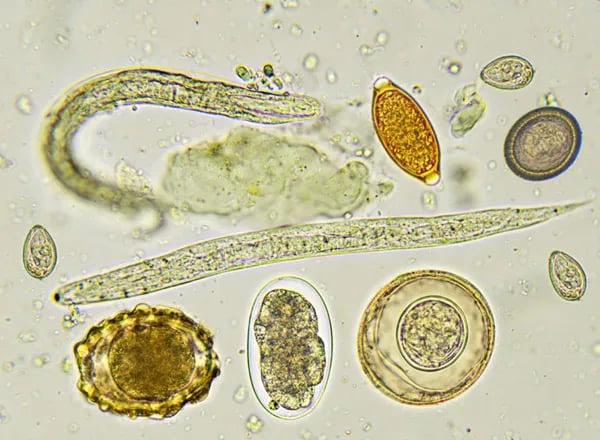 Figure: Eggs (ova) of various intestinal parasites
Two preparations made simultaneously:
- Saline wet mount: Detects motile trophozoites; identifies helminth eggs by morphology
- Iodine (Lugol's) wet mount: Stains glycogen mass and nuclei of protozoan cysts — makes internal structure visible; kills motility
Limitations: Not sensitive for low-density infections; protozoan cysts can be missed; cannot reliably identify all protozoan species by morphology alone (trichrome stain required for definitive speciation).
→ For full saline wet mount procedure: Saline Wet Mount for Intestinal Parasites
Step 3: Concentration Techniques
Concentration physically accumulates parasites from a large stool sample into a small volume — dramatically increasing sensitivity for low-burden infections.
Two methods available:
Formal-Ether (Formalin-Ethyl Acetate) Sedimentation
- Principle: Stool emulsified in formalin (fixes parasites), ethyl acetate added (dissolves fats), centrifuged → parasites sediment at bottom
- What it detects: Cysts, eggs, and larvae — all in one step
- Best for: Heavy or light infections with any intestinal parasite; the workhorse concentration technique
- Limitation: Protozoan trophozoites are destroyed by formalin fixation
→ Full procedure: Formal-Ether Sedimentation Technique
Kato-Katz Technique (Thick Smear)
- Principle: Large volume of stool (41.7 mg) pressed through a mesh screen onto a slide, covered with a glycerol-malachite green cellophane; helminth eggs cleared and stained
- What it detects: Helminth eggs only — highly sensitive for quantifying egg burden (eggs per gram of stool = EPG)
- Best for: Soil-transmitted helminths (Ascaris, hookworm, Trichuris), Schistosoma eggs; population-level surveys; assessment of infection intensity for treatment decisions
- Limitation: Does not detect protozoa; trophozoites and cysts not visible; slides must be read within 30–60 min (hookworm eggs clear and become unrecognizable after ~1 hour)
→ Full procedure: Kato-Katz Technique
Decision: Formal-Ether vs Kato-Katz?
| Scenario | Preferred concentration method |
|---|---|
| Suspected protozoa (Giardia, Entamoeba) | Formal-ether sedimentation |
| Survey for soil-transmitted helminths | Kato-Katz |
| Quantifying worm burden for treatment response | Kato-Katz (gives EPG) |
| Suspected mixed infection (protozoa + helminths) | Formal-ether (then permanent stain for protozoa) |
| Resource-limited setting, single method needed | Formal-ether (broader coverage) |
Step 4: Permanent Stained Smears
When direct wet mount and concentration fail to identify protozoa, or when formal morphological identification of a protozoan species is needed for treatment decisions, a permanent stained smear is prepared.
Trichrome Stain (Wheatley's)
The standard stain for intestinal protozoa. Three dyes differentiate cytoplasm (blue-green), nuclear material (red-purple), and background debris.
Best for: Definitive identification of Entamoeba histolytica vs E. dispar vs E. coli, Giardia cysts and trophozoites, Balantidium coli.
→ Full procedure: Trichrome Staining for Fecal Smears
Iron-Haematoxylin Stain
Older but highly precise stain; shows nuclear detail clearly. Used where trichrome reagents are unavailable.
Modified Acid-Fast (Ziehl-Neelsen or Kinyoun) Stain
Specifically for: Cryptosporidium parvum, Cyclospora cayetanensis, Cystoisospora belli — coccidian oocysts are acid-fast (stain pink/red against a blue background). Routine O&P examination misses these organisms unless a modified acid-fast stain is added.
Critical point for the exam: If a patient with HIV/AIDS or other immunocompromise has chronic diarrhoea and routine O&P is negative, always add a modified acid-fast stain for Cryptosporidium. Routine trichrome and wet mount will miss it.
Step 5 (Supplementary): Cellophane Tape Test (Pinworm)
This is NOT part of the standard O&P examination — it is a separate test for Enterobius vermicularis only.
Transparent adhesive tape is pressed against the perianal skin early in the morning (before bathing or defecation), then applied to a glass slide and examined microscopically for Enterobius eggs.
→ Full procedure: Cellophane Tape Test for Pinworm
Culture Methods
Culture is rarely the first-line diagnostic method in parasitology but has specific indications:
Harada-Mori filter paper technique: Used to recover and identify larvae of Strongyloides stercoralis and hookworm species. Stool is smeared on filter paper, placed in a tube with distilled water, and incubated at room temperature for 5–10 days. Larvae migrate from the stool into the water and can be identified by morphology. Particularly useful when direct smear is negative but clinical suspicion for Strongyloides is high (eosinophilia + possible hyperinfection risk before immunosuppression).
Robinson's or LJ medium (liquid culture): Used for Entamoeba histolytica and Balantidium coli. Higher sensitivity than a single direct wet mount but requires 2–3 days incubation. Rarely used in routine practice; reserved for reference laboratories.
NNN medium (biphasic): Used for Leishmania culture — not an intestinal pathogen but the medium name appears in discussions of parasitology culture methods. The intestinal diagnostic cultures use Robinson's and Harada-Mori, not NNN.
→ For NNN medium details: NNN Medium
Serology
Serology is used when:
- Routine O&P examination is negative but invasive parasitic infection is clinically suspected
- The parasite has disseminated to tissues where stool examination is unhelpful (liver abscess, cysticercosis, echinococcosis)
- The infection is below the detection threshold of microscopy
Serology by organism:
| Organism | Disease | Serological test used | Notes |
|---|---|---|---|
| Entamoeba histolytica | Amoebic liver abscess | ELISA, IHA | Serology positive in >90% of liver abscess; unreliable for intestinal amoebiasis alone |
| Giardia lamblia | Giardiasis | EIA antigen detection in stool | Antigen detection (not antibody) — highly sensitive |
| Taenia solium | Cysticercosis/neurocysticercosis | EITB (enzyme-linked immunoelectrotransfer blot); ELISA | For diagnosis of tissue disease; not reliable for intestinal tapeworm |
| Strongyloides stercoralis | Strongyloidiasis | ELISA | Sensitive; positive even when larvae not seen on smear |
| Schistosoma spp. | Schistosomiasis | ELISA, IHA | Used in non-endemic settings (travellers) |
| Cryptosporidium | Cryptosporidiosis | EIA antigen detection in stool | More sensitive than modified acid-fast stain alone |
Key limitation: Antibodies persist after cure; serology cannot distinguish active from past infection. Antigen detection (for Giardia and Cryptosporidium) is more specific for active infection.
Molecular Assays (PCR)
PCR and multiplex PCR panels (e.g., FilmArray GI Panel, BD MAX Enteric Parasite Panel) simultaneously detect multiple intestinal parasites from a single stool sample with very high sensitivity and specificity.
Advantages: Detects Cryptosporidium, Giardia, Entamoeba histolytica, Cyclospora, Cystoisospora, and some helminths simultaneously; does not require fresh specimen (works on preserved stool); identifies species where morphology is ambiguous (E. histolytica vs E. dispar vs E. moshkovskii).
Current limitations: Expensive; requires laboratory infrastructure; not widely available in LMIC settings; WHO does not yet recommend routine use over microscopy for resource-limited settings.
In practice: PCR is used in reference laboratories, for immunocompromised patients (HIV, transplant) where sensitivity is critical, for outbreak investigation, and for species identification when morphology is ambiguous.
When to Use Which Test: Decision Summary
| Clinical scenario | First-line test | Add if negative/inconclusive |
|---|---|---|
| Acute watery diarrhoea, endemic area | Direct saline wet mount (motile trophozoites) | Formal-ether; trichrome stain |
| Chronic diarrhoea, suspected Giardia | 3 stool specimens, formal-ether + trichrome | Stool antigen EIA (most sensitive) |
| Suspected amoebiasis (dysentery) | Direct wet mount (motile trophozoites + RBC ingestion) | Trichrome; serology if liver abscess suspected |
| School-age child, worm burden survey | Kato-Katz (EPG quantification) | — |
| Perianal itching, child | Cellophane tape test (not stool O&P) | — |
| HIV/AIDS patient, chronic diarrhoea | Modified acid-fast stain (Cryptosporidium, Cyclospora) | Stool antigen EIA; PCR panel |
| Eosinophilia + suspected Strongyloides | Harada-Mori culture; serology (ELISA) | Repeat 6 specimens; Baermann technique |
| Post-travel diarrhoea, returning traveller | Formal-ether + trichrome (3 specimens) | PCR multiplex if available; serology |
| Suspected neurocysticercosis | Serology (EITB/ELISA) + neuroimaging | Stool O&P for proglottids/eggs (may be negative) |
| Urinary symptoms, endemic Schistosoma area | Urine examination (terminal urine, midday) | Serology |
Where Students Actually Get Confused
1. "Stool examination will diagnose pinworm." Enterobius vermicularis eggs are rarely found in stool. The female migrates to the perianal skin at night to deposit eggs. The correct specimen is the cellophane tape test applied to the perianal region before the child bathes in the morning. Ordering a routine O&P on a child with perianal itching gives a high false-negative rate.
2. "One negative stool result excludes intestinal parasitic infection." Parasite shedding is intermittent, particularly for Giardia and Entamoeba. WHO and CDC guidelines recommend a minimum of three specimens collected on separate days before reporting negative. For giardiasis and amoebiasis, six specimens improve sensitivity further. A single negative O&P in a symptomatic patient is not a final answer.
3. "Trichrome stain detects all intestinal parasites." Trichrome is excellent for intestinal protozoa (Entamoeba species, Giardia, Balantidium), but it does NOT stain coccidian oocysts (Cryptosporidium, Cyclospora, Cystoisospora). These require a modified acid-fast stain. HIV patients with Cryptosporidium diarrhoea will have a negative trichrome and a negative routine wet mount — the diagnosis requires a specifically requested acid-fast stain.
4. "Formal-ether sedimentation detects trophozoites." No — formalin fixation kills trophozoites and destroys motility. Formal-ether concentrates cysts, eggs, and larvae. Trophozoite detection requires a direct saline wet mount from fresh stool processed within 30 minutes of passage.
5. "Kato-Katz detects protozoa." Kato-Katz is specific to helminth eggs — the glycerol-cellophane technique is optimised for clearing and staining eggs, and the staining characteristics do not work for protozoan cysts. Do not use Kato-Katz as the sole method when protozoa are suspected.
6. "Schistosoma haematobium is diagnosed from stool." S. haematobium causes urogenital schistosomiasis and its eggs are excreted in urine, not faeces. The correct specimen is terminal urine (last portion of a midday sample, when egg concentration is highest). S. mansoni and S. japonicum eggs are found in stool.
Key Exam Facts in One Table
| Fact | Detail | Memory hook |
|---|---|---|
| Standard O&P examination | Macroscopic → direct wet mount → concentration → permanent stain | 4-step sequence; each adds sensitivity |
| Liquid stool processing time | Within 30 minutes (trophozoites) | Motility dies fast |
| Formed stool processing time | Within 24 hours | Cysts and eggs stable |
| Number of specimens (general) | 3 specimens on separate days | 6 for Giardia/Entamoeba |
| Pinworm diagnosis | Cellophane tape test — NOT stool O&P | Perianal eggs, not stool eggs |
| Schistosoma haematobium specimen | Urine (not stool) | Urogenital schistosomiasis |
| Kato-Katz: detects | Helminth eggs only; quantifies EPG | NOT for protozoa |
| Formal-ether: detects | Cysts + eggs + larvae (NOT trophozoites) | Formalin kills motility |
| Modified acid-fast: needed for | Cryptosporidium, Cyclospora, Cystoisospora | Coccidia — missed by routine O&P |
| Trichrome stain: detects | Intestinal protozoa (Entamoeba, Giardia, Balantidium) | NOT acid-fast organisms |
| Harada-Mori culture | Strongyloides, hookworm larvae — 5–10 days | When smear negative but Strongyloides suspected |
| Stool antigen EIA | Giardia, Cryptosporidium — most sensitive | Detects active infection (not past) |
| PCR use | Immunocompromised, species ID, outbreak investigation | Not routine in LMIC |
| Reading Kato-Katz slides | Within 30–60 min — hookworm eggs clear and disappear | Time-critical slide |
| Giardia: stool appearance | Pale, frothy, fatty (steatorrhoea) | Fat malabsorption from villous flattening |
| Amoebic dysentery: stool | Bloody, mucoid; trophozoites with ingested RBCs | RBC ingestion = hallmark of E. histolytica invasion |
Self-Check Questions
- A child presents with perianal itching worse at night. You order a stool O&P examination — the result is negative. Have you excluded pinworm? What should you do next?
- A patient with AIDS has three weeks of profuse watery diarrhoea. Routine stool O&P (direct wet mount + formal-ether + trichrome) is negative. What additional stain must you request, and which organisms are you looking for?
- A school survey of a rural village finds high prevalence of Ascaris and hookworm. Which technique should be used to quantify egg burden and assess treatment response, and why?
- You receive a liquid stool sample from a patient with suspected amoebic dysentery. It arrives in the laboratory 2 hours after collection. Can you still detect trophozoites? What can you still reliably examine?
- Which of the following is correctly diagnosed from urine rather than stool: (a) Schistosoma mansoni (b) Schistosoma haematobium (c) Ascaris lumbricoides (d) Giardia lamblia?
- A clinician orders a single stool O&P for a patient with suspected giardiasis. The result is negative. What should you advise?
Answers:
1. No — pinworm eggs are rarely found in stool; the correct test is the cellophane tape test applied to the perianal skin early morning before bathing. Order this immediately.
2. Modified acid-fast stain; looking for Cryptosporidium parvum, Cyclospora cayetanensis, and Cystoisospora belli — coccidian oocysts missed by routine O&P.
3. Kato-Katz technique — it provides eggs per gram (EPG) of stool, allowing quantification of infection intensity and objective assessment of treatment efficacy. 4. No — trophozoite motility is lost after 30 minutes; at 2 hours, any trophozoites present have degenerated. Cysts and eggs (formal-ether concentration + iodine wet mount) can still be examined.
5. (b) Schistosoma haematobium — causes urogenital schistosomiasis with eggs in urine; S. mansoni and S. japonicum eggs are found in stool.
6. Advise repeat testing — giardiasis requires minimum 3 specimens on separate days (ideally 6) due to intermittent cyst shedding; a single negative does not exclude infection in a symptomatic patient.)
References and Further Reading
- Garcia, L. S. (2016). Diagnostic Medical Parasitology (6th ed.). ASM Press.
- Cheesbrough, M. (2006). District Laboratory Practice in Tropical Countries (2nd ed., Part 1). Cambridge University Press.
- World Health Organization. (2012). Bench aids for the diagnosis of intestinal parasites (2nd ed.). WHO. https://www.who.int/publications/i/item/9789241544764
- Sastry, A. S., & Bhat, S. (2014). Essentials of Medical Parasitology. Jaypee Brothers Medical Publishers.
- CDC – DPDx: Laboratory Identification of Parasites of Public Health Concern. https://www.cdc.gov/dpdx/index.html
- Verweij, J. J., & Stensvold, C. R. (2014). Molecular testing for clinical diagnosis and epidemiological investigations of intestinal parasitic infections. Clinical Microbiology Reviews, 27(2), 371–418. https://doi.org/10.1128/CMR.00122-13
- Dacal, E., Saugar, J. M., de Lucio, A., et al. (2018). Prevalence and molecular characterization of Strongyloides stercoralis, Giardia duodenalis, Cryptosporidium spp., and Blastocystis spp. isolates in school children in Cubal, Western Angola. Parasites & Vectors, 11, 67. https://doi.org/10.1186/s13071-018-2648-3
- WHO Bench aids for the diagnosis of intestinal parasites — available as a free downloadable pocket reference at the WHO link above; recommended for all bench laboratory staff.
Frequently Asked Questions
What is the O&P examination for intestinal parasites?
Why must liquid stool be examined within 30 minutes?
Why is pinworm not diagnosed from a routine stool O&P examination?
What stain is used to diagnose Cryptosporidium in stool?
What is the difference between formal-ether sedimentation and Kato-Katz technique?
How many stool specimens are needed to diagnose giardiasis?

Tankeshwar Acharya, MSc (Medical Microbiology)
Tankeshwar Acharya is an Assistant Professor in the Department of Microbiology at Patan Academy of Health Sciences (PAHS), Nepal, where he has been teaching and practicing clinical microbiology for over 14 years. He is the founder of Microbe Online, one of the leading free microbiology education resources on the web, covering bacteriology, mycology, parasitology, immunology, and clinical laboratory diagnostics written from direct experience in both the classroom and the diagnostic laboratory.