
Leprosy (Hansen's Disease): Why the Immune Response Decides Tuberculoid vs Lepromatous
How Mycobacterium leprae causes leprosy, why the strength of cell-mediated immunity decides whether disease is tuberculoid or lepromatous, how it damages nerves, and how the slit-skin smear and lepromin test are used.

On this page
A young adult notices a pale patch on the skin that has lost feeling: a pin or heat touched to it is not felt. Nearby, a nerve at the elbow is thickened and can be rolled under the fingers. There is no pain, and that loss of sensation is the clue. This is leprosy, an infection that targets the skin and the peripheral nerves, and its lasting harm comes not from the organism directly but from the nerve damage it causes.
Leprosy, or Hansen's disease, is one of the oldest recorded diseases and still occurs across South Asia. What makes it distinctive is that its two very different forms, and the disability that follows, are decided largely by one thing: how strongly the patient's own cell-mediated immunity responds to the organism. This page is about Mycobacterium leprae, how the immune response shapes the disease, and how leprosy is diagnosed.
Introduction
Leprosy (Hansen's disease) is a chronic infection caused by Mycobacterium leprae, an acid-fast bacillus. It mainly affects the skin, the peripheral nerves, and the cooler surfaces of the body (the upper respiratory mucosa, earlobes, and the cooler skin), because M. leprae grows best at about 30°C, below core body temperature. This preference for cool sites explains the distribution of the lesions.
M. leprae belongs to the genus Mycobacterium, whose waxy, mycolic-acid wall makes all its members acid-fast and slow-growing; that shared biology is covered on the Mycobacterium overview. Two features set M. leprae apart from the rest of the genus, and both shape the whole disease: it is the slowest-growing human bacterial pathogen (doubling time about 14 days), and it cannot be grown on any artificial medium or cell culture, uniquely even among mycobacteria.
In 1873, Gerhard Hansen identified the bacterium in leprosy lesions, showing that leprosy is an infectious disease, not a hereditary condition or a punishment, a finding that began to counter centuries of stigma.
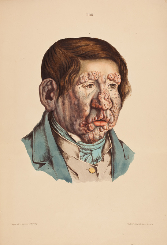 Figure: A man with leprosy (image by J. L. Losting.)
How leprosy spreads
Leprosy is not highly contagious. Most people have natural immunity and never develop disease even after exposure. Transmission needs prolonged, close contact with an untreated lepromatous (multibacillary) patient, who sheds large numbers of M. leprae in nasal secretions and from skin lesions. The main route is thought to be respiratory (nasal droplets), with skin contact playing a lesser role.
Two facts explain why leprosy is hard to trace. The incubation period is very long, around 5 years on average, and symptoms can take up to 20 years to appear, so the source of infection is usually untraceable. And a patient becomes non-infectious quickly once multidrug therapy starts. The nine-banded armadillo is a natural animal reservoir in parts of the Americas.
How Mycobacterium leprae causes disease
Leprosy is unusual: the same organism produces two almost opposite diseases, and which one a patient gets is decided by the strength of their cell-mediated (T-cell) immunity, not by the organism. This single idea is the key to the whole disease.
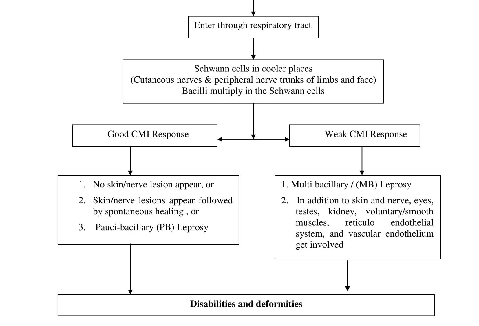 Figure: Pathogenesis of Leprosy
It is an intracellular organism that targets Schwann cells. M. leprae survives and multiplies inside host cells, especially macrophages (histiocytes) and, crucially, the Schwann cells that wrap peripheral nerves. Its ability to invade Schwann cells is what makes leprosy a nerve disease, and the nerve damage is the source of the disability that defines leprosy.
The immune response decides the form. After infection, the outcome depends on how the patient's cell-mediated immunity reacts:
- If cell-mediated immunity is strong, the body mounts a vigorous granulomatous response that kills most organisms. This limits the infection to a few lesions with very few bacilli, but the strong immune reaction itself damages the nerves it fights in. This is tuberculoid leprosy (paucibacillary): few organisms, strong immunity, positive lepromin test.
- If cell-mediated immunity is weak or absent against M. leprae, the organism multiplies almost unchecked inside macrophages, producing many lesions teeming with bacilli across the cooler skin. This is lepromatous leprosy (multibacillary): huge numbers of organisms, poor immunity, negative lepromin test.
- Between these two poles lies a spectrum of borderline forms (below), and patients can shift along it.
How nerves are damaged, in both forms. In tuberculoid leprosy, the strong immune (granulomatous) reaction destroys nerves as it attacks the organism within them. In lepromatous leprosy, the sheer bacillary load and slow damage affect nerves more diffusely. Either way, damage to peripheral nerves causes loss of sensation (so injuries and burns go unnoticed and lead to ulcers and deformity), muscle weakness, and autonomic changes. The disability of leprosy comes from this nerve damage, not from the organism destroying tissue directly.
Putting it together
M. leprae enters, grows slowly in the cool skin and in Schwann cells, and the patient's own cell-mediated immunity then decides the disease. Strong immunity contains the organism but produces the localized, nerve-damaging lesions of tuberculoid leprosy; weak immunity lets the organism multiply into the widespread, bacilli-rich lesions of lepromatous leprosy. In both, damage to peripheral nerves produces the anesthesia and deformity that define the disease. Understanding leprosy is understanding this immune spectrum.
Clinical features
The two poles look very different:
- Tuberculoid: one or a few well-defined, hypopigmented (pale) or reddish patches with loss of sensation, and often a nearby thickened peripheral nerve. Because immunity is strong, lesions are few and organisms scarce.
- Lepromatous: many symmetrical skin lesions, nodules, and diffuse thickening of the skin. Involvement of the face can cause the loss of the eyebrows (madarosis) and, in advanced disease, the flattening of facial features. Nasal involvement causes chronic stuffiness and destruction of the septum. Nerve damage is widespread.
The earliest and most important sign in either form is a skin patch with loss of sensation, or a thickened peripheral nerve, which is what should prompt testing.
Leprosy reactions. During the disease or after starting treatment, sudden immune-driven episodes called lepra reactions (type 1 reversal reactions and type 2 erythema nodosum leprosum) can flare, worsening skin and nerve inflammation. They are important because they cause much of the acute nerve damage and are medical urgencies.
Ridley and Jopling Classification
Ridley and Jopling (1966) have introduced a scale for classifying the spectrum of leprosy into five groups:
- Tuberculoid (TT)
- Borderline Tuberculoid (BT)
- Borderline (BB)
- Borderline Lepromatous (BL)
- Lepromatous (LL)
WHO Classification of Leprosy
The WHO simplified the classification into paucibacillary (few bacilli, up to 5 skin lesions) and multibacillary (many bacilli, more than 5 lesions) specifically to guide treatment in field settings where slit-skin smears are not available.
Comparison of tuberculoid and lepromatous leprosy
Feature | Tuberculoid | Lepromatous |
Type of lesion | One or few lesions with little tissue destruction | Many lesions with marked tissue destruction |
Number of acid-fast bacilli | Few | Many |
Likelihood of transmission | Low | High |
Cell-mediated response to M. Leprae | Present | Reduced or latent |
Lepromin skin test | Positive | Negative |
Laboratory diagnosis
Sample
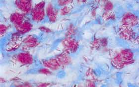 Figure: M. leprae in stained smear
Ideally, at least six sites can be sampled, including earlobes, eyebrows, elbow, knees, nasal mucosa, and skin lesions. Skin biopsy from edges of active patches and nerve biopsy from thickened nerves can also be collected.
Taking a skin smear for the diagnosis of Leprosy
Traditionally smears used to be taken from four or even six sites, but two sites are now considered adequate in most cases. Skin smear is taken from one ear lobe and one lesion. Smear should be taken from the edges of the most active lesions (active lesions are raised and reddish in color). If there is no suitable skin lesion, a second smear can be taken from another ear lobe.
To take a skin smear, the following equipment will be needed
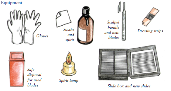 Figure: Equipment needed to take skin smear
- Gloves
- Swab and spirit
- Scalpel handle and new blades
- Dressing strips
- Safe disposal for used blades
- Spirit lamp
- Slide box and new slides
Procedure
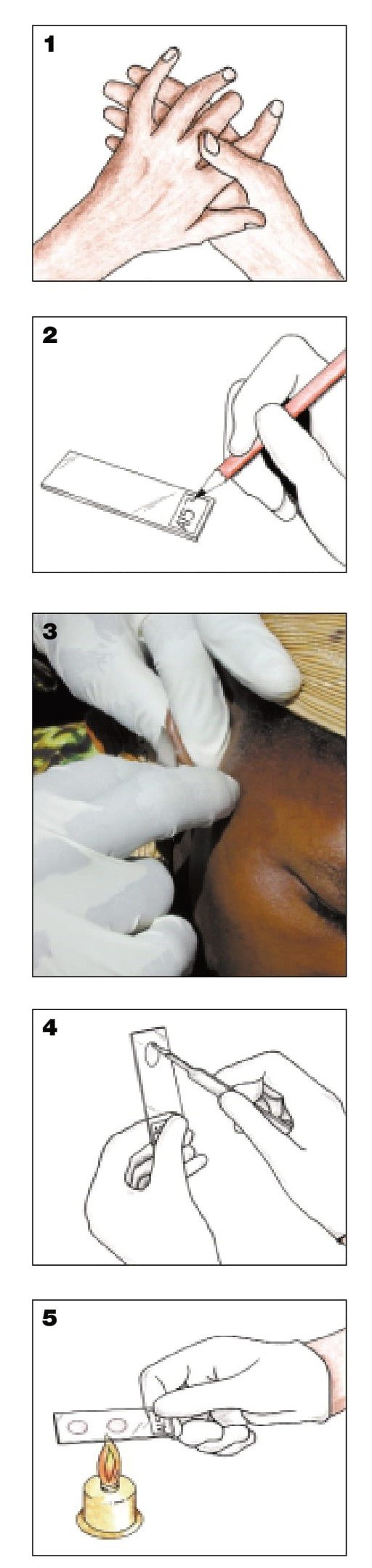 - Wash your hands (1) and put on gloves.
Take a new, clean, unscratched microscope slide. Using a slide marker, write the patient identification (ID) number at the bottom of the slide (2). This number must be on the request form.
Clean the skin at the smear sites with a cotton wad drenched in alcohol. Let it dry.
Light the spirit burner.
Put a new blade on the scalpel handle. If you put the scalpel down, make sure the blade does not touch anything.
Pinch the skin firmly between your thumb and forefinger; maintain pressure to press out the blood.
Make an incision in the skin about 5 mm long and 2 mm deep (3). Keep on pinching to make sure the cut remains bloodless. If bleeding, wipe the blood away with cotton wad.
Turn the scalpel 90° and hold it at a right angle to the cut.
Scrape inside the cut once or twice with the side of the scalpel, to collect tissue fluid and pulp. There should be no blood in the specimen, as this may interfere with staining and reading.
Stop pinching the skin and absorb any bleeding with a wad of cotton. Spread the material scraped from the incision onto the slide, on the same side as the ID number. Spread it evenly with the flat of the scalpel, making a circle 8 mm in diameter (4).
Rub the scalpel with a cotton wad drenched in alcohol. Pass the blade through the flame of the spirit burner for 3 to 4 seconds. Let it cool without touching anything. Repeat the steps above for the second site. Spread this smear next to, but not touching, the first one.
Discard the scalpel blade safely. Dress the wounds and thank the patient.
Let the slide dry for 15 minutes at room temperature, but not in direct sunlight.
Fix the smears by passing the slide, with the smears upwards, slowly through the flame of a spirit burner, 3 times (5). Do not overheat. The slide should not be too hot to touch.
Put the slide in a slide box and send it to the laboratory with the skin smear request form.
Microscopy
Slit-skin smear is stained with a modified Ziehl Neelsen stain (5% sulphuric acid or 1% v/v acid alcohol is used as a decolorizing agent). Find details about Ziehl-Neelsen stain procedure here. Stained slides are observed under a microscope (100x objective lens using immersion oil).
M. leprae is a slightly curved filament 3–10 μm in length containing irregular arrangements of dense material sometimes in the shape of rods. Acid-fast bacilli appear as fine red rods against a blue background. They can be straight or curved, and the red color can be uniformly distributed (solid bacilli) or unevenly distributed (fragmented and granulated bacilli). Clumps of bacilli are called globi. Solid bacilli may suggest the presence of viable organisms and may be seen in new, untreated cases or in relapse cases. Bacilli which stain irregularly are probably dead and degenerating.
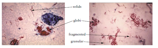 Figure: AFB in skin smear
- In lepromatous leprosy: lipid-laden macrophages called "foam cells” containing many acid-fast bacilli are seen in the skin.
- In tuberculoid leprosy: Very few acid-fast bacilli are seen and the appearance of typical granulomas is sufficient for diagnosis.
Based on the number of M. leprae and their morphology in the stained slides Bacteriological index (BI) and morphological index (MI) can be calculated. Bacteriological index (BI) is an expression of the extent of bacterial loads whereas morphological index (MI) is calculated by counting the numbers of solid-staining acid-fast rods (viable during sample collection).
BI and MI are useful in assessing the amount of infection, the viability of the organisms, and also the progress of the patient under treatment.
Grading | Bacteriological Index (BI) |
0 | Absence of AFB in 100 fields |
1+ | 1-10 AFB/100 fields |
2+ | 1-10 AFB/10 fields |
3+ | 1-10 AFB/field, on average, in each field |
4+ | 10-100 AFB, on average, in each field |
5+ | 100-1000 AFB, on average, in each field |
6+ | >1000 AFB, on average, in each field |
According to WHO, a more accurate and reliable index of the bacillary content of a lesion is given by the logarithmic index of biopsies (LIB). These indices help to assess the state of patients at the beginning of the treatment and to assess progress.
Patients with lepromatous leprosy may give false-positive results in the nonspecific serologic tests for syphilis, such as VDRL and RPR.
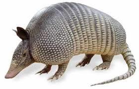 Figure: Nine-banded armadillo
Culture
M. leprae has not yet been successfully cultured in vitro (either on artificial media or in cell culture) but it can be grown in the laboratory by injection into the footpads of mice or nine-banded armadillo. It is a slow-growing pathogen with a doubling time of 14 days.
Serology
A serologic test for IgM against phenolic glycolipid-1 is useful in the diagnosis of lepromatous leprosy but not useful in the diagnosis of tuberculoid leprosy.
Molecular diagnosis
Polymerase Chain Reaction (PCR) can be used as a means of diagnosis of leprosy and also as a tool for drug assessment.
Lepromin skin test
The lepromin test is not a diagnostic test. Injecting M. leprae extract intradermally and reading the response weeks later shows whether the patient can mount a cell-mediated response against the organism. It is positive in tuberculoid leprosy (strong immunity) and negative in lepromatous leprosy (weak immunity). So it does not tell you whether someone has leprosy (a healthy immune person also reacts); it tells you which form they have and helps assess prognosis. It classifies and prognosticates; it does not diagnose.
Treatment
Leprosy is curable with multidrug therapy (MDT), and the principle is what matters. Because M. leprae grows so slowly and single drugs breed resistance, treatment combines several drugs for a prolonged period. The WHO regimen uses dapsone, rifampicin, and clofazimine, with the combination and duration set by the type: multibacillary disease is treated longer and with all three drugs, paucibacillary with fewer. Treatment renders the patient non-infectious quickly. Early treatment matters above all to prevent nerve damage, because established nerve damage and its disability are not reversed by killing the organism. Specific doses and durations are clinical decisions and are not covered here.
How to remember
One bug, two diseases, decided by your immunity. The whole of leprosy: strong cell-mediated immunity gives tuberculoid (few lesions, few bacilli, lepromin positive); weak immunity gives lepromatous (many lesions, many bacilli, lepromin negative). The immune response, not the organism, decides which disease you get.
Tuberculoid = few, lepromatous = loads. Tuberculoid is paucibacillary (few bacilli); lepromatous is multibacillary (loads of bacilli). The names track the bacterial count, which tracks the immune strength.
It loves the cool. M. leprae grows best at 30°C, so it settles in cool sites: skin, earlobes, nose, peripheral nerves. Cool places, not the warm core.
The damage is nerve damage. Leprosy disables through peripheral nerve damage, loss of sensation leads to unnoticed injury, ulcers, and deformity. Treating the organism does not undo established nerve damage, which is why early treatment matters.
Can't grow it at all. M. leprae is the mycobacterium you cannot culture on anything, not even cell culture. It's grown only in mouse footpads and armadillos. "Cannot be cultured" = M. leprae.
Lepromin classifies, it doesn't diagnose. The lepromin test tells you which form (tuberculoid vs lepromatous), not whether the person has leprosy. Prognosis, not diagnosis.
Key exam facts
| Fact | Detail |
|---|---|
| Organism | Mycobacterium leprae, acid-fast bacillus |
| Disease | Leprosy (Hansen's disease) |
| Growth | Slowest human bacterial pathogen (doubling ~14 days); cannot be cultured in vitro |
| Optimal temperature | ~30°C (cool sites: skin, nerves, earlobes, nose) |
| Target cells | Macrophages and Schwann cells (peripheral nerves) |
| Transmission | Prolonged close contact; mainly nasal droplets; low infectivity |
| Incubation | ~5 years (up to 20) |
| Determinant of form | Strength of cell-mediated immunity |
| Tuberculoid (paucibacillary) | Few lesions, few bacilli, strong CMI, lepromin positive |
| Lepromatous (multibacillary) | Many lesions, many bacilli, weak CMI, lepromin negative |
| Classification scales | Ridley-Jopling (TT–BT–BB–BL–LL); WHO (paucibacillary/multibacillary) |
| Diagnosis | Slit-skin smear (modified ZN), skin/nerve biopsy, clinical (anesthetic patch, thickened nerve) |
| Staining note | Modified ZN with weaker decolorizer (less acid-fast than M. tuberculosis) |
| Indices | Bacteriological index (load), morphological index (viable bacilli) |
| Lepromin test | Prognostic/classifying, NOT diagnostic |
| Treatment | Multidrug therapy: dapsone + rifampicin + clofazimine (regimen by type) |
| Key complication | Peripheral nerve damage and disability |
Where students get confused
One organism causes two opposite diseases. Tuberculoid and lepromatous leprosy look completely different, but the difference is the patient's immune response, not the organism. Strong immunity gives tuberculoid, weak gives lepromatous.
The lepromin test does not diagnose leprosy. It shows whether a person can mount immunity against M. leprae, which classifies the form and gives prognosis. A healthy person can be lepromin-positive without having leprosy.
Paucibacillary vs multibacillary is about bacterial load. Paucibacillary (tuberculoid end) has few bacilli; multibacillary (lepromatous end) has many. This tracks the immune strength and determines treatment.
The disability is from nerve damage, not the bug eating tissue. Loss of sensation from peripheral nerve damage leads to unnoticed injuries, ulcers, and deformity. This is why early treatment (to prevent nerve damage) is the priority, and why killing the organism does not reverse existing disability.
M. leprae can't be cultured, even in cell culture. Unlike M. tuberculosis (slow but culturable on LJ), M. leprae grows on no laboratory medium at all. It is propagated only in animals.
Modified ZN, not standard ZN. Because M. leprae is less strongly acid-fast, the smear uses a weaker decolorizer than for M. tuberculosis. Using the standard strong decolorizer would wash the stain out.
References
- Britton, W. J., & Lockwood, D. N. J. (2004). Leprosy. The Lancet, 363(9416), 1209–1219. https://doi.org/10.1016/S0140-6736(04)15952-7
- Bhandari, J., Awais, M., Robbins, B. A., & Gupta, V. (2023). Leprosy. In StatPearls. StatPearls Publishing. https://www.ncbi.nlm.nih.gov/books/NBK559307/
- Tille, P. M. (2022). Bailey & Scott's Diagnostic Microbiology (15th ed.). Elsevier.
- World Health Organization. (current). Guidelines for the diagnosis, treatment and prevention of leprosy. WHO.
Frequently Asked Questions
What causes leprosy?
What causes leprosy?
Mycobacterium leprae, a slow-growing acid-fast bacterium. It mainly infects the skin and peripheral nerves, preferring the cooler parts of the body, and it cannot be grown on any artificial laboratory medium.
Why does leprosy have two very different forms?
Why does leprosy have two very different forms?
Because the form depends on the patient's cell-mediated immunity. Strong immunity produces tuberculoid leprosy (few lesions, few bacilli); weak immunity produces lepromatous leprosy (many lesions, many bacilli). The same organism causes both.
Is leprosy highly contagious?
Is leprosy highly contagious?
No. Most people are naturally immune. Transmission requires prolonged close contact with an untreated multibacillary patient, mainly through nasal droplets, and patients become non-infectious soon after starting treatment.
Why is nerve damage the main problem in leprosy?
Why is nerve damage the main problem in leprosy?
Because M. leprae infects the Schwann cells of peripheral nerves, and the resulting nerve damage causes loss of sensation. Injuries and burns then go unnoticed, leading to ulcers, deformity, and disability. Treating the infection does not reverse established nerve damage, so early treatment is essential.
How is leprosy diagnosed?
How is leprosy diagnosed?
Mainly clinically (a skin patch with loss of sensation, or a thickened peripheral nerve) supported by a slit-skin smear stained with a modified Ziehl-Neelsen method to show acid-fast bacilli, and by skin or nerve biopsy. M. leprae cannot be cultured, so culture is not used.
What is the lepromin test used for?
What is the lepromin test used for?
To determine the type of leprosy and assess prognosis, not to diagnose it. It is positive in tuberculoid leprosy (strong immunity) and negative in lepromatous leprosy (weak immunity).
How is leprosy treated?
How is leprosy treated?
With multidrug therapy combining dapsone, rifampicin, and clofazimine, given for a period that depends on the type of leprosy. It is curable, and early treatment prevents the nerve damage that causes long-term disability.
Why can't Mycobacterium leprae be grown in the laboratory?
Why can't Mycobacterium leprae be grown in the laboratory?
It is so dependent on host cells that it grows on no artificial medium or cell culture. It is propagated only in the footpads of mice or in nine-banded armadillos, and it divides very slowly (about once every 14 days).

Tankeshwar Acharya, MSc (Medical Microbiology)
Tankeshwar Acharya is an Assistant Professor in the Department of Microbiology at Patan Academy of Health Sciences (PAHS), Nepal, where he has been teaching and practicing clinical microbiology for over 14 years. He is the founder of Microbe Online, one of the leading free microbiology education resources on the web, covering bacteriology, mycology, parasitology, immunology, and clinical laboratory diagnostics written from direct experience in both the classroom and the diagnostic laboratory.
Comments
No comments yet. Be the first to share your thoughts.
Leave a comment
All comments are reviewed before they appear.