
IgA Antibodies: Structure, Secretory IgA, and Mucosal Immunity
IgA, the antibody of mucosal immunity: serum monomer vs secretory dimer, the J chain and secretory component, how it crosses into secretions, and why IgA protects the newborn gut. For micro and health-science students.

On this page
Most antibodies patrol the blood. IgA works somewhere harder to defend: the wet surfaces where the body meets the outside world, the gut, the airways, the eyes, the mouth. These mucosal surfaces are the single largest entry point for pathogens, and IgA is their dedicated guard. It is the most abundant antibody in the body by production, yet most of it is never in the blood at all. It sits in saliva, tears, mucus, and breast milk, stopping pathogens at the door before they can attach. This article covers how IgA is built for that job, and why the secretory form is the key to understanding it.
Immunoglobulin A (IgA) is the second most abundant class of immunoglobulin next to IgG, constituting about 10-15% of total serum immunoglobulin and it is the predominant immunoglobulin class in external secretions.
IgA can exist as a monomer, dimer, trimer, or tetramer. IgA in serum (also called serum IgA) is predominantly in monomeric form. Secretory IgA, which is a dimeric form of IgA, is the predominant antibody found in body secretions such as breast milk, saliva, tears, and mucus of the intestinal and respiratory and genitourinary tract.
Structure of Immunoglobulin A (IgA)
IgA is built on the standard antibody plan, covered in the article on immunoglobulin structure. What is distinctive about IgA is that it exists in two very different forms, a simple monomer in serum and a specialized dimer in secretions, and that difference is the key to the whole class.
Immunoglobulin A (IgA) consists of two α heavy chains and two κ or two λ light chains with molecular formula (α2κ2)n or (α2λ2)n, where n =1, 2, 3 or 4. In humans, there are two subclasses of α chains-α1 and α2 and thus two subclasses, IgA1 and IgA2. IgA1 is present mainly in the serum (about 85% of serum IgA) and secretions of mucosal glands & upper intestine whereas IgA2 predominates in the secretions of the large intestine and female genital tract.
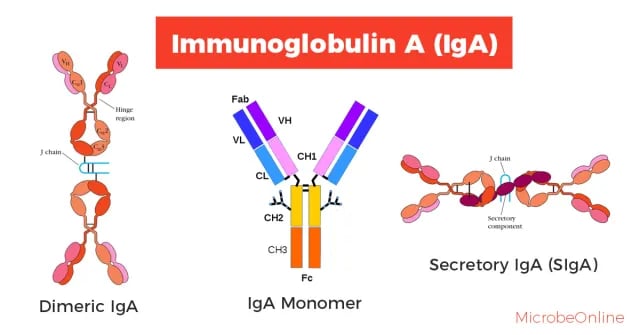 Figure: Various forms of Immunoglobulin A (IgA)
IgA monomer contains three constant-region domains (CH1, CH2, and CH3) and a hinge region. Secretory IgA consists of at least two IgA molecules, which are covalently linked to each other through a J chain. In addition, there is another joining segment present between two IgA molecules called secretory component. Secretory component helps the dimeric IgA to cross the epithelial surface (transcytosis) to reach the lumen. It also protects IgA from denaturation by bacterial proteases.
The two faces of IgA: serum vs secretory
IgA exists in two forms, and almost everything about the class makes sense once you separate them.
Serum IgA is mostly a monomer, floating in the blood like other antibodies. Its role there is surprisingly unclear, and it is not a major player in the blood's antibody defense (it does not activate complement well, for instance). Most IgA1.
Secretory IgA (SIgA) is a dimer, and it is the important form. It is the most abundant antibody at mucosal surfaces, made locally by plasma cells in the lining of the gut and airways, and it is what guards saliva, tears, mucus, and breast milk. When people talk about what IgA does, they almost always mean secretory IgA.
The difference between them is not just size. Secretory IgA carries two extra parts that the serum monomer lacks, and those two parts are what let it survive and work at mucosal surfaces. The next section explains them.
Two extra parts that make secretory IgA work
Secretory IgA is a dimer of two IgA monomers plus two added components:
The J chain joins the two monomers together. It is a small polypeptide, the same joining chain used to link IgM into a pentamer, and it is added by the plasma cell before secretion.
The secretory component is added during transport, and it comes from an unexpected source. To get from inside the mucosa out into secretions, dimeric IgA binds a receptor called the polymeric immunoglobulin receptor (pIgR) on the mucosal cell. This receptor carries the IgA across the cell (transcytosis) and, at the surface, is cleaved. Part of the receptor stays attached to the IgA, and that leftover piece is the secretory component. So the secretory component is not a separate molecule the cell makes for IgA; it is a piece of the transport receptor that comes along for the ride.
The secretory component does two jobs: it completes the transport across the cell, and it then protects the IgA from being digested by enzymes in the harsh mucosal environment. This protection matters because mucosal surfaces are full of proteases, including ones made by bacteria.
Functions of Immunoglobulin A (IgA)
IgA has a variety of functions. The main functions of IgA is carried by SIgA while the function of circulating IgA is less clear.
Mucosal Immunity
Immunoglobulin A (IgA) is the most prevalent antibody class in exocrine secretions where it provides the first line of immune defense against foreign microorganisms. sIgA present in mucosal surfaces can bind with bacterial and viral surface antigens forming sIgA-Ag complexes which are eliminated by the ciliated epithelial cells of the respiratory tract or by peristalsis of the gut. SIgA thus prevents attachment of the pathogens to the mucosal cells, inhibiting subsequent colonization and infection.
Some respiratory pathogens, including Streptococcus pneumoniae, Haemophilus influenzae, and Neisseria, fight back against IgA by secreting IgA proteases that cleave the antibody in its hinge region, disabling it. This is why the two IgA subclasses matter: IgA1 has a long hinge and is the main target of these proteases, while IgA2 has a shorter hinge and resists them. This is also why IgA2, the protease-resistant subclass, is the more common form in the large intestine and genital tract, where bacterial proteases are abundant.
Secretory IgA has been shown to provide an important line of defense against bacteria such as Salmonella, Vibrio cholerae, and Neisseria gonorrhoeae and viruses such as polio, and influenza.
IgA has several notable "does nots" that distinguish it from other classes. It does not activate the classical complement pathway efficiently, does not cross the placenta, and does not trigger mast cell degranulation the way IgE does. This restrained profile fits its job: at mucosal surfaces, the goal is to block and clear pathogens quietly, not to trigger inflammation that would damage delicate mucosal tissue.
Passive immunity to newborn babies
Breast milk (especially colostrums) is rich in secretory IgA and many other molecules that help to protect the newborn against infections during the first few months of life. Because the immune system of infants is not fully functional, breastfeeding plays an important role in maintaining the health of newborns by protecting the immunologically immature infant gut.
How to remember
IgA = the mucosal guard. It defends the wet surfaces where the body meets the world: gut, airways, eyes, mouth. Most IgA is in secretions, not blood.
Two forms: serum monomer (unclear job), secretory dimer (the real work). When IgA matters, it is secretory IgA.
SIgA = dimer + J chain + secretory component. Two monomers joined by the J chain, plus the secretory component picked up from the transport receptor on the way out.
The secretory component is a hand-me-down. It is a leftover piece of the pIgR transport receptor, and it protects IgA from proteases at the surface.
IgA1 long hinge = protease target; IgA2 short hinge = protease-resistant. This is why IgA2 dominates in the protease-rich large intestine.
Colostrum is liquid IgA. Breast milk, especially colostrum, is rich in secretory IgA and protects the newborn gut.
Key exam facts in one table
| Fact | Detail |
|---|---|
| Abundance in serum | Second most abundant (~10–15%) |
| Most abundant where | Mucosal secretions (by total production) |
| Serum form | Monomer (mostly IgA1) |
| Secretory form | Dimer (secretory IgA) |
| Heavy chain | α (alpha); three constant domains, has hinge |
| Subclasses | IgA1 (long hinge), IgA2 (short hinge) |
| J chain | Joins the two monomers |
| Secretory component | From the pIgR transport receptor; protects from proteases |
| Transport across epithelium | Transcytosis via pIgR |
| Crosses placenta | No |
| Activates complement | Not efficiently |
| Newborn protection | Via breast milk / colostrum |
| Bacterial evasion | IgA proteases cleave IgA1's long hinge |
Where students get confused
"IgA is mainly a blood antibody like IgG." No. Although it is the second most abundant in serum, IgA's real job is at mucosal surfaces as secretory IgA. The serum monomer's role is much less clear.
"The secretory component is a special molecule the cell makes to protect IgA." Not quite. It is a leftover piece of the pIgR transport receptor, left attached after the receptor carries IgA across the cell. It then happens to protect the IgA.
"IgA activates complement strongly like IgM." No. IgA does not activate the classical complement pathway efficiently. Its restraint suits its mucosal job, where triggering inflammation would harm delicate tissue.
"IgA1 and IgA2 are basically the same." They differ where it counts. IgA1 has a long hinge and is vulnerable to bacterial IgA proteases; IgA2 has a short hinge and resists them, which is why IgA2 dominates in protease-rich sites like the large intestine.
"A newborn makes its own IgA from birth." No. Newborns get protective IgA from breast milk, especially colostrum, while their own mucosal immunity is still developing.
References and further readings
- Abbas AK, Lichtman AH, Pillai S. Cellular and Molecular Immunology. 10th ed. Elsevier; 2022.
- Punt J, Stranford SA, Jones PP, Owen JA. Kuby Immunology. 8th ed. W.H. Freeman; 2019.
- Delves PJ, Martin SJ, Burton DR, Roitt IM. Roitt's Essential Immunology. 13th ed. Wiley-Blackwell; 2017.
Frequently Asked Questions
What is the main function of IgA?
What is the main function of IgA?
IgA is the antibody of mucosal immunity. As secretory IgA, it guards the wet surfaces of the body (gut, airways, eyes, mouth) by binding pathogens and blocking them from attaching, stopping infection at the entry point.
What is the difference between serum IgA and secretory IgA?
What is the difference between serum IgA and secretory IgA?
Serum IgA is mostly a monomer in the blood with an unclear role. Secretory IgA is a dimer found in secretions like saliva, tears, and breast milk, and it is the form that does IgA's important mucosal defense work.
What is the secretory component?
What is the secretory component?
It is a piece of the transport receptor (pIgR) that carries IgA across mucosal cells. After transport, part of the receptor stays attached to the IgA and protects it from being digested by enzymes at the surface.
Why do some bacteria make IgA proteases?
Why do some bacteria make IgA proteases?
To disable IgA at mucosal surfaces. These enzymes cleave IgA1's long hinge region. IgA2, with a shorter hinge, resists them, which is why IgA2 is more common in protease-rich sites like the large intestine.
Why is breast milk important for a newborn's immunity?
Why is breast milk important for a newborn's immunity?
Breast milk, especially the early colostrum, is rich in secretory IgA. This protects the newborn's gut against infection while the infant's own mucosal immune system is still developing.
Does IgA cross the placenta?
Does IgA cross the placenta?
No. IgA does not cross the placenta (only IgG does). Newborns receive IgA after birth through breast milk instead.

Tankeshwar Acharya, MSc (Medical Microbiology)
Tankeshwar Acharya is an Assistant Professor in the Department of Microbiology at Patan Academy of Health Sciences (PAHS), Nepal, where he has been teaching and practicing clinical microbiology for over 14 years. He is the founder of Microbe Online, one of the leading free microbiology education resources on the web, covering bacteriology, mycology, parasitology, immunology, and clinical laboratory diagnostics written from direct experience in both the classroom and the diagnostic laboratory.
Comments
No comments yet. Be the first to share your thoughts.
Leave a comment
All comments are reviewed before they appear.