
Blood Grouping (ABO and Rh): Forward vs Reverse Typing and How to Interpret

On this page
The test is almost never wrong. The transfusion still kills.
A fatal ABO-incompatible transfusion is one of the most feared events in medicine, and the striking thing about it is where the error usually lies. It is rarely the blood group test that fails. The patient was typed correctly, the unit was labeled correctly, and the incompatibility was created afterward, when the right blood reached the wrong patient: a mislabeled sample, two patients confused at the bedside, a unit hung without a final identity check.
When group A blood enters a group O patient, the recipient's own anti-A antibodies attack the transfused cells within minutes. The result can be fever, back pain, dark urine, collapsing blood pressure, kidney failure, and death, from a test that gave the correct answer every step of the way.
This is why blood grouping is built the way it is: two independent checks that must agree (forward and reverse), controls on every run, and identity verification at every handoff. The point of the method is not just to name a blood group. It is to make sure the right blood reaches the right person, because the biology is unforgiving of the moment it does not. The sections below cover how the groups are determined, how the built-in cross-checks work, and where the process is most easily, and most dangerously, gotten wrong.
“Blood group” refers to the system that comprises specific red blood cell (RBC) antigens from a series of allelic or closely-linked genes in the same chromosome. Blood type is the particular reaction pattern when testing the antisera within a given system.
Karl Landsteiner discovered the blood grouping system in 1901, which helped understand the reason for clumping, sometimes observed when the blood of two individuals was mixed. He gave the concept of the two clinically most crucial blood grouping systems; ABO and Rh system of blood grouping.
Blood grouping is, at its core, an agglutination test: known antibodies (or known cells) are mixed with the sample, and visible clumping reveals which antigens or antibodies are present. Everything from a bedside tile test to a blood-bank crossmatch rests on reading that clumping correctly.
Screening of blood groups of donors and receivers before transfusion is critical as incompatibility during a transfusion can lead to the death of the receivers. Besides the ABO and Rh systems, there are numerous other blood grouping systems, including the Lewis, H-antigen, Lutheran, and MNS systems.
Blood Grouping System
As of 2024, the International Society of Blood Transfusion (ISBT) recognizes 47 blood group systems containing over 360 red cell antigens. Of these, the ABO and Rh systems are by far the most important for transfusion.
Some of the blood grouping systems are discussed below:
ABO Blood Group System
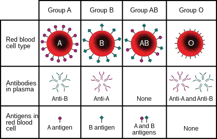 Figure: ABO blood grouping
| Genotype | Phenotype |
|---|---|
| AA or AO | Blood group A |
| BB or BO | Blood group B |
| AB | Blood group AB |
| OO | Blood group O |
It is the most important type of blood grouping system for screening blood because people above six months possess clinically significant anti-A or anti-B antibodies in their serum. The ABO blood group relies on the three genes, A, B, and O, located on chromosome 9. The genotype and phenotype of all the genes are as follows:
- The person with AO or AA genotype has A antigen in their RBC (red blood cell) and antibodies against B (anti-B) in their serum.
- The person with BO or BB genotype has B antigen in their RBC and anti-A in their serum.
- The person with genotype AB has both A and B antigens in their RBC. But neither anti-A nor anti-B in the serum.
- The person with the OO genotype has neither A nor B antigen in their RBC. But has anti-A and anti-B in their serum.
Rh Blood Group System
The Rh system is encoded chiefly by two genes, RHD and RHCE, which give the D, C, c, E, and e antigens. Of these, the D antigen is by far the most immunogenic and clinically important, and its presence or absence defines Rh-positive or Rh-negative status. (Note there is no true "d" antigen; "d" simply denotes the absence of D.)
But the D gene plays a crucial role in the Rh grouping. RBC of any individual may or may not have an Rh factor or immunogenic D-antigen. The presence of antigens and antibodies determines a person as Rh positive or Rh negative.
- Rh positive: Has D-antigen in the RBC.
- Rh negative: Lacks D-antigen in the RBC.
Unlike the ABO system, anti-D does not circulate in those that lack D-antigen but forms in the body if transfusion of Rh positive blood group occurs in Rh negative person.
Lewis Blood Group System
In the Lewis blood grouping system, the Lewis antigen is formed in the tissues and secreted in body fluids which are then absorbed by the RBC membrane. Since the Lewis antigen is secreted as bodily secretion, it is also known as secretor antigens. Lewis antibodies are usually IgM and only occasionally cause transfusion reactions; the system is of limited clinical significance.
MNS Antigen Blood Group System
Landsteiner and Levine described MNS antigen system in 1927. This system is based on two genes, glycophorin A and glycophorin B. An autosomal locus on chromosome 4 and a pair of alleles LM and LN control this blood group. The anti-M and anti-N are usually IgM and are rarely associated with transfusion reactions.
Lutheran Grouping System
The Lutheran grouping system has four allelic antigens representing single amino acid substitution in the Lutheran glycoprotein at chromosome 19. It is not clinically significant as antibodies against the blood group are rare.
Forward (cell) grouping and reverse (serum) grouping
ABO typing is done two ways at once, and the two must agree.
Forward (cell) grouping tests the patient's red cells with known anti-A and anti-B sera. It asks: which antigens are on the cells?
Reverse (serum) grouping tests the patient's serum against known A and B cells. It asks: which antibodies are in the serum?
The power of ABO typing is that these two are built-in checks on each other. A person with group A has A antigen on their cells (forward) and anti-B in their serum (reverse). A group O person has neither antigen (forward) and both anti-A and anti-B (reverse). Because everyone over about six months of age makes predictable anti-A and/or anti-B antibodies against the antigens they lack, forward and reverse results should always mirror each other.
| Blood group | Forward: cells + anti-A | Forward: cells + anti-B | Reverse: serum + A cells | Reverse: serum + B cells |
|---|---|---|---|---|
| A | Agglutination | No reaction | No reaction | Agglutination |
| B | No reaction | Agglutination | Agglutination | No reaction |
| AB | Agglutination | Agglutination | No reaction | No reaction |
| O | No reaction | No reaction | Agglutination | Agglutination |
Why forward and reverse must agree
When forward and reverse grouping disagree, it is called an ABO discrepancy, and it is a signal to stop and investigate, not to guess. Common causes include weak or subgroup A antigens, cold autoantibodies, recent transfusion, age (infants and the very elderly make weak antibodies), and rare phenotypes. Reporting a blood group from forward typing alone, without the reverse check, is unsafe, which is exactly why reverse grouping exists.
The Bombay phenotype: the "O that isn't"
A rare but critical pitfall. People with the Bombay phenotype (Oh) lack the H antigen, the precursor on which A and B are built, so their cells carry no A, B, or H antigen. On forward grouping they look exactly like group O. But their serum contains anti-A, anti-B, and also anti-H, and that anti-H will agglutinate ordinary O cells (which are rich in H). This is caught only when reverse grouping includes pooled O cells: a "group O" patient whose serum agglutinates O cells is not O, but Bombay. It matters because a Bombay patient can be transfused only with Bombay blood; give them ordinary O and they will have a severe hemolytic reaction. This single example is the clearest argument for never skipping reverse grouping.
Requirements for ABO Blood Grouping
- Patient's blood sample (EDTA anticoagulated or clotted, as available)
- Monoclonal anti-A antiserum (conventionally dyed blue) and anti-B antiserum (conventionally dyed yellow)
- Known group A and group B reagent red cells (for reverse grouping)
- Normal saline, glass tubes or a clean grouping tile, applicator sticks
- Positive and negative controls
Procedure for ABO Blood Group Determination
ABO grouping should be done by both forward and reverse methods, and the two results must agree before a group is reported. Reverse grouping is not performed on infants below about 4 months of age, because they have not yet made their own anti-A and anti-B.
Forward (cell) grouping: tube method
- Prepare a 3 to 5 percent suspension of the patient's washed red cells in saline.
- Label two tubes anti-A and anti-B. Place 1 drop of the corresponding antiserum in each (antiserum first, so you can confirm it was added).
- Add 1 drop of the patient's red cell suspension to each tube.
- Mix gently, centrifuge at low speed for about 1 minute.
- Gently resuspend the button and read for agglutination, macroscopically first, then microscopically only if the tube looks negative.
Forward (cell) grouping: tile/slide method
- Place 1 drop of anti-A on one marked area of a clean tile and 1 drop of anti-B on another.
- Add 1 drop of a well-mixed 20 percent red cell suspension (or a small drop of well-mixed blood) beside each antiserum, using a separate stick or corner for each.
- Mix each pair over a circle about 2 cm across and rock gently for up to 2 minutes.
- Read agglutination with the naked eye. The tile method is a rapid screen; equivocal or negative-looking results should be confirmed by the tube method.
Reverse (serum) grouping: tube method
- Label two tubes A cells and B cells. Add 2 drops of the patient's serum or plasma to each.
- Add 1 drop of known group A reagent cells (3 to 5 percent) to the first tube and 1 drop of known group B reagent cells to the second.
- Mix, centrifuge at low speed for about 1 minute.
- Resuspend gently and read for agglutination, macroscopically first, then microscopically if negative.
Result Interpretation
Read forward and reverse together and confirm they mirror each other, using the reaction table in the "Forward and reverse grouping" section above. A valid result shows the expected opposite pattern: the antigen present on the cells and the antibody absent from the serum, and vice versa.
If forward and reverse do not match, do not report a group. Treat it as an ABO discrepancy and investigate before any transfusion (see "Why forward and reverse must agree" above). Common causes include weak or subgroup A antigens, cold autoantibodies, recent transfusion, very young or very old patients with weak antibodies, and rare phenotypes such as Bombay.
Quality control
Include a known positive and a known negative control with each grouping session, and check that antisera and reagent cells are within date and have been brought to room temperature. Weak or expired antisera are a common cause of false-negative or discrepant results, especially in the tile method.
Determination of Rh Factor
Rh or Rhesus factor is present in the RBC as antigens. Landsteiner and Wiener first discovered it in the “Rhesus” monkey. Since antigen D is more antigenic than other Rh antigens, agglutination reaction to antiserum D gives a positive test result.
Principle
Among all the genes responsible for producing Rh factor, the D gene secreting antigen D in the RBC of humans is more prominent. So, during the determination of the Rh factor, the presence of the D antigen is considered Rh-positive. In contrast, the absence of D antigen after agglutination reaction with antiserum D is Rh negative.
Requirement
- Patient’s blood
- Monoclonal antisera D (color-coded red)
- Tile/Tube
Procedure
Like ABO typing, the Rh factor determination is carried out in tile as well as tube.
The procedure is similar to the ABO grouping system.
Tube method
- Place 1 drop of anti-D (antisera D) in a labeled glass tube.
- Add 1 drop of a 3 to 5 percent washed red cell suspension of the patient's cells.
- Centrifuge at low speed for 1 minute.
- Observe the agglutination with the naked eye or for confirmation under a microscope.
Tile method
- Place a drop of the patient’s blood at the end of the tile.
- Gently place a drop of antisera D on top of the blood drop.
- Mix thoroughly using a toothpick or mixer.
- Observe agglutination after 2 minutes.
Result
- If agglutination is seen, Rh is positive.
- If agglutination is absent, Rh is negative.
Importance of Blood Grouping
Performing blood grouping before transfusion is essential to avoid the complication of transfusion reactions. The following are some of the importance of blood grouping:
- Knowing your blood type: In case of an emergency, knowing your blood group can be critical because you may require a blood transfusion or be eligible to donate to a suitable person.
It also allows you to donate blood if you are fit and healthy. Typical eligibility is being roughly 18 to 65 years old and weighing at least 50 kg, though the exact age and weight limits vary by country and blood service. - Cross-matching during transfusion: Transfusion should only be performed between compatible persons. If the blood group is not compatible, it may lead to complications as blood can hemolyze and cause the person’s death.
- During pregnancy: Rh factor plays a critical role in the pregnancy, so blood grouping is always advised for pregnant mothers. Rh-negative mothers should be given Rh-negative blood; if the child is Rh-positive, there is a risk of Rh sensitization. Rh sensitization can lead to hemolytic disease of the fetus and newborn, which is the reason the Rh status of every pregnant woman is checked.
An Rh-negative mother is given anti-D immunoglobulin (RhIg) at about 28 weeks of pregnancy, and again within 72 hours of delivery if the baby is Rh-positive. The antenatal dose covers silent sensitization during pregnancy, and the postpartum dose covers the larger exposure at delivery. This is the dose most often missed, and it is the more critical of the two.
Transfusion Reactions Due to Incompatibility
Transfusion reactions are adverse reactions in the recipient’s body due to the transfusion of incompatible or mismatched blood. A reaction occurs between the donor’s RBC and the recipient’s plasma during the incompatible transfusion. It happens only when the amount of agglutinins against the donor’s RBC is high in the recipient’s plasma. The reaction can be mild with hives and slight fever or severe, leading to renal failure, shock, and death. So, it is categorized into two subheadings; non-hemolytic and hemolytic transfusion reaction.
Transfusion reactions are broadly divided into hemolytic reactions, in which red cells are destroyed, and non-hemolytic reactions, in which they are not. It is important not to confuse the two, because the most dangerous reactions are hemolytic and are caused by ABO incompatibility.
Hemolytic Transfusion Reactions
These are caused by antibody-mediated destruction of red cells and are the reactions that kill.
Acute hemolytic transfusion reaction is most often caused by ABO incompatibility, usually from a clerical or identification error (the wrong unit given to the wrong patient) rather than a typing mistake. The recipient's naturally occurring anti-A or anti-B rapidly destroys the transfused donor cells, often within minutes. Symptoms include fever, chills, back or flank pain, pain at the infusion site, a burning sensation, red or dark urine (hemoglobinuria), a falling blood pressure, and in severe cases disseminated intravascular coagulation, acute renal failure, shock, and death. The transfusion must be stopped immediately.
Delayed hemolytic transfusion reaction typically occurs 1 to 14 days after transfusion, most often from Rh or other minor blood group antibodies in a previously sensitized patient (through earlier transfusion or pregnancy). Hemolysis is slower and milder than the acute ABO type, releasing hemoglobin into the plasma and often producing jaundice and a falling hemoglobin over days.
Non-hemolytic Transfusion Reactions
These do not destroy red cells and are not caused by ABO incompatibility. The two common types are the febrile non-hemolytic reaction (fever and chills from recipient antibodies against donor white cells, or from cytokines in the stored unit) and the allergic reaction (hives and itching, occasionally severe, from plasma proteins in the donor unit). They are usually far less dangerous than an acute hemolytic reaction, but a severe allergic (anaphylactic) reaction is an exception and can be life-threatening.
How to Remember
- Forward reads cells, reverse reads serum, and they mirror. Forward grouping asks what antigens are on the red cells; reverse grouping asks what antibodies are in the serum. Because you make antibodies against the antigens you lack, the two are always opposite and always confirm each other. Group A: A antigen on cells, anti-B in serum. If they don't mirror, something is wrong.
- Bombay is the O that fights O. A Bombay patient looks like group O on forward typing but their serum attacks ordinary O cells, because they lack the H antigen that O cells carry in abundance. If a "group O" serum agglutinates O cells, think Bombay. It is the reason reverse grouping uses O cells.
Key exam facts in one table
| Point | What to remember |
|---|---|
| Basis of the test | Agglutination of red cells by antibody |
| Blood group systems | 47 recognized by ISBT (2024); ABO and Rh most important |
| ABO antibodies | Naturally occurring, IgM, present from ~6 months of age |
| Forward (cell) grouping | Patient cells + known anti-A/anti-B; detects antigens |
| Reverse (serum) grouping | Patient serum + known A/B cells; detects antibodies |
| Reciprocal rule | Forward and reverse must agree; disagreement = ABO discrepancy, investigate |
| Group A | A antigen on cells, anti-B in serum |
| Group O | No A/B antigen, both anti-A and anti-B in serum; universal red cell donor |
| Group AB | Both antigens, no ABO antibody; universal plasma donor |
| Rh key antigen | D antigen; present = Rh positive. There is no "d" antigen (only absence of D) |
| Anti-D | Not naturally occurring; forms only after exposure (transfusion or pregnancy) |
| Bombay phenotype | Lacks H antigen; looks like O but serum agglutinates O cells; needs Bombay blood |
| Pregnancy | Rh-negative mother, Rh-positive fetus: risk of sensitization; anti-D Ig given ~28 weeks |
Where students get confused
"You can determine blood group from cell (forward) typing alone." Unsafe. Reverse (serum) grouping is a required cross-check. When the two disagree, there is an ABO discrepancy that must be resolved before any transfusion. The whole design of ABO typing is built on the two halves confirming each other.
"A Bombay person is group O." They look like O on forward typing but they are not. They lack the H antigen, and their serum contains anti-H that agglutinates ordinary O cells. Transfusing them with normal O blood causes a severe hemolytic reaction. Only reverse grouping with O cells reveals it.
"There is a 'd' antigen for Rh-negative." There is not. Rh-negative means the absence of the D antigen. Lowercase "d" is only a notation for "no D," not a real antigen you can test for.
"Anti-D works like anti-A and anti-B, always present." No. Anti-A and anti-B occur naturally without exposure. Anti-D does not: an Rh-negative person makes anti-D only after being exposed to D-positive cells, through transfusion or pregnancy. This difference is the entire basis of Rh sensitization and of giving anti-D immunoglobulin in pregnancy.
"Group O is the universal donor for everything." O is the universal donor for red cells (no A/B antigens to be attacked), but AB is the universal donor for plasma (no anti-A/anti-B). Mixing up the red-cell and plasma directions is common.
References
- Cheesbrough, M. (2006). Blood transfusion practice. In District Laboratory Practice in Tropical Countries, Part 2 (2nd ed., pp. 348–369). Cambridge University Press.
- Mitra, R., Mishra, N., & Rath, G. P. (2014). Blood group systems. Indian Journal of Anaesthesia, 58(5), 524–528.
- International Society of Blood Transfusion. Red cell immunogenetics and blood group terminology (2024).
- Dean, L. (2005). Blood Groups and Red Cell Antigens. National Center for Biotechnology Information (NCBI).
- NHS. (2023). Blood groups. https://www.nhs.uk/conditions/blood-groups/
Frequently Asked Questions
What is the difference between forward and reverse blood grouping?
Why is reverse grouping necessary if forward grouping already gives the blood group?
What is the Bombay blood group?
Is there a "d" antigen in the Rh system?
Why is anti-D not naturally present in Rh-negative people?
Which blood group is the universal donor?

Tankeshwar Acharya, MSc (Medical Microbiology)
Tankeshwar Acharya is an Assistant Professor in the Department of Microbiology at Patan Academy of Health Sciences (PAHS), Nepal, where he has been teaching and practicing clinical microbiology for over 14 years. He is the founder of Microbe Online, one of the leading free microbiology education resources on the web, covering bacteriology, mycology, parasitology, immunology, and clinical laboratory diagnostics written from direct experience in both the classroom and the diagnostic laboratory.
Comments
No comments yet. Be the first to share your thoughts.
Leave a comment
All comments are reviewed before they appear.