
Hfr Conjugation: How Bacteria Transfer Chromosomal DNA and Map Genes by Time

On this page
The experiment that used a kitchen blender to map a genome
By 1953, William Hayes had identified something odd about certain E. coli donor strains: when mated with F– recipients, they produced far more genetic recombinants, sometimes a thousand times more, than ordinary F+ donors did. He called these "high frequency of recombination" strains, or Hfr. But why they behaved so differently, and what exactly they were handing over to the recipient, was still unclear.
A few years later, François Jacob and Elie Wollman designed an experiment to find out, using nothing more exotic than a kitchen blender. They mated Hfr donor cells with F– recipients, let the mating pairs conjugate for a set number of minutes, and then dropped the mixture into a blender. The violent agitation physically ripped the mating pairs apart, mid-transfer, at whatever point they happened to have reached.
By varying exactly how long they waited before blending, from a few minutes up to well over an hour, and then checking which donor genes had successfully made it into the recipient at each interruption point, Jacob and Wollman discovered something remarkable: genes didn't just transfer, they transferred in a fixed order, over time, like a train timetable. A gene located near the start of the transfer route showed up in recipients interrupted after just a few minutes. A gene located further along only appeared if the mating pair was left undisturbed for much longer.
This single experiment did two things at once. It explained exactly what makes an Hfr strain "high frequency," it transfers a portion of the chromosome itself, not just a plasmid, and it gave bacterial geneticists an entirely new tool: they could map the order of genes on the E. coli chromosome simply by timing how many minutes of uninterrupted mating it took for each gene to arrive. The result became known as the "time-of-entry map," an early genetic map of E. coli built entirely from a blender and a stopwatch.
Conjugation Overview
Conjugation is the process of transfer of genetic material from one bacterium to another. It requires close, direct contact between the two cells. In its most common form, F plasmid conjugation, an F+ donor transfers a self-contained plasmid to an F– recipient. Hfr conjugation is a variant of this same basic process, but with a very different outcome: instead of transferring a plasmid, an Hfr donor transfers a portion of its own chromosome.
F plasmid terminology (a quick, self-contained reference)
| Strain | Definition |
|---|---|
| F+ strains | Bacteria carrying a free, unintegrated F plasmid; act as donors |
| F– strains | Bacteria lacking the F plasmid entirely; act as recipients |
| Hfr strains | Arise when the F plasmid in an F+ cell integrates into the bacterial chromosome at one of several possible sites, becoming part of the chromosome itself |
| F′ (F-prime) strains | Arise when an integrated F plasmid later excises imprecisely from an Hfr chromosome, carrying a fragment of chromosomal DNA out with it as it re-forms a free plasmid |
(For the full F+ × F– plasmid-only conjugation mechanism, see Mechanism of Conjugation in Bacteria: The Transfer of F Plasmid.)
An F+ strain in which the F plasmid has integrated into the chromosome as described above produces more than a thousand times the number of genetic recombinants seen in an ordinary F+ × F– mating. This is exactly why such a donor is called a high frequency of recombination (Hfr) strain, the name refers to the frequency of recombinants seen in the recipient population after mating, not to any property of the donor's own genome.
Mechanism of chromosomal DNA transfer by Hfr strains
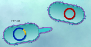 Figure: An Hfr cell, with the F plasmid integrated into the bacterial chromosome
- Because the F plasmid is now embedded in the chromosome, initiating transfer at the plasmid's origin of transfer (oriT) means the transfer machinery pulls the chromosome through the mating bridge, not just the plasmid.
- The sex pilus first contacts the F– recipient cell and draws the two cells together, forming a conjugation bridge, exactly as in ordinary F plasmid conjugation.
- The donor's chromosome is transferred as single-stranded DNA, starting at the origin of transfer within the integrated plasmid. The gene closest to the origin transfers first, and every subsequent gene follows in a fixed order, determined entirely by its physical distance from that starting point.
- Because transfer proceeds continuously over time, and mating pairs commonly break apart spontaneously well before transfer finishes, the amount of chromosome that actually reaches the recipient depends on how long the mating pair stays connected. This is exactly what Jacob and Wollman exploited with their blender: interrupting mating earlier delivers less chromosome; letting it continue longer delivers more.
- A complete E. coli chromosome takes roughly 100 minutes to transfer in full under laboratory conditions, and mating pairs virtually always separate before that much time elapses.
- Segments of the integrated F plasmid sit at both the very beginning and the very end of the region being transferred (the leading segment first, the trailing segment last). Only the leading segment reliably transfers.
- Since the mating pair typically separates long before the entire chromosome, and therefore the trailing plasmid segment, has crossed over, the recipient almost never receives a complete copy of the F plasmid.
- Once inside the recipient, the transferred single-stranded chromosomal DNA is converted to double-stranded DNA and integrates into the recipient's own chromosome by homologous recombination.
- The recipient cell ends up carrying the transferred chromosomal genes but remains F–, since it never received the complete plasmid needed to become a donor. The donor cell, meanwhile, remains Hfr, since it retains the original integrated plasmid within its own chromosome throughout the process.
Why This Matters
- The origin of modern bacterial genetic mapping. The time-of-entry technique pioneered by Jacob and Wollman was used to build detailed maps of gene order on the E. coli chromosome (traditionally expressed in minutes rather than base pairs) long before genome sequencing existed, and it established the basic principle, physical distance on a chromosome corresponds to transfer time, that still underlies how students are taught to think about gene order and operon organization today.
- Why Hfr conjugation rarely spreads plasmid-borne antibiotic resistance directly. Because Hfr transfer very rarely completes the full chromosome (and therefore the trailing plasmid segment), it is a poor vehicle for spreading the F plasmid itself, which is why F+ × F– conjugation, not Hfr × F– conjugation, is the dominant real-world route for plasmid-mediated antibiotic resistance spread (see the F Plasmid article for that clinical detail).
- Chromosomal gene transfer between related strains. Hfr-type transfer can, less commonly than plasmid conjugation, move chromosomally located virulence genes between closely related strains of the same species, a reminder that not all clinically relevant horizontal gene transfer is plasmid-mediated.
The train timetable analogy: picture the Hfr chromosome as a single, very long train, with the origin of transfer as the departure platform. Genes near the platform board (transfer) within the first few minutes; genes far down the track only make it aboard if the train keeps running long enough. Jacob and Wollman's blender was simply a way of stopping the train at a precise, chosen minute and checking who had boarded.
Why the recipient never becomes F+: the F plasmid's own genes are split at the very front and very back of the transferred region, like the engine at one end of the train and the caboose at the other. The recipient reliably gets the engine (leading segment) but the mating pair almost always separates before the caboose (trailing segment) ever arrives, so the recipient never receives a complete, functional plasmid.
Mnemonic for the whole family — "F Has Fully Prepared":
- F+: has a free plasmid, is a donor
- Hfr: the plasmid has settled into the chromosome
- F– : has no plasmid at all, is a recipient
- Prime (F′): the plasmid packed up carelessly on its way back out of an Hfr chromosome, taking a bit of chromosome with it
Anchor for the name "Hfr": the name describes what happens in the recipient population after mating, a high frequency of recombinants, not some special property of the Hfr cell's own DNA.
Key exam facts in one table
| Fact | Detail |
|---|---|
| Definition | An F+ strain in which the F plasmid has integrated into the bacterial chromosome, enabling transfer of chromosomal genes during conjugation |
| Discovered by | William Hayes (1953); mechanism explained by Jacob and Wollman's interrupted mating ("blender") experiment |
| What transfers | A portion of the donor chromosome, starting at the plasmid's origin of transfer (oriT), not the intact plasmid |
| Order of gene transfer | Fixed and time-dependent: genes closest to the origin transfer first |
| Full chromosome transfer time | Approximately 100 minutes in E. coli; mating pairs almost always separate before this completes |
| Historical application | Time-of-entry mapping — genetic maps of E. coli built by timing when each gene arrived in interrupted matings |
| Recipient outcome | Gains transferred chromosomal genes by homologous recombination; remains F– |
| Donor outcome | Remains Hfr; retains its integrated plasmid |
| Why recipient doesn't become F+ | The plasmid's leading segment transfers first, its trailing segment transfers last; mating pairs typically separate before the trailing segment arrives |
| Contrast with F+ × F– mating | F+ × F– transfers the whole plasmid quickly and converts the recipient to F+; Hfr × F– transfers chromosomal genes slowly and the recipient stays F– |
Where Students Get Confused
- Assuming the recipient becomes F+ after Hfr conjugation. It doesn't, and this is the most commonly reversed fact in the whole conjugation topic. Only F+ × F– plasmid transfer converts the recipient to F+; Hfr × F– transfer almost never does, because the trailing plasmid segment almost never arrives before the mating pair separates.
- Misreading what "high frequency of recombination" refers to. The name describes the frequency of recombinant genotypes observed in the recipient population after mating, not a property of the Hfr donor cell replicating or recombining its own genome more often.
- Direction of the time-distance relationship. Genes closer to the origin of transfer arrive sooner; genes farther away take longer and are the first to be lost if mating is interrupted early. Students sometimes reverse this.
- Confusing Hfr with F′. Both involve the F plasmid interacting with the chromosome, but in opposite directions: Hfr is the plasmid moving into the chromosome (integration); F′ is the plasmid moving back out of the chromosome, imprecisely, carrying chromosomal genes with it.
- Assuming Hfr conjugation is the main way antibiotic resistance plasmids spread. It isn't. Because it so rarely transfers a complete plasmid, ordinary F+ × F– conjugation is the dominant real-world route for plasmid-borne resistance spread; Hfr conjugation is mainly significant for chromosomal gene transfer and genetic mapping.
References
- Sana, T. G., Laubier, A., & Bleves, S. (2014). Gene transfer: conjugation. Methods in Molecular Biology (Clifton, N.J.), 1149, 17–22. https://doi.org/10.1007/978-1-4939-0473-0_3
- Llosa, M., Gomis-Rüth, F. X., Coll, M., & de la Cruz Fd, F. (2002). Bacterial conjugation: a two-step mechanism for DNA transport. Molecular Microbiology, 45(1), 1–8. https://doi.org/10.1046/j.1365-2958.2002.03014.x
- Hayes, W. (1953). The mechanism of genetic recombination in Escherichia coli. Cold Spring Harbor Symposia on Quantitative Biology, 18, 75–93.
- Wollman, E. L., Jacob, F., & Hayes, W. (1956). Conjugation and genetic recombination in Escherichia coli K-12. Cold Spring Harbor Symposia on Quantitative Biology, 21, 141–162.
Frequently Asked Questions
What is an Hfr strain?
Why is it called "high frequency of recombination"?
What order do genes transfer in during Hfr conjugation?
What was the "blender experiment"?
Does the recipient become F+ after Hfr conjugation?
How long does it take to transfer the entire E. coli chromosome by Hfr conjugation?
How is Hfr conjugation different from ordinary F plasmid conjugation?
What is the difference between an Hfr strain and an F' strain?

Tankeshwar Acharya, MSc (Medical Microbiology)
Tankeshwar Acharya is an Assistant Professor in the Department of Microbiology at Patan Academy of Health Sciences (PAHS), Nepal, where he has been teaching and practicing clinical microbiology for over 14 years. He is the founder of Microbe Online, one of the leading free microbiology education resources on the web, covering bacteriology, mycology, parasitology, immunology, and clinical laboratory diagnostics written from direct experience in both the classroom and the diagnostic laboratory.
Comments
No comments yet. Be the first to share your thoughts.
Leave a comment
All comments are reviewed before they appear.