
Chromogenic Culture Media: Principle, Composition, Examples, and Advantages

On this page
A urine culture from a hospitalized patient grows two distinct colony types on a single chromogenic UTI agar plate: one bright pink and one dark blue. Without any further testing, the laboratory can report a presumptive result: E. coli (pink) and Enterococcus faecalis (blue), a mixed infection. The clinician receives this within 24 hours.
On conventional media, the same result would require primary plating onto MacConkey agar (for Gram-negative identification) and blood agar (for Gram-positive recovery), followed by subculture of individual colonies for biochemical identification, a two-day process minimum. Chromogenic media compress this workflow into a single plate and a single overnight incubation because they exploit a principle fundamentally different from conventional media: instead of observing pH change, they detect the specific enzymatic activity of each target organism.
Chromogenic culture media are used to isolate, identify, and differentiate specific microorganisms from a heterogeneous population. The medium contains chromogenic substrate which is utilized by the microorganisms to give colored colonies that is specific for each microorganism. Depending on the color of the result, the presence or absence of the target organism is determined and also accurately differentiated from others.
Classical culture media are based on the principle of change in color of the pH indicator whereas Chromogenic media are based on enzymatic utilization of chromogenic substrates.
Principle of Chromogenic Media
Chromogenic media contains soluble colorless molecules called chromogens. Chromogens are composed of two parts: a substrate (which is the target of specific enzymatic activity of the microorganism) and a chromophore.
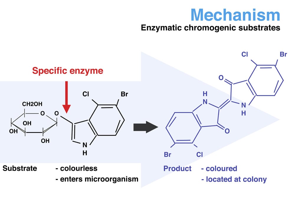 Figure: Principle of Chromogenic Agar Medium (source: chromagar.com)
When the bond between the substrate and chromophore is split by a specific enzyme produced by the target microorganism, chromophore is released. In its unconjugated form, the chromophore shows distinctive color. Due to reduced solubility, chromophore forms a precipitate that imparts unique color to the colony.
How X-Glucuronide produces the blue color of E. coli on chromogenic UTI media:
One of the most widely used chromogenic substrates is 5-bromo-4-chloro-3-indolyl-β-D-glucuronide (X-Gluc):
- E. coli produces the enzyme β-glucuronidase: a highly specific enzyme that cleaves glucuronide bonds
- β-glucuronidase cleaves X-Gluc at the bond between the indolyl chromophore and the glucuronide substrate
- The released 5-bromo-4-chloro-3-indoxyl is initially colorless
- In the presence of oxygen, two indoxyl molecules spontaneously dimerize to form 5,5'-dibromo-4,4'-dichloro-indigo: an intensely blue, insoluble compound
- This blue indigo precipitates within and around the colony, producing blue colonies specific to E. coli
The selectivity is high: β-glucuronidase activity is found in >97% of E. coli strains and only rare other Enterobacteriaceae. A blue colony on X-Gluc-containing media is therefore a strong presumptive indicator of E. coli without any further testing.
The same chemistry applies across different chromogens, substrate specificity determines which organism is identified; the chromophore determines which color is produced.
The chromophore library: common substrates and their colors:
| Substrate | Enzyme detected | Color released | Common target organisms |
|---|---|---|---|
| X-Gluc (5-bromo-4-chloro-indolyl-glucuronide) | β-glucuronidase | Blue | E. coli |
| X-GAL (5-bromo-4-chloro-indolyl-galactoside) | β-galactosidase | Blue | Coliforms |
| Rose-Gal (Rose Bengal-galactoside) | β-galactosidase | Pink/red | Candida albicans (on CHROMagar Candida) |
| HexNAc (5-bromo-4-chloro-indolyl-N-acetyl-glucosaminide) | Glucosaminidase | Blue-green | Staphylococcus aureus |
| IPTG (isopropyl-β-D-thiogalactoside) | β-galactosidase inducer | No color, promotes expression | Used alongside chromogenic substrates |
| Esculin | Glucosidase / esculinase | Brown-black precipitate (with iron) | Enterococci, Group D streptococci |
Composition of Chromogenic Agar Media
Like traditional culture media, chromogenic media also contains nutrients such as peptones, amino acids, yeast extract, minerals, vitamins, and solidifiers (agar). Depending on the purpose chromogenic media may also contain inhibitors. Unlike traditional media, they contain chromogenic substrates or chromogens. These chromogenic substrates such as ONPG, X-Gal, or X-Glu, together with a specified selectivity of the medium, is the simple principle behind chromogenic media.
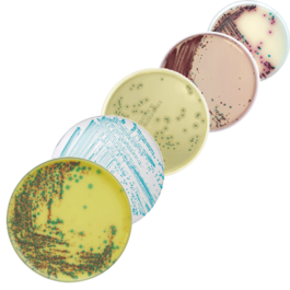 Figure: Chromogenic Agar Media (Source:biomerieux)
Advantages of Use of Chromogenic Agar Media
- Less Labor intensive and more economic: Though people perceive chromogenic media as an expensive alternative, the use of a single chromogenic medium rather two-three selective ones reduces the cost of sample processing. Chromogenic media may eliminate the need for subculture and further biochemical tests for the identification of the isolates.
- Less time consuming: As chromogenic media eliminate various steps of sample processing (e.g. subculturing, biochemical testing) results are available within 24 hours as compared to 48 hours or more by conventional methods. Timely diagnosis not only ensures a better outcome for the patients but also helps in the prevention and spread of infections.
- Easy identification: On chromogenic agar medium, target colonies of specific microorganisms can be recognized by their color at a glance. No specialized equipment needed.
Examples of Chromogenic Media: Colony Colors and Clinical Uses
| Target organism | Medium examples | Colony color of target | Clinical use |
|---|---|---|---|
| Candida albicans | CHROMagar Candida, Brilliance Candida, HiCrome Candida | Green (C. albicans); pink (C. tropicalis); blue-grey (C. krusei) | Urine/genital cultures; mixed Candida species identification in a single plate |
| E. coli and UTI pathogens | CHROMagar Orientation, UriSelect, Chromogenic UTI medium | E. coli pink/red; Klebsiella metallic blue; Enterococcus blue-green; Proteus brown/beige halo | Urine culture, rapid species-level ID and colony count in 24 hrs |
| MRSA | CHROMagar MRSA II, Brilliance MRSA | MRSA: mauve/pink on pink background; MSSA: suppressed or different color | ICU admission screening; MRSA surveillance |
| Carbapenem-resistant Enterobacteriaceae (CRE) | Brilliance CRE Agar | CRE organisms: cream/pink; non-CRE: inhibited or different color | CRE surveillance and outbreak screening |
| E. coli O157:H7 | CHROMagar O157, Rainbow Agar O157 | O157: mauve/pink; non-O157: blue-green | Food safety testing; HUS outbreak investigation |
| VRE (Vancomycin-resistant Enterococcus) | CHROMagar VRE | VRE: pink/mauve; VSE: inhibited | VRE hospital screening |
| Salmonella spp. | CHROMagar Salmonella | Salmonella: mauve/pink | Food safety; clinical stool culture |
| Listeria spp. | ALOA (Agar Listeria Ottaviani & Agosti), CHROMagar Listeria | L. monocytogenes: blue-green with opaque halo; other Listeria: blue without halo | Food safety; neonatal/pregnancy screening |
| Pseudomonas aeruginosa | ChromID Pseudomonas | P. aeruginosa: pink | Wound infection monitoring; CF sputum |
| Bacillus cereus | Bacillus cereus ChromoSelect Agar | B. cereus: blue-green with surrounding precipitate | Food safety — contaminated dairy, rice |
Note on color specificity: Colony colors vary between manufacturer formulations. Always consult the specific product insert for color interpretation. The colors above are representative of common formulations but are not universal across all brands.
Limitations of Chromogenic Media
Despite their advantages, chromogenic media have important limitations that students and laboratory staff must understand:
1. Higher cost than conventional media Chromogenic substrates are expensive to manufacture. A single chromogenic plate typically costs 3–10 times more than an equivalent conventional selective medium. In resource-limited settings, this cost differential restricts routine use.
2. Manufacturer-dependent color interpretation Colony colors for the same organism may differ between CHROMagar, Brilliance (Oxoid), ChromID (bioMérieux), HiCrome (HiMedia), and other manufacturers. A laboratory switching manufacturers must re-validate color interpretation. Color variations also occur with inoculum size, incubation time, and temperature.
3. Not all strains produce the expected color Chromogenic reactions depend on enzyme expression, which can be variable. Some strains of target organisms lack the specific enzyme and produce atypical or absent color. For example, approximately 2–5% of E. coli strains are β-glucuronidase-negative and will not produce the expected blue color on glucuronide chromogenic media.
4. Cannot replace biochemical confirmation for definitive identification Chromogenic media provide presumptive identification only. For clinical reporting of significant pathogens, biochemical confirmation (API, MALDI-TOF, or automated identification systems) or molecular testing is still required in most laboratory protocols.
5. Sensitive to storage and preparation conditions Chromogenic substrates can degrade with improper storage (exposure to light, incorrect temperature) or over-incubation beyond recommended timepoints. Degraded substrates produce incorrect or absent colony colors.
6. Limited pathogen coverage in a single medium A chromogenic medium is designed for one or a few target organisms. It cannot replace a full battery of conventional media for specimens where multiple unknown pathogens are possible (e.g., wound cultures, blood cultures, CSF).
Chromogenic vs Conventional Media: Workflow Comparison
| Step | Conventional media workflow | Chromogenic media workflow |
|---|---|---|
| Day 1, Primary plating | Multiple plates (blood agar + MacConkey ± selective agars) | Single chromogenic plate (e.g., chromogenic UTI agar) |
| Day 2, Reading | Colony morphology only; cannot identify to species | Presumptive species-level ID by colony color |
| Day 2, Subculture | Select colonies for subculture onto identification media | Often unnecessary for common organisms |
| Day 3, Biochemical ID | Perform biochemical panel (API, VITEK, etc.) | Confirmatory testing only for atypical colors |
| Day 3, Report | Final species identification | Often possible 24 hours earlier |
| Cost per specimen | Lower media cost; higher labor cost | Higher media cost; lower labor cost |
| Best suited for | Low volume labs; uncommon pathogens; mixed specimens | High volume labs; common pathogens; screening programs |
How to Remember
The fundamental distinction, pH change vs enzymatic cleavage: Traditional media: organism grows → ferments sugar → acid produced → pH indicator changes color → all fermenters same color. Chromogenic media: organism grows → specific enzyme cleaved chromogen → colored product precipitates on colony → each organism its own specific color.
The specificity comes from the enzyme, not the acid. This is why chromogenic media can distinguish E. coli (blue) from Klebsiella (metallic) from Enterococcus (teal) on a single plate, while MacConkey can only separate fermenters (pink) from non-fermenters (colorless).
Two parts, one result: Every chromogen = substrate (what the enzyme acts on) + chromophore (what produces the color). Enzyme cuts substrate → releases chromophore → chromophore precipitates → color. Remove either part and no color is produced.
Clinical memory anchors:
- CHROMagar Candida: green = C. albicans (the one you most need to treat)
- Chromogenic UTI: pink = E. coli (most common UTI pathogen)
- CHROMagar MRSA: mauve/pink = MRSA (the one you most need to isolate and prevent)
In each case, the target pathogen with the most clinical significance gets the most visible, distinctive color.
References
- Perry J. D. (2017). A Decade of Development of Chromogenic Culture Media for Clinical Microbiology in an Era of Molecular Diagnostics. Clinical microbiology reviews, 30(2), 449–479. https://doi.org/10.1128/CMR.00097-16
- Garcia, B. L. N., Fidelis, C. E., Freu, G., Granja, B. M., & Dos Santos, M. V. (2021). Evaluation of Chromogenic Culture Media for Rapid Identification of Gram-Positive Bacteria Causing Mastitis. Frontiers in veterinary science, 8, 662201. https://doi.org/10.3389/fvets.2021.662201
- Tille, P. M. (2017). Bailey and Scott's Diagnostic Microbiology (14th ed.). Elsevier.
Frequently Asked Questions
How does a chromogenic medium produce a specific colour for one organism but not another growing on the same plate?
What are the main limitations of chromogenic media that prevent them from replacing conventional media entirely?
Why are chromogenic media particularly valuable for detecting mixed infections, and what is the clinical example?
What is the significance of chromogenic culture media?

Tankeshwar Acharya, MSc (Medical Microbiology)
Tankeshwar Acharya is an Assistant Professor in the Department of Microbiology at Patan Academy of Health Sciences (PAHS), Nepal, where he has been teaching and practicing clinical microbiology for over 14 years. He is the founder of Microbe Online, one of the leading free microbiology education resources on the web, covering bacteriology, mycology, parasitology, immunology, and clinical laboratory diagnostics written from direct experience in both the classroom and the diagnostic laboratory.
Comments
No comments yet. Be the first to share your thoughts.
Leave a comment
All comments are reviewed before they appear.