Capillary Electrophoresis: Principle and Application
Capillary Electrophoresis: Principle and Application
Electrophoresis is a method of separation where the movement of ions under the influence of electricity helps separate components. There are various types of electrophoresis. Gel electrophoresis, cellular acetate electrophoresis, and capillary electrophoresis are some of the commonly used types of electrophoresis.
Capillary electrophoresis is a liquid-based separation technique that uses a capillary as a separation channel under the influence of an electric field. It is an analytical technique where the electroosmotic flow helps separately charged ions. It is applicable in various areas like analysis of chemical substances, DNA analysis, identifying specific proteins, and separating coenzymes.
Principle of Capillary Electrophoresis
The velocity of the analyte when migrating under the influence of an electric field of intensity “E” is given by the analyte’s electrophoretic mobility and the buffer’s electroosmotic mobility inside the capillary.
The electrophoretic mobility of solute depends on the different characteristics of the solute, like electric charge, molecular size, and shapes, and the properties of the buffer, like electrolyte’s ionic strength, pH, viscosity, and additives. The following equation gives the electrophoretic velocity (Vep) of the solute:
Vep= μepE = (q/6πηr) (V/L) where η is the viscosity of the electrolyte solution, V is the applied voltage, L= the length of the capillary, r is the Stoke’s radius of the solute, μep is the electrophoretic mobility of the solute, and q is the effective charge of the solute.
When applied through the capillary filled with the buffer, the electric field generates a solvent flow called the electroosmotic flow inside the capillary. The velocity of which depends on the electrophoretic mobility. Electrophoretic mobility depends on the charge density of the capillary’s internal wall and the buffer’s properties. The following equation gives the electroosmotic velocity (Veo):
Veo = μeoE = (ε𝜁/η) (V/L) where ε= buffer’s dielectric constant, 𝜁= zeta potential of the capillary surface, η is the viscosity of the electrolyte solution, V is the applied voltage, μeo is the electrophoretic mobility, and L is the length of the capillary.
The solute’s velocity (V) is given by: V= Vep+Veo.
The electroosmotic and electrophoretic mobility of the analyte can act in the same or opposite direction based on the solute’s charge. In the case of normal capillary electrophoresis, anions will move in the opposite direction of electroosmotic flow with velocities smaller than the electroosmotic velocity. Whereas, the cations will migrate in the same direction of the electroosmotic flow with velocities higher than the electroosmotic velocity. In conditions with a fast electroosmotic rate compared to the electrophoretic rate of the solutes, the separation of both cations and anions occurs in the same run. The time is taken (t) for the solute to migrate to the detection point from the injection end of the capillary(effective capillary length) is given by:
t = l/ Vep+Veo = l(L)/ V( Vep+Veo)
Generally, uncoated fused silica capillaries above pH three that have a negative charge are used where the electroosmotic flow occurs from the anode to the cathode. The electroosmotic flow must be constant for each run for good reproducibility in the migration velocity of the solutes. For some experiments, reduction or suppression of electroosmotic glow by changing the inner wall of the capillary, the concentration, composition, or pH of the buffer solution might be necessary.
After adding the sample, an independent zone of each analyte ion of the sample forms. The zone formation is due to the migration within the background electrolyte. The spreading of each solute band occurs as a result of different phenomena. Under ideal conditions, the only phenomenon contributing to the zone’s broadening is molecular diffusion of the solute inside the capillary. Here, the efficiency of the zone is given as the number of theoretical plates (N), which is given by:
N= (μep+μeo) (Vl)/ 2DL, where D is the molecular diffusion coefficient of the solute buffer.
In general practice, other phenomena like the length of the injection plug, detector cell size, unleveled buffer reservoirs, mismatched conductivity between sample and buffer, sample adsorption onto the capillary wall, and heat dissipation, play a significant role in band dispersion. The separation between two bands is gained by modifying the analytes’ electrophoretic mobility, the electroosmotic mobility induced in the capillary, and increasing each analyte’s efficiency for the band as follows;
Rs= N(μepb-μepa)/ 4(μaep+μeo); where μepa and μepb= the two analytes’ electrophoretic mobilities, μaep is the average electrophoretic mobility of the two analytes calculated by: μaep= ½ ( μepb+μepa).
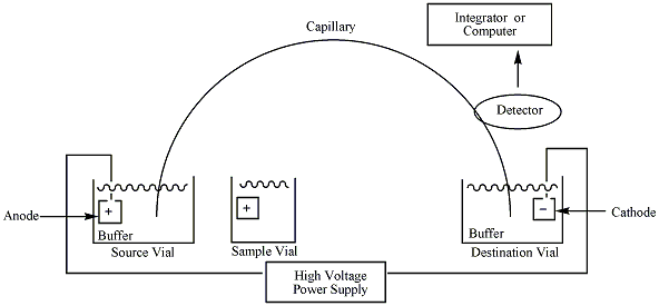
Instruments Used in Capillary Electrophoresis
- Power supply: Capillary electrophoresis requires a high voltage controllable direct current power supply.
- Buffer reservoirs: Capillary electrophoresis requires two buffer reservoirs held at the same level that contain specified anodic and cathodic solutions.
- Electrodes: CE needs two electrodes (cathode and anode) immersed in the buffer reservoirs connected to the power supply.
- Capillary: A capillary made up of fused silica, usually with a diameter of fewer than 100 microns.
- Viewing window: CE requires an optical viewing window that is aligned to the detector.
- Injection system: CE requires a suitable injection system for adding sample and buffer into the capillary. The method of injection can be automated for precision. The common mode of injecting samples is gravity, pressure or vacuum, or electrokinetic.
- Detector: CE requires a detector for monitoring the amount of substance that passes through the capillary at a given time based on conductimetric, fluorimetry, absorption spectrophotometry (UV and visible), amperometric, or mass spectrometric detection.
- Thermostatic system: CE requires a thermostatic system for maintaining a constant temperature inside the capillary.
- Recorder: CE requires a recorder that records the data after the completion of electrophoresis.
- Suitable integrator or computer: CE also needs the right integrator or computer to convert data digitally.
Types of Capillary Electrophoresis Method
There are six types of capillary electrophoresis methods commonly used; CZE (capillary zone electrophoresis), CGE (capillary gel electrophoresis), Micellar electro kinetic capillary chromatography (MEKC), capillary electrochromatography (CEC), capillary isoelectric focusing (CIEF), and capillary isotachophoresis (CITP).
Capillary zone electrophoresis (CZE)
The separation in this type of electrophoresis requires a capillary with only a buffer without any anticonvective medium. The analytes are separated into bands whose velocity depends on the electrophoretic mobility and electroosmotic flow. The electroosmotic flow moves toward the cathode, and when the polarity has reversed, the analytes with electroosmotic mobilities higher than the electroosmotic force will pass the outlet. The coated capillaries help increase the separation capacity of the substances that adhere to the fused-silica surfaces. The CZE is applicable in analyzing small to large (<2000 molecular weight to <100,000 molecular weight). It is a very efficient form of capillary electrophoresis.****
Capillary gel electrophoresis (CGE)
The separation in capillary gel electrophoresis occurs in a capillary filled with a gel acting as a molecular sieve quite similar to gel electrophoresis. Molecules are separated according to molecular size. The smaller molecules move more freely through the gel network; hence these migrate faster than larger molecules. Capillary gel electrophoresis is used to separate proteins, DNA fragments, and other biological macromolecules with similar charge-to-mass ratios based on their molecular size. ****
Micellar electrokinetic capillary chromatography (MEKC)
The separation occurs in an electrolytic solution that consists of a surfactant with a concentration above the critical micellar concentration (CMC). This technique is a hybrid of electrophoresis and chromatography. The method is applicable to both neutral and charged solutes. The solute molecules get distributed between the pseudo-stationary phase made up of micelles and aqueous buffer based on the solute’s partition coefficient. The commonly used surfactant in MKEC is sodium dodecyl sulfate, cetyl trimethyl, and ammonium salts. In neutral and alkaline pH, the electroosmotic flow is strong, and the separation buffer ions move toward the direction of the cathode.
In case sodium dodecyl sulfate (SDS) is used as a surfactant, the anionic micelle’s electrophoretic migration is in the order of the anode, slowing down the overall micelle migration velocity in the bulk flow of the electrolytic solution. Suppose neutral solutes are present; the analyte partitions between the aqueous buffer and the micelle and lacks electrophoretic mobility. In such cases, the analyte’s migration velocity depends on the partition coefficient between the aqueous buffer and the micelle. Because of this, the separation in neutral and weakly ionized solutes in MKEC is chromatographic. In the case of charged solutes, migration velocity depends on the electrophoretic mobility of the solute without a micelle and the partition coefficient between the aqueous buffer and the micelle.
Capillary electrochromatography (CEC)
Capillary electrochromatography is a hybrid separation technique that uses the principle of both capillary electrophoresis and chromatography, especially high-performance liquid chromatography (HPLC). Analytes separate based on the differences in the partition ratio between the mobile and stationary phase or due to electrophoretic mobility. The mobile phase is run across the chromatographic bed with the help of electroosmotic force instead of pressure. CEC is more advantageous than HPLC and capillary electrophoresis. It applies to enantiomeric separation, separating amino acids, proteins, peptides, and carbohydrates. It is mainly used in pharmaceutical industries to identify acidic and basic drugs and in the industrial sector that involves analyzing polymers. ****
Capillary isoelectric focusing (CIEF)
The charged molecules migrate due to the electric field in the isoelectric focusing in a pH gradient obtained from the ampholytes having pI (isoelectric point) value in a wide range that is dissolved in the separation buffer. There are three steps in CIEF; loading, focusing, and mobilization. The introduction of the ampholytes and separation buffer is loaded in the capillary in the loading step. In the focusing step, the voltage is provided in the capillary, causing the ampholytes to move to their respective electrode based on their net charges and creating a pH gradient where the molecules migrate until they reach a pH corresponding to their pI (isoelectric point). The mobilization can be required for detection, which is obtained by either focusing under the influence of electroosmotic flow, applying positive pressure after focusing, or adding salt to the anode or cathode reservoir, depending on the direction of mobilization.
Capillary isotachophoresis (CITP)
The technique was developed to separate the serum lipoproteins. Compared to other electrophoretic processes, it has negligible molecular sieve effects, requires no gel casting, applicable for whole serum, and can easily differentiate lipoprotein subfractions. Here, the total serum lipoproteins are pre-stained in a free-flow capillary system (with an internal diameter of 0.5 mm) having a discontinuous buffer system for 30 minutes at 4℃ before starting the process. The lipoproteins are separated into HDL (high-density lipoproteins) and LDL (low-density lipoproteins) based on electrophoretic mobility. The method is commonly used in clinical laboratories for determining the lipoprotein level in human serum.
Application of Capillary Electrophoresis
Capillary electrophoresis is commonly applied in various fields. Analyzing food contents, genetic differentiation, and clinical diagnosis are typical applications of capillary electrophoresis. The following is a detailed explanation of the area of applications of capillary electrophoresis:
- **Food technology:**Capillary electrophoresis is applied to analyze different food, like beverages and fermented food, to detect flavonoids, vitamins, carbohydrates, proteins, pigments, and color. CE is also applicable for detecting DNA and RNA from bacteria and viruses, which indicates contamination.
- Molecular analysis: The DNA, RNA, and amino acids are detected using capillary electrophoresis, especially in the microbiology laboratory and forensic science.
- Disease diagnosis: Lipid profile analysis using CITP is critical for detecting cholesterol levels in clinical laboratories. Likewise, the human serum’s analysis of vitamins and minerals is performed using capillary electrophoresis.
- Environmental monitoring: Capillary electrophoresis has been applied to track inorganic ions in rivers and water. (4) Likewise, nano-capillary electrophoresis is applied to detect environmental pollutants at the nano level. (5)
- Pharmaceutical laboratories: Capillary electrophoresis is used in the analysis of small molecules in drugs during preparatory stages and standard solutions. (6)
Advantages of Capillary Electrophoresis
Capillary electrophoresis has many advantages over other methods of electrophoresis and chromatography due to the use of tiny capillaries for separating. The following is a detailed explanation of the benefits of capillary electrophoresis:
- Highly efficient: Heat dissipation occurs efficiently due to using a small diameter of the capillary. Some articles claim the efficiency of capillary electrophoresis is quite similar to that of liquid chromatography. (7)
- Less time-consuming: If the person is familiar with computer software, it takes no time to read the result of separation. Likewise, the entire process takes only one hour, from setup to separation. So, capillary electrophoresis carries more advantages as compared to other separation techniques.
- Wide area of application: Capillary electrophoresis is used in analyzing macromolecules like proteins, DNA, different drugs, and RNA. So, CE is applied widely from pharmaceutical to environmental monitoring.
- Has the possibility of automation: The injection method, data analysis, buffer, and sample uploading steps can be automated in capillary electrophoresis.
Disadvantages of Capillary Electrophoresis
Although capillary electrophoresis is highly advantageous, it has some disadvantages, which are as follows:
- Changing capillaries is challenging: Due to the size of the capillaries, removing and replacing capillaries can be challenging at times. So, expertise is required for changing capillaries to experiment.
- Less robust: Small changes like the size of capillaries and choice of analytes can be crucial in capillary electrophoresis. So CE is less robust than other methods.
References
- Capillary electrophoresis. (n.d.). Retrieved December 16, 2022, from https://www.usp.org/sites/default/files/usp/document/harmonization/biotechnology/harmonization-september-2019-m859.pdf
- Borst, C., Belal, F., & Holzgrabe, U. (2013). Possibilities and limitations of capillary electropherosis in pharmaceutical analysis. Die Pharmazie, 68(7), 526–530.
- Robert, F., Bouilloux, J. P., & Denoroy, L. (1991). L’électrophorèse capillaire: principe et applications [Capillary electrophoresis: principle and applications]. Annales de biologie clinique, 49(3), 137–148.
- Sirén, H., & Väntsi, S. (2002). Environmental water monitoring by capillary electrophoresis and result comparison with solvent chemistry techniques. Journal of chromatography. A, 957(1), 17–26. https://doi.org/10.1016/s0021-9673(02)00218-2
- Ali, I., Alharbi, O. M. L., & Marsin Sanagi, M. (2016). Nano-capillary electrophoresis for environmental analysis. Environmental chemistry letters, 14(1), 79–98. https://doi.org/10.1007/s10311-015-0547-x
- Ali, I., Alharbi, O. M. L., & Marsin Sanagi, M. (2016). Nano-capillary electrophoresis for environmental analysis. Environmental chemistry letters, 14(1), 79–98. https://doi.org/10.1007/s10311-015-0547-x
- Masár, M., Hradski, J., Schmid, M. G., & Szucs, R. (2020). Advantages and Pitfalls of Capillary Electrophoresis of Pharmaceutical Compounds and Their Enantiomers in Complex Samples: Comparison of Hydrodynamically Opened and Closed Systems. International journal of molecular sciences, 21(18), 6852. https://doi.org/10.3390/ijms21186852

Tankeshwar Acharya, MSc (Medical Microbiology)
Tankeshwar Acharya is an Assistant Professor in the Department of Microbiology at Patan Academy of Health Sciences (PAHS), Nepal, where he has been teaching and practicing clinical microbiology for over 14 years. He is the founder of Microbe Online, one of the leading free microbiology education resources on the web, covering bacteriology, mycology, parasitology, immunology, and clinical laboratory diagnostics written from direct experience in both the classroom and the diagnostic laboratory.