Electrophoresis: Principles, Types, and Uses
Electrophoresis separates charged molecules such as proteins and DNA by moving them through a gel in an electric field. Learn the principle, the factors that control mobility, the main types, and how serum protein electrophoresis detects multiple myeloma.
A 58-year-old man comes to the clinic with four months of nagging low back pain and fatigue that rest does not fix. His hemoglobin is low. His ESR is strikingly high. His total serum protein is elevated, yet his albumin is low, which is an odd pairing: if albumin is down, what is holding the total up?
The laboratory runs a serum protein electrophoresis. A drop of his serum is placed on a supporting medium, a voltage is applied, and over the next hour his serum proteins sort themselves into bands according to how fast each one travels. Albumin sprints furthest. The globulins trail behind in groups.
In a healthy person, the gamma region at the far end is a low, broad hill, because normal immunoglobulins are made by thousands of different plasma cell clones, each producing a slightly different protein that migrates at a slightly different speed. In this man's tracing, the hill has been replaced by a tall, narrow spike.
That spike is a monoclonal band, an M band. One plasma cell clone has multiplied out of control and is pouring a single identical immunoglobulin into his blood. Thousands of identical molecules, identical charge, identical size, all arriving at the same place at the same time. The diagnosis is multiple myeloma, and the back pain is from lytic bone lesions.
No machine in that laboratory "detected myeloma." All the electrophoresis did was separate charged molecules by how fast they move in an electric field. The clinical answer was visible in where the proteins stopped. That is the whole reason this technique is worth understanding: master what governs a molecule's speed, and you can read a gel.
**What is electrophoresis?**
Electrophoresis is a separation technique in which charged molecules are made to migrate through a supporting medium under an applied electric field. Molecules separate because each one travels at its own speed, determined mainly by its net charge, its size and shape, and the properties of the medium and buffer.
The principle in one line: negatively charged molecules (anions) migrate toward the positive electrode (anode) and positively charged molecules (cations) migrate toward the negative electrode (cathode), and among molecules moving the same direction, those with more charge per unit size travel fastest.
Electrophoresis is a simple and sensitive separation technique in clinical and research laboratories. Since its discovery, it has been an essential tool used by biologists and chemists to separate mixtures, especially proteins and nucleic acids.
Electrophoresis consists of two words; electro, meaning electricity, and phoresis, meaning movement. Thus, it implies the migration and separation of a charged particle (ions) through a solution under the influence of an electric field.
It was first described in 1807 by Ferdinand Frederic Reuss, who applied an electric field to a clay-water mixture and observed clay particles migrating toward the anode. It has improved from the initial crude paper electrophoresis to the modern automated system. Various improved versions are available, which apply in miniaturization, precision engineering, etc. The benefit of advancement is meeting the requirement for faster and better resolution of results.
Principle of Electrophoresis
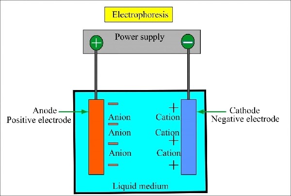 Figure: General Principle of Gel ElectrophoresisFigure source:https://www.smacgigworld.com/blog/principle-of-electrophoresis.php
Biological molecules, like amino acids, peptides, proteins, nucleic acids, and nucleotides, possess ionizable groups. These molecules exist in solution as electrically charged species, cations (+), or anions (-) at any given pH. Thus, the electric field allows the migration of the negatively charged molecule towards the anode (a positive terminal). In contrast, the positively charged molecule migrates towards the cathode (a negative terminal).
The separation of the molecules, ions, or colloidal particles suspended in the matrix occurs due to the force of an electric field. The molecules move through a sieve-like compound based on the molecular mass and charge ratio.
It is an incomplete form of electrolysis as the electric field is removed before the molecules reach the electrode, yet the molecules separate due to electrophoretic mobilities. Nucleic acids have negative phosphate backbones. Hence they move towards the anode in DNA electrophoresis.
Proteins are ampholytes, meaning they carry both positive and negative groups, and their net charge depends on the pH of the buffer relative to their isoelectric point (pI), the pH at which their net charge is zero. When the buffer pH is above the protein's pI, the protein is net negative and migrates toward the anode. When the buffer pH is below its pI, the protein is net positive and migrates toward the cathode. At exactly its pI, the protein does not migrate at all. This is why the choice of buffer pH is not a technical detail in protein electrophoresis. It decides which way each protein travels.
The speed at which a molecule travels in a given electric field is its electrophoretic mobility. Conceptually:
electrophoretic mobility ∝ net charge ÷ (size × buffer viscosity)
A molecule with more net charge is pulled harder by the field. A larger molecule meets more frictional drag and is slowed further by the sieving matrix. Two molecules separate only if their mobilities differ. This is why the M band in our patient's tracing is a sharp spike rather than a broad hill: every molecule in that clone is identical, so every molecule has identical mobility and they all arrive together.
Factors Affecting Electrophoretic Mobility
The velocity of ions depends on both inherent factors and the external environment.
Inherent factors
The inherent factors that affect the velocity of ions are:
- Charge density
- Molecular weight
- The net charge of the molecule
- Size and shape of the molecule
External factors
The external factors affecting the rate of movement of ions are:
- Electrical parameters, like current, voltage, and power
- Viscosity and pore size of supporting medium
- Temperature
- The pH of the buffer
Electrophoresis Instrument
Modern electrophoresis equipment and systems vary based on its types and forms. However, every electrophoretic system shares the same essential components:
- Power pack: Power supply drives the movement of ionic species in the medium and allows adjustment and control of either the current or the voltage.
- An electrophoresis unit: An electrophoretic system depends on its type but essentially consists of two electrodes of opposite charge (anode and cathode), connected by a conducting medium called an electrolyte. In addition, a supportive medium is present in electrophoretic systems like gel and paper electrophoresis.
- Buffer (Electrolyte): Buffers carry applied electric current and provide appropriate pH for the process. Conducting (running) buffers such as Tris-borate-EDTA (TBE) and Tris-acetate-EDTA (TAE) are commonly used.
- Supportive Medium: The supportive medium is the matrix (gel), in which biomolecules are separated. It can be in the slab or capillary form. The supporting media used include polysaccharide gels such as agarose and starch, the synthetic cross-linked polymer polyacrylamide, and cellulose acetate, which is used as a membrane strip rather than a gel.
The medium runs either vertical or horizontal gel systems in gel electrophoresis. Horizontal: agarose gel electrophoresis, and vertical: SDS-PAGE. The larger the pore size, the less the matrix impedes migration, so molecules travel faster. The trade-off is resolution: large pores separate large molecules well but allow small molecules to run through almost unhindered, so they are poorly resolved. Gel concentration is therefore selected to match the size range being separated.
General Procedure of Electrophoresis
The electrophoresis process has three main steps; separation, detection, and quantification.
Separation
The instrument set up is according to its type. In the gel electrophoresis, gels are prepared and cast. Then placed into the electrophoresis chamber. The supportive medium can be agarose gels or polyacrylamide gels. Then appropriate buffer solution is added to the system.
After the proper setup of the instrument, the sample is placed into the medium. Then the sample is run at a specific current, voltage, or power.
Detection and Quantification
Staining with a dye or autoradiography (for radioactive samples) helps in the detection of the separated components.
Quantification is done using a densitometer or by direct measurement using an optical detection system. For example, protein is fixed by precipitating in gel with acetic acid. Methanol helps prevent the diffusion of proteins from the gel during the staining process.
Forms of Electrophoresis
Based on the forms, it is of two types; zone and moving boundary electrophoresis.
In zone electrophoresis, the sample is applied as a narrow starting zone on a supporting medium. The components separate into discrete bands, and the medium prevents them from remixing by diffusion or convection. Paper, cellulose acetate strips, and gel electrophoresis are examples.
In moving boundary electrophoresis, charged molecules migrate in free solution with no supporting medium, and separation is seen as moving boundaries rather than discrete bands. The classical example is the Tiselius apparatus. Capillary electrophoresis also runs in free solution, although its most widely used mode is capillary zone electrophoresis, in which the sample is injected as a discrete zone.`
In the moving boundary electrophoresis, charged molecules migrate in a free-moving solution without a supporting medium. E.g., Capillary electrophoresis.
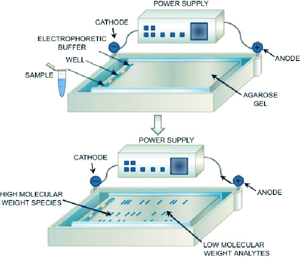 Figure: Figure source:https://www.sciencedirect.com/topics/pharmacology-toxicology-and-pharmaceutical-science/agarose-gel-electrophoresis
Types of Electrophoresis
Based on the nature of the supporting medium, it is of the following types:
- Agarose gel electrophoresis, the standard method for separating DNA fragments.
- Polyacrylamide gel electrophoresis (PAGE), used mainly for proteins and small nucleic acids.
- Cellulose acetate electrophoresis, a membrane-based method used for serum proteins and hemoglobin variants.
- Starch gel electrophoresis, now largely of historical interest.
Depending on the mode of technique, it has the following types:
- Paper electrophoresis
- Isoelectric focusing electrophoresis
- Two-dimensional Polyacrylamide gel electrophoresis
- Pulsed-field gel electrophoresis (PFGE)
- Zymography
- Immunoelectrophoresis
- Capillary electrophoresis
- High voltage electrophoresis
- Isotachophoresis
- Microchip electrophoresis
Uses of Electrophoresis
It is applied for routine laboratory experiments, disease diagnosis, research-oriented separations and identification. Similarly, it is used in various other fields, like forensics, agriculture, pharmaceutical, foods, etc. Some of its applications are described below:
DNA Analysis and DNA Fragmentation
Gel electrophoresis is the core technique for genetic analysis and purification of nucleic acids for further studies or disease diagnosis.
Identifying Specific protein
- The rate of movement of macromolecules in an electric field is a helpful parameter to know any changes in amino acids regarding their charge.
- Quantitative analysis of specific serum protein classes such as gamma globulins and albumins
- It allows identification and quantitation of hemoglobin variants such as HbA, HbA2, HbF, HbS, and HbC, which is the basis of screening for sickle cell disease and thalassemia.
- It identifies monoclonal protein in serum or urine. This is the M band described at the start of this article. A sharp, narrow spike in the gamma region, replacing the normal broad hill, indicates that a single plasma cell clone is overproducing one identical immunoglobulin, as occurs in multiple myeloma. The same principle applied to urine detects free light chains (Bence Jones protein).
- Likewise, it helps in the separation and quantitation of significant lipoprotein classes.
- Immunoelectrophoresis helps to analyze several kinds of protein’s existence and how they behave chemically in different environments.
- It is also helpful in purifying proteins for different purposes.
- Similarly, it is useful in determining the molecular weights of protein.
Isoenzyme Separation
Many enzymes exist as isoenzymes, which are different molecular forms of the same enzyme produced by different tissues. Because these forms differ in net charge, electrophoresis separates them, and the pattern reveals which tissue released the enzyme.
Creatine kinase separates into CK-MM (skeletal muscle), CK-MB (cardiac muscle), and CK-BB (brain). A rise in the CK-MB fraction points to myocardial injury.
Lactate dehydrogenase separates into five isoenzymes, LDH-1 through LDH-5, with different tissue distributions.
Alkaline phosphatase separates into liver, bone, intestinal, and placental isoenzymes, which helps determine the source of a raised total ALP.
Note the terminology carefully. These are enzymes, and electrophoresis separates them into isoenzymes. They are not coenzymes. Coenzymes are small organic cofactors such as NAD+ and coenzyme A.`
Analysis of chemical compounds
- It helps analyze compounds, such as water, soil, air quality or contamination, food quality, processing hygiene, and medical forensic analysis.
- It also helps in analyzing transition metals.
- Likewise, it helps to analyze organic compounds.
- Similarly, it helps in analyzing components of pesticides.
References
- Wilson K, Walker J. Principles and Techniques of Biochemistry and Molecular Biology. 8th ed. Cambridge: Cambridge University Press; 2018. Chapter: Electrophoretic Techniques.
- Westermeier R. Electrophoresis in Practice: A Guide to Methods and Applications of DNA and Protein Separations. 5th ed. Weinheim: Wiley-VCH; 2016.
- Westermeier R. Gel Electrophoresis. In: eLS. Chichester: John Wiley & Sons; 2005. doi:10.1038/npg.els.0005335
- Rifai N, Chiu RWK, Young I, Burnham CD, Wittwer CT, editors. Tietz Textbook of Laboratory Medicine. 7th ed. St. Louis: Elsevier; 2022. [VERIFY edition and year]
- Vesterberg O. History of electrophoretic methods. Journal of Chromatography. 1989;480:3-19. [VERIFY]
- Fritsch R, Krause I. Electrophoresis. In: Encyclopedia of Food Sciences and Nutrition. 2nd ed. Amsterdam: Elsevier; 2003. p. 2055-2062. doi:10.1016/b0-12-227055-x/01409-7
Frequently Asked Questions
What is the basic principle of electrophoresis?
Why does DNA always move toward the anode?
Which way does a protein move in electrophoresis?
Why is electrophoresis called an incomplete form of electrolysis?
What is the difference between zone and moving boundary electrophoresis?
What are the main factors affecting electrophoretic mobility?
How is electrophoresis used to diagnose multiple myeloma?
Does electrophoresis separate molecules by size or by charge?
Why is a larger pore size not always better?

Tankeshwar Acharya, MSc (Medical Microbiology)
Tankeshwar Acharya is an Assistant Professor in the Department of Microbiology at Patan Academy of Health Sciences (PAHS), Nepal, where he has been teaching and practicing clinical microbiology for over 14 years. He is the founder of Microbe Online, one of the leading free microbiology education resources on the web, covering bacteriology, mycology, parasitology, immunology, and clinical laboratory diagnostics written from direct experience in both the classroom and the diagnostic laboratory.