
Biuret Test: Principle, Procedure, and Uses
Biuret test: how copper(II) detects peptide bonds, violet vs pink results, false positives, and when to use it over Lowry, Bradford, or BCA. For micro and lab-science students.

On this page
A serum sample comes to the bench with a total protein of 9.5 g/dL. The biuret method is what most clinical analyzers use to measure it. But if you ran the same method on a purified protein fraction from the research lab next door, you might get a falsely high reading, and a dilute cerebrospinal fluid sample might read as almost no protein at all when protein is actually present. The biuret test is simple to run. Knowing when to trust its result, and when to reach for a different method, is the part that takes judgment.
The biuret test is a colorimetric method that detects peptide bonds. Because it responds to the peptide bond itself and not to individual amino acids, it measures total protein rather than any single amino acid, which is exactly what makes it useful for whole samples like serum, urine, and other body fluids.
The biuret test is a colorimetric test that helps detect specific proteins or peptide bonds in given analytes. It is followed by spectrophotometry for quantification.
The test requires the use of a biuret reagent. This reagent is a solution that consists of hydrated copper (II) sulfate, sodium hydroxide, and potassium sodium tartrate.
The use of copper (II) ions present in the biuret reagent results in the formation of purple coloration if peptides are present. The intensity of the purple color is measured using a spectrophotometer.
Principle of Biuret Test
Biuret test requires testing the analytes with biuret reagents. The reagent is a mixture of potassium sodium tartrate (KNaC4H4O6 or C4H4KNaO6), copper (II) sulfate or cupric sulfate (CuSO4), and sodium hydroxide (NaOH).
Sodium hydroxide makes the solution alkaline, and potassium sodium tartrate is the chelating agent. The potassium sodium tartrate helps stabilize the cupric ions in the mixture and maintains the alkaline solution’s solubility.
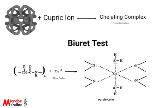 Figure: Principle of Biuret Test
Under alkaline conditions, the copper (II) ion (Cu²⁺) binds to the nitrogen atoms of the peptide bonds. This binding displaces the peptide hydrogens. The nitrogen atoms then donate their lone pairs of electrons to the copper ion to form coordinate covalent bonds. One copper (II) ion coordinates with four to six nearby peptide bonds, forming a colored chelate complex that absorbs light at 540 nm and appears violet.
The copper stays as copper (II) in this reaction. The violet color comes from the coordination complex, not from a change in the copper's oxidation state. This is the point students most often get wrong. The reduction of copper (II) to copper (I) does happen, but it is the basis of the later Lowry and BCA methods, not the source of the biuret violet color itself.
The intensity of the violet color is directly proportional to the number of peptide bonds present, which is why the method can be used to quantify total protein with a spectrophotometer.
The test depends on peptide bonds, not on individual amino acids. A molecule needs at least two peptide bonds to react, so tripeptides and larger proteins give a positive result, while free amino acids and dipeptides do not. Because it reads the peptide bond directly and is not affected by free amino acids, the method works well on whole samples with a high protein concentration, such as serum.
Samples prepared by ammonium sulfate precipitation are a known problem. The ammonium ions interfere with the reaction and can give a falsely high reading, so the biuret method is not suitable for protein fractions purified this way.
Reagents and Materials Required
The reagents and materials required for the biuret test are as follows:
- The reagent required for performing a the test is biuret reagent. To prepare biuret reagent, dissolve 1 g of CuSO₄ in 100 mL of distilled water to make a 1% solution, then add 1.2 g of potassium sodium tartrate. Prepare a 10% NaOH solution separately by dissolving 10 g of NaOH in distilled water and making the volume up to 100 mL. Add the NaOH solution to the copper-tartrate mixture. The final reagent is blue because of the copper (II) sulfate.
- Other equipment required for the test are test tubes, a dropper or apipette, a test tube holder, and a stand.
Procedure of Biuret Test
The following steps are followed to perform the biuret test:
- Take three clean and dry test tubes.
- In the first tube, add 1-2 ml test sample. Likewise, add 1-2 ml of egg albumin in the second one; in the third tube, add 1-2 ml of distilled water. The egg albumin is a positive control, whereas distilled water is a negative control for this test.
- Then, add 1-2 ml biuret reagent in all three tubes.
- After that, properly shake all the tubes to mix the reagent and samples/analytes. Then, let the mixture in the tubes stand for at least 5 minutes.
- Finally, observe the color change.
Precautionary measures
- Use test tube holders when holding the tubes with a solution.
- When preparing biuret reagent, handle NaOH carefully as it is a strong base that might cause corrosion when exposed to the skin.
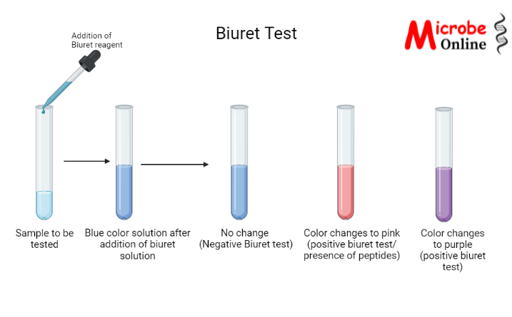 Figure: Biuret Test
Result Interpretation
The results of the biuret test are interpreted as follows:
| Observation | Interpretation |
|---|---|
| 1. The color of the first solution changes to purple. 2. The color of the first solution changes to pink . 3. No change in the color of the first solution. | 1. Presence of proteins. (Positive biuret test) 2. Presence of peptides (Positive biuret test) 3. Absence of proteins or peptides (Negative biuret test) |
| The color of the second tube changed to purple. | Positive biuret test (Positive control) |
| No change was seen in the color of the solution in the third tube.. | Negative biuret test ( Negative control) |
How to read the color: violet, pink, or blue
The color is not just positive or negative. The shade tells you something about what is in the tube.
A violet or purple color means proteins are present. These have many peptide bonds, and a large number of peptide bonds shifts the color toward violet.
A pink color means short peptides are present. Short chains have fewer peptide bonds, and fewer bonds give a pink color rather than a full violet. So a pink result is still positive, it just points to smaller peptides rather than intact protein.
No color change, meaning the solution stays blue, means no peptide bonds are available to react. This is either a true absence of protein, or a sample that contains only free amino acids or dipeptides, which cannot react.
This gradient is the reason the biuret test can give a rough sense of peptide chain length, not only a yes-or-no answer for protein.
Uses of Biuret Test
The biuret test is used mainly for diagnostic purposes, like determining serum proteins. Other applications of this test are as follows:
- The test helps in determining the type of proteins in unknown samples.
- It is used for quantification of protein by using a spectrophotometer alongside.
- It can help determine proteins in the urine, CSF, and other body fluids.
- The test helps determine the presence of specific proteins during food analysis.
Advantages of Biuret Test
The advantages of the biuret test are as follows:
- The test is simple and inexpensive.
- It responds to the peptide bond itself, so it measures total protein rather than any single amino acid.
- Very few components interfere with the test.
- The color is stable, so it causes less deviation.
- It is also a rapid test.
- It detects any protein or peptide with at least two peptide bonds, so it works across a wide range of proteins.
Disadvantages of Biuret Test
- Samples rich in the amino acid histidine can give a false positive, because histidine side chains also coordinate copper.
- If the buffer used to purify proteins has ammonium and magnesium salts, it can hinder the test.
- The presence of carbohydrates and fats can also hinder the test.
- This test alone cannot help in quantifying the protein in the sample; spectrophotometric analysis is required for quantification.
- Its sensitivity is low compared with the Lowry, Bradford, and BCA methods, which detect protein in the microgram range. Biuret works in the milligram range, so it is not suitable for dilute samples.
- Only soluble proteins are helpful in this test, and different proteins give different colors, so standardization of colors is required for known proteins.
Biuret vs Lowry vs Bradford vs BCA: which method and when
Students often learn these as four separate tests. In practice they are a family, and the choice between them is a bench decision based on how much protein you have and what else is in the sample.
| Method | Basis | Sensitivity (approx. range) | Best for | Main interference |
|---|---|---|---|---|
| Biuret | Cu²⁺ coordinates with peptide bonds, violet at 540 nm | Low, about 1–10 mg/mL | High-protein samples: serum, total protein on analyzers | Ammonium salts, some buffers |
| Lowry | Biuret reaction plus reduction of copper and Folin reagent reacting with tyrosine and tryptophan | High, about 5–150 µg | Dilute samples, following purity during purification | Many: reducing agents, detergents, buffers |
| Bradford | Coomassie dye binds basic amino acid residues, blue | High, low µg range | Fast routine assays, dye-based | Detergents such as SDS, Triton, Tween |
| BCA | Copper reduced to Cu⁺, then chelated by BCA, purple at 562 nm | High, µg range | Samples containing detergents | Reducing agents, chelators such as EDTA |
The logic in one line: reach for biuret when you have plenty of protein and want a simple, cheap, interference-resistant readout. Reach for Lowry, Bradford, or BCA when the sample is dilute and you need sensitivity, accepting that each of those brings its own interfering substances.
Both Lowry and BCA are built on the biuret reaction. They add a second step that reduces copper and amplifies the signal, which is what makes them far more sensitive. So understanding biuret first is what makes the other three make sense.
Where students get confused
"The copper gets reduced, and that is what makes the violet color." No. The violet biuret color is a copper (II) coordination complex. The copper stays as Cu²⁺. Copper reduction to Cu⁺ is the basis of the Lowry and BCA methods that came later, which is a different reaction with different chemistry.
"The test detects amino acids." No. It detects peptide bonds. A tube of free amino acids will stay blue no matter how concentrated it is. You need at least two peptide bonds, so a tripeptide is the smallest thing that reacts.
"A negative (blue) result always means no protein." Not necessarily. A sample of free amino acids also stays blue. A negative biuret result means no peptide bonds are available, which is not the same as no nitrogen and not the same as no amino acids.
"Biuret is a sensitive test." It is not. Biuret has low sensitivity, with a working range of roughly 1 to 10 mg/mL of protein. This is a feature, not a flaw: it is why the test is chosen for high-protein samples like serum, where sensitive methods would need heavy dilution.
"The reagent is called biuret because it contains biuret." No. The reagent contains no biuret. It is named after the biuret molecule because that molecule has the same peptide-like bonding that the test detects.
How to Remember
"Biuret needs a Bond, not a Building block." The test detects peptide bonds, not amino acids (the building blocks). Free amino acids stay blue.
"Two to react." A minimum of two peptide bonds is needed, so a dipeptide (one bond) stays blue and a tripeptide (two bonds) is the smallest thing that turns the tube.
Violet vs pink: more bonds, more violet. Think of it as a dimmer switch, not an on-off switch. Long protein chains push the color all the way to violet; short peptides only get to pink.
Copper keeps its number. In biuret, copper stays 2+. The moment copper drops to 1+, you are talking about Lowry or BCA, not biuret.
Key exam facts in one table
| Fact | Detail |
|---|---|
| What it detects | Peptide bonds (not amino acids) |
| Minimum to react | Two peptide bonds (tripeptide or larger) |
| Reagent | CuSO₄ + NaOH + potassium sodium tartrate |
| Role of NaOH | Provides the alkaline conditions |
| Role of tartrate | Chelating agent; keeps Cu²⁺ soluble in alkali |
| Copper oxidation state in the color | Stays Cu²⁺ (coordination complex, not reduction) |
| Positive color | Violet (protein) or pink (short peptides) |
| Absorbance measured at | 540 nm |
| Copper coordination | One Cu²⁺ binds four to six peptide bonds |
| Sensitivity | Low, about 1–10 mg/mL |
| Positive control | Egg albumin |
| Negative control | Distilled water |
| Common false positive | Ammonium salts, histidine-rich samples |
| Built on the biuret reaction | Lowry and BCA methods |
References
- Nelson DL, Cox MM. Lehninger Principles of Biochemistry. 8th ed. W.H. Freeman; 2021. (Protein quantitation methods.)
- Bianchi-Bosisio A. Proteins: Physiological Samples. In: Worsfold P, Townshend A, Poole C, eds. Encyclopedia of Analytical Science. 2nd ed. Elsevier; 2005:357–375. https://doi.org/10.1016/B0-12-369397-7/00494-5
- Thermo Fisher Scientific. Chemistry of Protein Assays. Pierce Protein Methods. (For the biuret, Lowry, and BCA reaction chemistry and relative sensitivity.)
- Burtis CA, Bruns DE. Tietz Fundamentals of Clinical Chemistry and Molecular Diagnostics. 8th ed. Elsevier; 2019. (Total serum protein measurement by biuret.)
Frequently Asked Questions
Why does the biuret test detect peptide bonds and not amino acids?
Why does the biuret test detect peptide bonds and not amino acids?
The violet color forms when copper (II) coordinates with the nitrogen atoms of peptide bonds. A free amino acid has no peptide bond, so there is nothing for the copper to coordinate with in that way. This is why a tube of free amino acids stays blue.
What is the minimum number of peptide bonds needed for a positive result?
What is the minimum number of peptide bonds needed for a positive result?
Two. That means a tripeptide is the smallest molecule that reacts. A dipeptide, which has only one peptide bond, does not react.
Why is the reagent blue before the test?
Why is the reagent blue before the test?
The blue color comes from the copper (II) sulfate in the reagent. When peptide bonds are present, the copper forms a new complex that shifts the color to violet.
Does the copper change oxidation state in the biuret test?
Does the copper change oxidation state in the biuret test?
No. In the classic biuret reaction the copper stays as copper (II). The violet color is a coordination complex. Reduction of copper to copper (I) is the basis of the related Lowry and BCA methods, not of the biuret color itself.
Why is the biuret test used for serum protein if it is not very sensitive?
Why is the biuret test used for serum protein if it is not very sensitive?
Serum has a high protein concentration, so low sensitivity is not a problem. The low sensitivity is actually an advantage here, because more sensitive methods would need the sample to be heavily diluted first. Biuret is also cheap and resists interference from many substances.
What causes a false positive in the biuret test?
What causes a false positive in the biuret test?
Ammonium salts, such as those left over from ammonium sulfate precipitation, and samples rich in histidine can both give falsely high readings.

Tankeshwar Acharya, MSc (Medical Microbiology)
Tankeshwar Acharya is an Assistant Professor in the Department of Microbiology at Patan Academy of Health Sciences (PAHS), Nepal, where he has been teaching and practicing clinical microbiology for over 14 years. He is the founder of Microbe Online, one of the leading free microbiology education resources on the web, covering bacteriology, mycology, parasitology, immunology, and clinical laboratory diagnostics written from direct experience in both the classroom and the diagnostic laboratory.
Comments
No comments yet. Be the first to share your thoughts.
Leave a comment
All comments are reviewed before they appear.