
Electroporator: How Electrical Pulses Open Cell Membranes, Principle, Parts, and Uses

On this page
A student sets up an electroporation to transform E. coli with a new plasmid, sets the voltage high to be sure the DNA gets in, delivers the pulse, and plates the cells. The next day the plates are nearly empty. Almost nothing grew. The pulse worked exactly as intended: it opened the membranes. It just opened them too far. Instead of transient pores that reseal, the cells got holes they could not recover from, and most of them died before they could take up and express the plasmid. The entire skill of electroporation lives in this narrow margin: enough field to open the membrane, not so much that the cell cannot close it again.
An electroporator is an instrument that applies a controlled electrical pulse to cells to make their membranes temporarily permeable, allowing molecules such as DNA, RNA, or proteins to enter. The technique is called electroporation (or electropermeabilization). It is one of the main physical methods used to introduce foreign DNA into cells during transformation, alongside chemical methods such as heat-shock.
Once inside, a plasmid usually remains separate from the chromosome and replicates on its own or is expressed transiently; stable integration into the genome is a separate, much rarer event. Electroporators range from small benchtop units to larger automated systems.
Principle of Electroporator
An electroporator works by storing electrical charge in a capacitor, then discharging it through the cell suspension as a brief, controlled pulse. This pulse creates the electric field that raises the voltage across the cell membrane and opens the pores. The main adjustable parameters are the field strength (voltage divided by the electrode gap, in V/cm or kV/cm), the pulse duration, and the number of pulses. The five steps below break down what happens to the cell.
Electroporation proceeds in five stages: cell membrane permeabilization, electrical pulses, pore formation, uptake of genetic material, and cellular recovery.
- Cell membrane permeabilization: The cell membrane is normally impermeable to large molecules like RNA, DNA, and proteins. The applied pulse briefly disrupts the membrane, opening transient pores that let these molecules enter.
- Electrical pulses: Electroporation applies short, high-voltage pulses across the cell suspension. The pulse creates an electric field, which raises the voltage across the cell membrane. Once this transmembrane voltage passes a threshold, the lipid bilayer becomes unstable and temporary pores form.
- Pore formation: The raised transmembrane voltage makes the lipid bilayer unstable, and lipid molecules reorient to line small aqueous channels, the transient pores.
- Uptake of genetic material: While the pores are open, DNA enters through them, driven partly by the electric field pushing the negatively charged DNA and partly by diffusion.
- Cellular recovery: If the pulse stayed within the survivable range, the pores reseal once the field is removed, and the cells are placed in recovery medium before selection.
However, the successful completion of electroporation depends on factors like pulse voltage, duration, and number, the type of cells used, and the introduced genetic material’s size and type. The optimization of these parameters is critical to achieve efficient electroporation and ensure cell viability.
How the pulse opens the membrane, and reversible vs. irreversible
A cell membrane is a lipid bilayer that normally blocks large, charged molecules like DNA. Electroporation works by exploiting the fact that the membrane is also an electrical insulator separating two conductive solutions, inside and outside the cell.
Opening the pores. When a brief, strong electric field is applied, charge builds up across the membrane, raising the transmembrane voltage. Once this exceeds a threshold (roughly a few hundred millivolts across the membrane), the lipid bilayer becomes unstable and transient aqueous pores form. For a short window the membrane is permeable, and DNA is driven through these pores, partly by the field pushing the negatively charged DNA and partly by diffusion.
The reversible window. If the pulse is within the right range of strength and duration, the pores reseal after the field is removed and the cell survives, now carrying the introduced DNA. This is reversible electroporation, and it is what every transformation and transfection protocol aims for. Too weak, and no pores form, so nothing enters. Too strong or too long, and the pores grow, the membrane cannot reseal, and the cell dies. That is the failure in the hook.
Irreversible electroporation. Pushing deliberately past the reversible threshold kills the cell, and this is not always a mistake. Irreversible electroporation (IRE) is used clinically to ablate tumors: a series of strong pulses permanently permeabilizes tumor-cell membranes and kills the cells without the heat damage of thermal ablation, which helps spare nearby structures such as blood vessels and ducts. The same physics that ruins a transformation is, at higher intensity, a targeted tissue-destruction tool.
The single idea to carry away: electroporation is a dose. The instrument's job is to deliver a field strong enough to open the membrane but controlled enough to stay on the survivable side of the line, unless killing the cell is the goal.
Parts of Electroporator
An electroporator is a complex laboratory instrument that performs electroporation to introduce foreign materials into the cells by forming temporary pores. Based on different companies, the parts of the electroporator can vary widely.
However, some of the common parts of an electroporator include a control panel, electrical pulse generator, cuvette chamber, electrodes, pulse controller, high-voltage supply, safety features, cooling systems, and adapters.
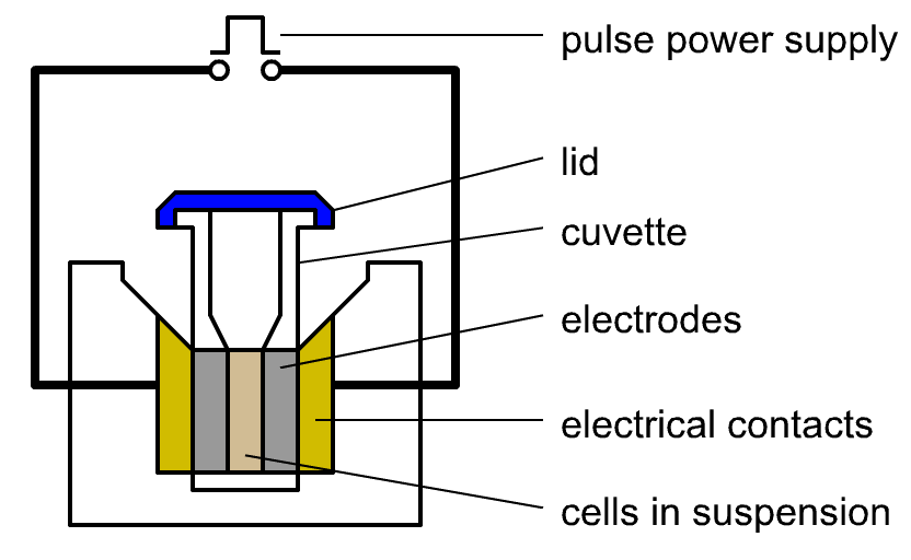 Figure: A diagram of the main components of an electroporator with cuvette loaded
Control Panel
The control panel is the area in the laboratory instrument where the user inserts instructions for the electroporation process. It is also referred to as an interface. This part helps in controlling voltage, pulse duration, and pulse number. It may have a display screen for viewing the progress of the procedure.
Electrical Pulse Generator
This part of the laboratory instrument generates electrical pulses required for electroporation. It produces high-voltage pulses, which are delivered to the cell.
Cuvette Chamber
This holds the electroporation cuvette, a small vessel with two parallel electrodes on its sides. The cell-and-DNA suspension sits between the electrodes. Cuvettes come in fixed electrode gaps, commonly 1, 2, or 4 mm, and the gap matters: field strength equals the voltage divided by the gap, so the same voltage produces a stronger field in a narrower cuvette. Choosing the correct gap for a protocol is therefore as important as setting the voltage.
Electrodes
The instrument has two electrodes, one at each end of the cuvette chamber. The electrodes provide electrical pulses to the cell sample. The electrodes come in different formats: cuvette electrodes (built into disposable cuvettes), plate electrodes (for cells in multiwell plates), and specialized electrodes such as needle arrays used for in vivo applications.
High-voltage Supply
The high-voltage power supply provides the electrical energy for the pulses and ensures they are delivered at the set voltage.
Safety Features
The main safety features are electrical isolation and interlocks. Electrical isolation protects the user from shock and provides overcurrent protection, while interlocks prevent the instrument from firing unless it is correctly set up.
User Manual and Software
The electroporators come with a user manual that provides instructions on operation and maintenance. Modern electroporator has software for programming and controlling the instrument.
Miscellaneous Parts
- Adapters: Some electroporators may include holders or adapters. These adapters ensure the cuvettes or samples are appropriately placed for electroporation.
- Pulse Controller: Some electroporators have a separate component or module for controlling the different parameters of electrical pulse like duration, number, and voltage. This part helps in obtaining precise control of the electroporation conditions.
- Cooling Systems: Various electroporators have a cooling system for dissipating generated heat. It is crucial when dealing with multiple samples or high-throughput applications.
- Data Logging and Connectivity: New generation electroporators may have data recording capabilities for recording the parameters and outcomes of each electroporation test. Some models of electroporators may also have connectivity options like USB ports or data transferring software for data retrieval and analysis.
- Trigger: Some electroporators consist of trigger or footswitch mechanisms. This mechanism allows the user to initiate the electrical pulse delivery manually.
Although these are some of the commonly present parts of an electroporator, the specific features and details can vary between different models and brands. So, studying the user manual is vital before operating the electroporator.
Uses of Electroporator
Electroporators are widely useful laboratory instruments in various molecular biology, genetic engineering, and biotechnology fields. Across these fields, the primary use of the device is to deliver genetic material into cells.
- Electroporators help in gene therapy research, functional genomics, and gene expression studies in mammalian cells by the process called transfection. It is also helpful in producing viral vectors by introducing viral genomes into producer cells for gene therapy and gene delivery applications.
- It is used to introduce plasmid DNA or other genetic material into bacterial and yeast cells, that is, to transform them, a routine step in molecular biology and biotechnology.
- It is used to generate genetically modified organisms by delivering genes or gene-editing components into cells.
- Likewise, this technique is used to produce recombinant proteins by introducing the genes that encode them into host cells. This introduction helps in expressing the desired proteins.
- Electroporators are also helpful in gene editing technologies like CRISPR-Cas9 for delivering CRISPR components into the cells for editing specific genomes.
- In some cases, electroporation is useful in fusing cells, which applies to hybridoma technology producing monoclonal antibodies. It also applies to drug delivery mechanisms in pharmacology research and drug discovery.
- Electroporation is helpful in cancer research for drug screening, gene therapy, and delivery of therapeutic agents in cancer cells. Similarly, this technique is also applicable in vaccine development by enhancing the delivery of DNA-based vaccines to cells, which helps in improving immune response.
- The instrument helps introduce genes into stem cells, essential in stem cell research. In plant biotechnology, this instrument helps introduce foreign genes into plant cells for improving crops, disease resistance, and developing genetically modified crops.
- Beyond gene delivery, irreversible electroporation (IRE) is used clinically to destroy tumor tissue. Strong pulses permanently permeabilize tumor-cell membranes and kill the cells without heat, which helps spare nearby blood vessels, nerves, and ducts.
Types of Electroporator
Electroporators are grouped in two useful ways: by the shape of the electrical pulse they deliver, which is the most important technical distinction, and by their intended application or format. The pulse waveform determines how gentle and how controllable the pulse is, which in turn decides what cells the instrument suits.
By pulse waveform
- Exponential-decay electroporators: A charged capacitor discharges through the sample, so the voltage starts high and falls away exponentially. This is the simple, standard waveform for transforming bacteria and yeast, which tolerate a sharp pulse.
- Square-wave electroporators: The electronics hold the voltage constant for a set time, then cut it off, and can deliver several controlled pulses. This is gentler and more precise, and it is preferred for fragile mammalian cells, primary cells, and stem cells, where survival matters as much as delivery.
By application and format
- Yeast and Bacterial Electroporators: These instruments transform yeast and bacterial cells. The bacterial electroporators are used in molecular biology research for cloning, genetic engineering, and protein expression of bacteria. These electroporators also have specialized cuvettes and parameters optimized for bacterial cells.
- Plant and Mammalian Cell Electroporators: The mammalian electroporators provide the necessary flexibility and control to work on mammal cells. These instruments are helpful in transfecting mammalian cells with DNA, RNA, or other nucleic acids. These are significant in gene expression studies, gene therapy research, and gene editing. Plant electroporators are crucial for genetically modifying plant cells and tissues and can accommodate larger volumes and use specialized electrodes or cuvettes suitable for plant materials.
- High-throughput electroporators: These can process multiple samples simultaneously, giving high throughput. These are applicable in drug screening or large-scale protein expression studies.
- Benchtop Electroporators: These are compact instruments suitable for small-scale experiments and individual laboratories. Despite their compact size, they are versatile and commonly used for various electroporation applications.
- Automated Electroporators: Various other processes within electroporation get automated in this type of electroporator, like cell handling, dispensing, and data collection. These help in high-throughput and automation of laboratories like drug discovery and genomics.
- Flow Electroporators: These help enable continuous and efficient cell electroporation in a fluid stream. These are highly efficient in cell therapy and other applications requiring high cell throughputs.
- Clinical and Portable Electroporators: These are used in clinical trials and potentially therapeutic purposes for gene delivery to patient’s cells during gene therapy. Some electroporators are small, portable, and designed for field applications like point-of-care diagnostics and on-site genetic modification experiments in remote locations.
How to Remember
Electroporation is a dose, not a switch. Too little field, nothing opens. Too much, the cell dies. The reversible window in between is the whole game. When a transformation gives no colonies, ask first whether the pulse was too strong, not too weak.
Field strength, not voltage, does the work. Field = voltage ÷ electrode gap. The same 2000 V means a much stronger field in a 1 mm cuvette than a 4 mm one. Remember the gap, or you will misjudge the pulse.
Reversible builds, irreversible destroys. Same phenomenon, two intensities. Reversible electroporation puts DNA into living cells (transformation). Irreversible electroporation kills cells on purpose (tumor ablation). The line between them is how hard you pulse.
Exponential for bugs, square for mammalian. Exponential-decay pulses are the workhorse for bacteria and yeast. Square-wave pulses are gentler and preferred for fragile mammalian cells. Match the waveform to how much the cell can take.
Key exam facts in one table
| Concept | Fact to remember |
|---|---|
| Principle | A brief high-voltage pulse raises the transmembrane voltage past a threshold, forming transient pores that let DNA and other molecules enter |
| Reversible electroporation | Pores reseal and the cell survives; the goal of all transformation and transfection |
| Irreversible electroporation | Pores do not reseal and the cell dies; used deliberately for non-thermal tumor ablation (IRE) |
| What matters most | Field strength (voltage ÷ electrode gap, in V/cm or kV/cm), not voltage alone |
| Cuvette gap | Common gaps 1, 2, or 4 mm; a narrower gap gives a stronger field at the same voltage |
| Exponential-decay type | Capacitor discharge, voltage decays exponentially; standard for bacteria and yeast |
| Square-wave type | Constant voltage for a set time; gentler, preferred for mammalian and fragile cells |
| Main uses | Transformation and transfection (introducing DNA, RNA, protein), and IRE for tissue ablation |
| Common failure | Over-strong pulse kills cells, giving few or no transformants |
Where Students Get Confused
"Higher voltage means better transformation." No. Beyond the reversible threshold, a stronger pulse kills more cells and gives fewer transformants. There is an optimum, not a "more is better" rule.
Voltage versus field strength. The cell responds to the field across it, which is voltage divided by the electrode gap. The same voltage produces a stronger field in a narrow cuvette. Always think in field strength (V/cm), not raw volts.
Reversible versus irreversible. They are the same physical process at different intensities. Reversible electroporation keeps the cell alive to take up DNA; irreversible electroporation destroys the cell on purpose. A killed transformation is accidental irreversible electroporation.
Electroporation is not the only way to transform cells. It is one physical method. Chemical transformation (heat-shock of competent cells) does the same job differently. Electroporation is often more efficient but needs low-salt samples, because high salt causes arcing.
Why arcing happens. If the sample conducts too well (high salt) or has bubbles, the pulse discharges as a spark (an arc) instead of a controlled field, which destroys the sample. This is why cells for electroporation are washed into low-ionic-strength media.
References
- Brown TA. Gene Cloning and DNA Analysis: An Introduction. 8th ed. Wiley-Blackwell; 2021.
- Alberts B, Heald R, Johnson A, Morgan D, Raff M, Roberts K, Walter P. Molecular Biology of the Cell. 7th ed. New York: W. W. Norton; 2022.
- Schmitt MA, Friedrich O, Gilbert DF. Portoporator: a portable low-cost electroporation device for gene transfer to cultured cells in biotechnology, biomedical research and education. Biosensors and Bioelectronics. 2019;131:95-103. doi:10.1016/j.bios.2019.02.024
Frequently Asked Questions
What is the principle of an electroporator?
What is the principle of an electroporator?
It applies a brief, high-voltage electrical pulse to a cell suspension. The pulse raises the voltage across the cell membrane past a threshold, which forms transient pores in the lipid bilayer. While the pores are open, DNA or other molecules enter the cell. If the pulse is within the survivable range, the pores reseal and the cell lives, now carrying the introduced material.
What is the difference between reversible and irreversible electroporation?
What is the difference between reversible and irreversible electroporation?
In reversible electroporation the pores reseal after the pulse and the cell survives, which is the goal of transformation and transfection. In irreversible electroporation the pulse is strong enough that the pores do not reseal and the cell dies. Irreversible electroporation is used deliberately in medicine to ablate tumors without heat.
Why did my electroporation kill the cells?
Why did my electroporation kill the cells?
Most often the pulse was too strong or too long, pushing the membrane past the reversible threshold into irreversible electroporation. Arcing from high-salt samples or air bubbles also kills cells. Lowering the field strength and washing cells into low-salt medium usually fixes it.
Why does the electrode gap of the cuvette matter?
Why does the electrode gap of the cuvette matter?
Because the cell responds to field strength, which is the voltage divided by the gap between the electrodes. The same voltage gives a stronger field in a 1 mm cuvette than in a 4 mm one. Using the wrong gap for a protocol changes the actual field the cells experience.
What is the difference between exponential-decay and square-wave electroporators?
What is the difference between exponential-decay and square-wave electroporators?
An exponential-decay electroporator discharges a capacitor, so the voltage starts high and falls exponentially; it is standard for bacteria and yeast. A square-wave electroporator holds the voltage constant for a set time then stops; it is gentler and better for fragile mammalian cells.
Is electroporation better than chemical (heat-shock) transformation?
Is electroporation better than chemical (heat-shock) transformation?
Electroporation is usually more efficient and works with a wide range of cells, but it needs specialized equipment and low-salt samples to avoid arcing. Chemical transformation is simpler and needs no instrument but is generally less efficient. The choice depends on the cell type, the efficiency needed, and the equipment available.

Tankeshwar Acharya, MSc (Medical Microbiology)
Tankeshwar Acharya is an Assistant Professor in the Department of Microbiology at Patan Academy of Health Sciences (PAHS), Nepal, where he has been teaching and practicing clinical microbiology for over 14 years. He is the founder of Microbe Online, one of the leading free microbiology education resources on the web, covering bacteriology, mycology, parasitology, immunology, and clinical laboratory diagnostics written from direct experience in both the classroom and the diagnostic laboratory.
Comments
No comments yet. Be the first to share your thoughts.
Leave a comment
All comments are reviewed before they appear.