Voges-Proskauer (VP) Test: Principle, Procedure, Results & VP-Positive Organisms
VP test principle, the α-naphthol-then-KOH procedure, and how to tell a true positive from the copper-color false positive that trips up most students.
The Voges-Proskauer (VP) test determines whether an organism produces acetylmethylcarbinol (acetoin) from glucose fermentation. It's the third test in the IMViC panel, and it's always run alongside its mirror-image partner, the Methyl Red test; same broth, opposite logic.
Why It Matters
Recall the fork from the MR test: every Enterobacteriaceae member ferments glucose to pyruvate the same way, but what happens next splits the family into two camps. VP is reading the other branch of that same fork:
- Mixed acid pathway → stable acid end products → MR positive, VP negative (this is E. coli)
- 2,3-Butanediol pathway → pyruvate converted to neutral acetoin → MR negative, VP positive (this is the Klebsiella-Enterobacter-Hafnia-Serratia group)
This reciprocal pattern is the classic way to separate E. coli from the Klebsiella-Enterobacter group on paper — but VP isn't limited to Enterobacteriaceae. It also separates clinically important Gram-positive groups: among the viridans streptococci, the Streptococcus mitis group is VP-negative while most other viridans strep are VP-positive (except S. vestibularis, which is variable). That distinction matters when viridans strep shows up in a blood culture and you need to narrow down which subgroup you're dealing with.
Principle
Pyruvic acid (the central junction of glucose fermentation) is converted to acetoin (acetylmethylcarbinol) via the butylene glycol pathway in VP-positive organisms. In the presence of α-naphthol, strong alkali (40% KOH), and atmospheric oxygen, acetoin is oxidized to diacetyl, which then condenses with guanidine-containing compounds in the broth's peptone to form a pinkish-red polymer.
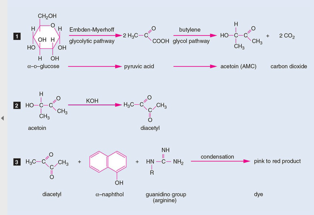 Figure: Voges Proskauer (VP) Test Principle
Procedure of Voges Proskauer (VP) Test
VP uses the same MR/VP broth as the Methyl Red test — only the reagents and reading window differ.
- Inoculate an MR/VP broth tube with a pure culture of the test organism.
- Incubate at 35°C for 24 hours.
- Aliquot 1 mL of broth into a clean tube.
- Add 0.6 mL of 5% α-naphthol, then 0.2 mL of 40% KOH — in that exact order.
- Shake gently to expose the medium to atmospheric oxygen, then let it stand undisturbed for 10–15 minutes.
- Read within the next hour.
Where students actually get confused
- Why does VP say 24 hours when MR says 48? Both tests use the same broth, but they're read on different timelines: VP can be screened as early as 24 hours because acetoin accumulates relatively quickly. If it's negative at 24 hours, you incubate the same broth out to 48 hours and repeat the test before calling it truly negative. MR, by contrast, needs the full 48 hours minimum before its first reading — it's measuring stable acid accumulation, which takes longer. They're not on the same clock, and that's expected, not a mistake.
- Order matters — α-naphthol before KOH, always. This isn't a minor detail: Barritt added α-naphthol specifically to intensify and stabilize the color reaction. Reverse the order and you risk a weak-positive or false-negative result. (This is one of the most repeated questions readers ask about this exact test.)
- A pale or faint pink color is not automatically positive. One reader described getting a pale pink at 10 minutes and being told by classmates it should have been read as positive — but a true positive is a clear pink-red color, and timing matters as much as color: read between 10–15 minutes, never past 1 hour.
- Don't read past 1 hour. Negative cultures can develop a copper-like color over time purely from reagent interaction, which is easy to mistake for a weak positive. If you're not sure whether you're seeing "positive pink" or "copper false-positive," the read window has likely closed — repeat the test rather than guess.
- Excess KOH causes the same copper-color trap. Don't exceed 2 drops of KOH per 2 mL of medium — too much KOH reacting with α-naphthol alone can produce a weak positive that's really an artifact, not a true reaction.
Quality Control
| Organism | Expected result |
|---|---|
| Klebsiella pneumoniae ATCC 13883 | VP positive (red) |
| Escherichia coli ATCC 25922 | VP negative (no change) |
Result Interpretation
| Color | Interpretation |
|---|---|
| Pink-red, within 15 min | VP positive |
| No color change | VP negative |
| Copper-like color (after >1 hr or excess KOH) | Not a true positive — repeat the test |
VP-positive organisms
Enterobacteriaceae: Klebsiella species, Enterobacter species, Hafnia species, Serratia species
Gram-positive cocci: most viridans group streptococci, including the Streptococcus anginosus and S. salivarius groups. The S. mitis group is VP-negative, and S. vestibularis is variable. This is the distinction referred to in "Why It Matters" above.
Other: Bacillus species are characteristically VP-positive, which is one feature separating them from Clostridium.
Same exceptions as MR: Hafnia alvei and Proteus mirabilis can show both a positive MR and a positive VP reaction, with the VP reaction often delayed. If your reciprocal expectation breaks, check for these two before assuming a technical error.
Limitations
- Prolonged incubation (>3 days) can let some VP-positive organisms acidify the medium, producing a weak-positive or false-negative result.
- Don't exceed 2 drops of KOH per 2 mL of medium — excess KOH can mimic a weak positive via the copper-color artifact.
- Don't read more than 1 hour after adding reagents — copper color can be misread as positive.
- Reagents must go in the specified order (α-naphthol, then KOH) — reversing it risks a weak-positive or false-negative result.
How to remember
VP and MR are a see-saw. Same broth, opposite answers. E. coli takes the mixed-acid path: stable acids, MR positive, VP negative. The Klebsiella-Enterobacter-Hafnia-Serratia group takes the butanediol path: neutral acetoin, VP positive, MR negative. If you know one result, you usually know the other. When the see-saw balances on both sides (both positive), suspect Hafnia alvei or Proteus mirabilis, the two organisms that break the reciprocal rule.
Reagent order: "A before K." Alpha-naphthol before KOH, alphabetically and chronologically. Barritt added the α-naphthol step in 1936 specifically to intensify the color, and it has to be in the tube first. Reverse them and a true positive can read as a weak positive or a false negative.
The copper trap. Ask yourself before reading: is it pink, or is it copper? Pink within 15 minutes is real. Copper, or anything appearing after an hour, is the reagents reacting with each other, not with acetoin. When in doubt, the window has closed. Repeat, do not guess.
References and further readings
- Leber AL, editor. Clinical Microbiology Procedures Handbook. 4th ed. Washington, DC: ASM Press; 2016. doi:10.1128/9781555818814
- Procop GW, Church DL, Hall GS, Janda WM, Koneman EW, Schreckenberger PC, Woods GL. Koneman's Color Atlas and Textbook of Diagnostic Microbiology. 7th ed. Philadelphia: Wolters Kluwer; 2017.
- MacFaddin JF. Biochemical Tests for Identification of Medical Bacteria. 3rd ed. Philadelphia: Lippincott Williams & Wilkins; 2000.
Frequently Asked Questions
I got a pale pink color — is that positive or negative?
Why does MR need 48 hours but VP only needs 24?
Why does the order of reagents matter?
Which organisms are VP-positive besides the Enterobacteriaceae?

Tankeshwar Acharya, MSc (Medical Microbiology)
Tankeshwar Acharya is an Assistant Professor in the Department of Microbiology at Patan Academy of Health Sciences (PAHS), Nepal, where he has been teaching and practicing clinical microbiology for over 14 years. He is the founder of Microbe Online, one of the leading free microbiology education resources on the web, covering bacteriology, mycology, parasitology, immunology, and clinical laboratory diagnostics written from direct experience in both the classroom and the diagnostic laboratory.