
Ultrasonication: How Cavitation Breaks Open Cells (Principle, Parts, Uses)

On this page
A student sonicates a bacterial suspension to release the protein they need, running the probe continuously for two minutes to be thorough. The cells lyse, but the protein is denatured and useless. The mistake was not the sonication; it was the heat.
Sonication works by collapsing microscopic bubbles inside the liquid, and each collapse releases an intense burst of local heat. Run continuously, that heat cooks the very molecules you are trying to extract. Understanding how ultrasound breaks a cell open, and why it also heats the sample, is what separates a clean lysate from a ruined one. It all comes down to a process called cavitation.
Ultrasonication, also known as sonication, is one of the homogenization techniques that use high-frequency sound waves (that is, above 20 kHz) to break large particles into smaller fragments or better uniform-sized particles in the base fluid.
Due to its versatility and effectiveness, this technique is commonly used in various fields, including chemistry, biology, materials science, and food processing. This technique is applicable for multiple purposes, such as cell disruption, nanoparticle dispersion, degassing, and cleaning of solid surfaces.
Principle
Ultrasonication uses high-frequency sound waves, above the range of human hearing (typically 20 kHz and higher), to disrupt cells and particles in a liquid. Following it as a chain, each step explains the next.
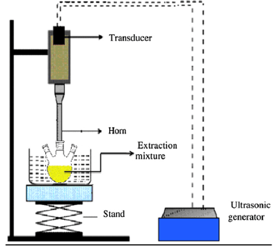 Figure: Experimental setup for ultrasonication
Why sound can do mechanical work. A sound wave is a pressure wave. As it travels through a liquid, it rapidly alternates the local pressure: a low-pressure phase where the liquid is pulled apart, and a high-pressure phase where it is pushed together. At ultrasonic frequencies this happens tens of thousands of times per second.
Why bubbles form (cavitation). During each low-pressure phase, the liquid is stretched so hard that it briefly tears apart, forming tiny vapor-filled bubbles. This formation of bubbles by sound is called acoustic cavitation. The bubbles grow slightly over successive cycles.

Why the bubbles collapse violently. When a bubble reaches an unstable size, the next high-pressure phase crushes it, and it implodes, collapsing in on itself almost instantly. This implosion is the actual working event of a sonicator.
Why the collapse breaks cells open. Each implosion is tiny but extreme. It generates powerful local shear forces and micro-jets of liquid, and for a fraction of a second an intense spot of local heat and pressure. Cells caught near a collapsing bubble are torn apart by these shear forces, their cell walls and membranes ruptured. Thousands of collapses per second across the sample add up to thorough disruption.
Why the sample heats up. The same implosions that break cells also dump energy into the liquid as heat. Over seconds of continuous sonication, the sample temperature climbs quickly. This is the central practical problem with sonication, covered below, because that heat can destroy the proteins, enzymes, or nucleic acids you are trying to release.
In short: sound makes bubbles, the bubbles implode, and the implosions tear cells apart while also heating the sample.
Why Heat Is the Enemy, and How to Control It
The heat generated by cavitation is the main reason sonication goes wrong. Controlling it is the core skill.
Pulse, do not run continuously. Sonicate in short bursts (for example a few seconds on, a few seconds off) rather than one long run. The off periods let the heat dissipate so the sample does not cook. Most protocols specify a pulse cycle for exactly this reason.
Keep the sample on ice. Hold the tube in an ice bath throughout. Heat-sensitive targets such as enzymes, proteins, and nucleic acids denature or degrade if the sample warms, so keeping it cold, near the refrigeration temperatures used to protect such samples (see refrigerator), preserves what you are extracting.
Do not over-sonicate. More sonication is not better. Once the cells are lysed, further bursts only add heat and can shear DNA into fragments or denature proteins. Use the minimum needed.
Avoid foaming. Excessive foaming means air is being drawn in, which reduces cavitation efficiency and denatures proteins at the air-liquid interface. Keep the probe tip below the surface and the power moderate.
Parts of Sonicator
The machine used to carry out ultrasonication is known as a sonicator. The significant parts of the sonicator are as follows:
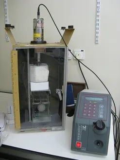 Figure: Sonicator
Generator
The generator supplies high-frequency electrical energy that drives the transducer. Its controls (keypads or knobs) set the parameters used during sonication, such as amplitude, pulse cycle, and total time.
Converter
It is also known as a transducer. Because of the properties of the intrinsic piezoelectric crystals, the converter converts electrical signals into high-frequency mechanical vibration (above 20 kHz). Furthermore, the generator and transducer are connected by high-voltage cable, and generated pulse is amplified and transmitted down the probe.
Probe
It is also known as a horn or tip. The probe is attached to the transducer and amplifies the ultrasonic vibrations generated by the transducer into a longitudinal vibration resulting in a cavity in the sample.
Sonication vessel
The sonication vessel or container holds the sample that needs to be processed. It is typically made of glass or plastic and can vary in size and shape depending on the application. The vessel should be compatible with the sonicator’s probe or horn for effective sample processing. The vessel itself is not soundproof; noise control comes from running the sonicator inside an enclosure or acoustic hood, covered under Precautions.
Probe vs. Bath Sonicator
Two forms are used in the laboratory, and the choice matters.
- Probe (horn) sonicator: a metal probe dips directly into the sample and delivers intense, focused energy. It lyses cells quickly and is the choice for tough samples, but it heats the sample fast (so pulsing and ice are essential), can contaminate between samples, and processes one tube at a time.
- Bath sonicator: samples in tubes sit in a water bath through which ultrasound passes. It is gentler and processes several tubes at once without direct contact (no cross-contamination), but it is slower and less powerful, better for mild disruption, degassing, and cleaning than for hard cell lysis.
The rule: reach for the probe when you need fast, forceful lysis of one sample, and the bath when you need gentle or parallel processing of several.
Applications of Ultrasonication
Ultrasonication is a technique that has many applications, which are as follows:
- It is used for homogenization and emulsification in food and beverage, pharmaceuticals, cosmetics, and paint manufacturing industries.
- It is used to clean rust surfaces, jewelry, and delicate laboratory equipment.
- It is used to reduce particles’ size and synthesize nanoparticles applicable in pharmaceuticals and nanotechnology.
- It is also an energy source in chemical reactions such as organic synthesis, wastewater treatment, and environmental remediation.
- It removes dissolved gases or bubbles in a liquid, a process known as degassing.
- It is also used in gene therapy, target therapy, and drug delivery.
- Cell lysis and extraction: sonication ruptures cells to release their contents, a common first step in extracting proteins, enzymes, and nucleic acids (see DNA extraction). This is its primary use in the microbiology and molecular biology laboratory.
Advantages of Ultrasonication
Ultrasonication has lots of benefits in various fields, which are as follows;
- It is an efficient and rapid processing method.
- It is simple to operate and requires minimal maintenance.
- It is a versatile technique applicable to various samples and materials.
- It effectively reduces the size of particles, leading to improved homogeneity and uniformity of materials.
Disadvantages of Ultrasonication
Ultrasonication also has some disadvantages, which are as follows;
- This technique has limited penetration depth.
- The intense mechanical forces generated during ultrasonication can cause sample degradation, particularly for delicate biological or sensitive compounds.
- It can generate heat that leads to sample heating, which may affect the stability of heat-sensitive compounds or induce unwanted reactions.
- High-quality ultrasonic equipment can be relatively expensive, especially for industrial-scale applications or specialized setups.
Precautions
- It should be carried out in a soundproof chamber to minimize noise and any potential hazards from the ultrasonic energy.
- Use appropriate personal protective equipment (PPE) to protect from high-frequency energy.
- Always carry out ultrasonication in a sonication vessel in order to avoid spillage of the sample.
How to Remember
Bubbles, not sound, break the cell. The counterintuitive core: ultrasound does not shake cells apart directly. It creates tiny bubbles that implode, and those implosions do the tearing. Picture a collapsing bubble as a microscopic explosion next to the cell. That image is the whole principle.
Every implosion is also a spark of heat. The same collapses that lyse the cells heat the sample. That is why you pulse and keep it on ice: you want the shear force without the cooked protein. Break, do not bake.
Key Exam Facts in One Table
| Fact | Detail |
|---|---|
| Working principle | Acoustic cavitation: sound forms bubbles that implode |
| Frequency | Ultrasonic, typically 20 kHz and above |
| What disrupts the cell | Shear forces and micro-jets from imploding bubbles, not the sound itself |
| Main use | Cell lysis to release proteins, enzymes, and nucleic acids |
| Main problem | Heat generated by cavitation denatures the target |
| Heat control | Pulse (on/off cycles) and keep the sample on ice |
| Probe sonicator | Direct, intense, fast; heats quickly; one sample at a time |
| Bath sonicator | Indirect, gentle; several samples; degassing and cleaning |
| Over-sonication | Shears DNA and denatures protein; use the minimum needed |
| Other uses | Degassing, mixing, cleaning, nanoparticle dispersion |
Where Students Get Confused
What actually breaks the cell. The sound waves do not directly shake cells apart. They create imploding bubbles (cavitation), and the shear forces from those implosions tear the cells open. Cavitation is the mechanism, not the sound alone.
Why the sample must stay cold. Sonication generates heat with every bubble collapse. Students who run it continuously denature the proteins they are trying to extract. Pulsing and an ice bath keep the sample cold enough to preserve the target.
Probe vs. bath sonicator. A probe goes into the sample for fast, forceful lysis but heats quickly and handles one tube. A bath is gentler, contact-free, and handles several tubes, but is weaker. The task decides which.
More is not better. Once cells are lysed, extra sonication only adds heat and shears DNA or denatures protein. The goal is the minimum disruption that does the job.
Reference
- Wilson, K., & Walker, J. (2018). Principles and Techniques of Biochemistry and Molecular Biology (8th ed.). Cambridge University Press.
- Tille, P. M. (2022). Bailey & Scott's Diagnostic Microbiology (15th ed.). Elsevier.
- Suslick, K. S. (1990). Sonochemistry. Science, 247(4949), 1439–1445. https://doi.org/10.1126/science.247.4949.1439
Frequently Asked Questions
What is the principle of ultrasonication?
What is the principle of ultrasonication?
Ultrasonication uses high-frequency sound waves (20 kHz and above) to create tiny bubbles in a liquid, a process called acoustic cavitation. These bubbles grow and then violently implode, and the shear forces from their collapse tear cells and particles apart. It is the imploding bubbles, not the sound directly, that do the work.
What is cavitation?
What is cavitation?
Cavitation is the formation and collapse of tiny vapor bubbles caused by the pressure changes of a sound wave in a liquid. The low-pressure phase of the wave forms the bubbles, and the high-pressure phase implodes them, releasing intense local shear forces and heat.
What is the difference between a probe and a bath sonicator?
What is the difference between a probe and a bath sonicator?
A probe (horn) sonicator dips directly into the sample and delivers intense, focused energy for fast cell lysis, but it heats the sample quickly and handles one tube at a time. A bath sonicator passes ultrasound through a water bath, which is gentler and processes several tubes at once without contact, better for degassing, cleaning, and mild disruption.
What is ultrasonication used for?
What is ultrasonication used for?
Its main laboratory use is cell lysis, rupturing cells to release proteins, enzymes, and nucleic acids for extraction. It is also used for degassing liquids, mixing and dispersing particles, preparing nanoparticles, and cleaning instruments.

Tankeshwar Acharya, MSc (Medical Microbiology)
Tankeshwar Acharya is an Assistant Professor in the Department of Microbiology at Patan Academy of Health Sciences (PAHS), Nepal, where he has been teaching and practicing clinical microbiology for over 14 years. He is the founder of Microbe Online, one of the leading free microbiology education resources on the web, covering bacteriology, mycology, parasitology, immunology, and clinical laboratory diagnostics written from direct experience in both the classroom and the diagnostic laboratory.
Comments
No comments yet. Be the first to share your thoughts.
Leave a comment
All comments are reviewed before they appear.