
Restriction Fragment Length Polymorphism (RFLP): Principle, Steps, and Uses
RFLP detects DNA differences by cutting DNA with restriction enzymes and comparing fragment sizes. Learn the principle, the steps, the sickle cell example, and how RFLP differs from PCR-RFLP.

On this page
Restriction Fragment Length Polymorphism (RFLP) is a method that detects differences in DNA between individuals by cutting the DNA with restriction enzymes and comparing the sizes of the fragments that result. The full form of RFLP is Restriction Fragment Length Polymorphism.
The idea rests on one fact. A restriction enzyme cuts DNA only where it finds its exact recognition sequence. If two people differ in that sequence, even by a single base, one of them may gain or lose a cut site. When a cut site is lost, two short fragments stay joined as one long fragment. When a cut site is gained, one long fragment is split into two short ones. Run the cut DNA on a gel, and that difference shows up as a different pattern of bands. The "polymorphism" in the name is exactly this: different people give different fragment lengths from the same enzyme.
In the early 1970s, scientists discovered restriction enzymes, also known as restriction endonucleases, which cut DNA at specific recognition sequences. In the late 1970s and early 1980s, researchers realized that the variations in DNA sequences could be detected by digesting DNA samples with restriction enzymes. Gel electrophoresis then separated the resulting fragments by size. This approach became known as Restriction Fragment Length Polymorphism (RFLP) analysis.
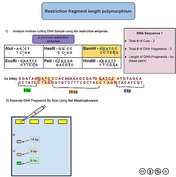 Figure: An example of DNA analysis using RFLP.
Principle of RFLP
A restriction enzyme is molecular scissors with a fixed target. It cuts DNA only at a specific short sequence, called its recognition site. For example, the enzyme EcoRI cuts only where it reads GAATTC. Give it a stretch of DNA, and it cuts at every GAATTC it finds and nowhere else. The number and spacing of those sites decide how many fragments you get and how long each one is.
Now change one base. If a mutation, insertion, or deletion changes the DNA so that a recognition site is destroyed or created, the cutting pattern changes with it.
There are two ways this happens:
- A cut site is lost. The enzyme no longer cuts at that spot. Two fragments that used to be separate now stay joined as one longer fragment.
- A cut site is gained. The enzyme now cuts at a new spot. One long fragment is split into two shorter fragments.
Either way, the total lengths of the fragments change. Because a gel separates DNA by size, these length changes appear as a changed pattern of bands. That changed pattern is the readout. You are not reading the DNA sequence directly. You are reading the sizes of the pieces the enzyme leaves behind, and using those sizes to infer what the sequence must have been.
This is why the difference must sit inside or very close to a recognition site to be seen. A mutation in the middle of a fragment, far from any cut site, does not change any fragment length, so RFLP cannot detect it. RFLP sees only the differences that add, remove, or move a cut site.
One more point that trips up many students. A "polymorphism" here is simply a normal, common difference in DNA sequence between individuals. It is not the same as a harmful mutation. Most RFLP differences fall in non-coding DNA and cause no disease at all. They are still useful, because a harmless difference that sits close to a disease gene can be tracked as a marker for that gene, even when the actual disease-causing change is unknown.
A worked example: sickle cell anemia and MstII
Sickle cell anemia is the cleanest example of RFLP reasoning, and it appears again and again in exams.
The disease comes from a single base change in the β-globin gene. At codon 6, an A is replaced by a T (GAG becomes GTG). This one change swaps glutamic acid for valine in the β-globin chain, and that is what produces hemoglobin S.
Here is the useful coincidence. That exact spot lies inside the recognition site of the restriction enzyme MstII, which cuts at the sequence CCTNAGG (N is any base). In the normal gene, MstII cuts here. The A-to-T change in the sickle allele destroys this cut site, so MstII no longer cuts at that position.
Follow the fragment sizes:
- Normal allele (A): MstII cuts at this internal site, giving two fragments of about 1.15 kb and 0.2 kb.
- Sickle allele (S): the internal cut site is gone, so those two fragments stay joined as one 1.35 kb fragment.
Now read the gel by genotype:
- A healthy person (AA) has two bands: 1.15 kb and 0.2 kb.
- A person with sickle cell anemia (SS) has one band: 1.35 kb.
- A carrier with sickle cell trait (AS) has all three bands: 1.35, 1.15, and 0.2 kb.
Because the carrier shows both patterns at once, the inheritance is called co-dominant at the DNA level. Reading the band pattern tells you the genotype directly, without needing a blood sample from the fetus. This was a major advance, because it allowed prenatal diagnosis from fetal DNA rather than from risky fetal blood sampling.
Two cautions worth remembering. First, the loss of the MstII site does not cause sickle cell disease. It is a marker that happens to travel with the mutation, because the mutation sits inside the site. Second, most modern sickle cell testing uses PCR-RFLP or direct sequencing rather than the original Southern blot method, but the reasoning about gained and lost cut sites is exactly the same.
Steps of Restriction Fragment Length Polymorphism
Restriction Fragment Length Polymorphism (RFLP) involves several steps:
- DNA Extraction: DNA is extracted from the biological sample of interest, such as blood, tissue, or saliva. The extracted DNA is then purified to remove proteins and other contaminants.
- Restriction Enzyme Digestion: The second step is cleaving of extracted DNA into fragments using restriction enzymes. These enzymes recognize particular DNA sequences and cleave the DNA at or near these recognition sites. Different restriction enzymes have different recognition sequences, allowing researchers to generate a specific pattern of DNA fragments.
- Gel Electrophoresis: Gel electrophoresis helps in the separation of the digested DNA fragments by size. The DNA fragments are loaded into wells in a porous gel, and an electric current is applied. The negatively charged DNA molecules move through the gel towards the positively charged electrode. Smaller DNA fragments move more quickly through the gel than larger fragments, resulting in separation based on size.
- Denaturation and Transfer (Optional): In some cases, the separated DNA fragments are denatured (separated into single strands) and transferred onto a solid support such as a nylon membrane in a process called Southern blotting. This step is optional and often facilitates further analysis, such as hybridization with DNA probes.
- Hybridization (Optional): If Southern blotting is performed, the membrane containing the transferred DNA fragments may be hybridized with DNA probes. These probes are single-stranded DNA molecules complementary to specific target sequences within the DNA fragments. Hybridization allows for the detection of specific DNA sequences of interest.
- Visualization and Analysis: The DNA fragments are visualized using staining techniques or autoradiography after gel electrophoresis or hybridization. The resulting pattern of DNA bands represents the unique fragment sizes present in the sample. The analysis of this pattern can help to identify genetic variations or polymorphisms among individuals.
- Data Interpretation: Interpreting the pattern of DNA fragments from RFLP analysis helps infer genetic information, such as the presence or absence of specific alleles, genetic mutations, or relationships between individuals.
Applications of RFLP
The value of RFLP comes from one property: the band pattern is inherited, stable, and specific to an individual or an allele. That makes it useful anywhere you need to track a piece of DNA through families, populations, or samples. The main uses are below.
- Genetic Mapping: RFLP analysis is applicable to construct genetic maps by identifying polymorphic markers (RFLPs) inherited along with genes of interest. By analyzing the segregation of these markers in families or populations, geneticists can determine the relative positions of genes on chromosomes.
- Linkage Analysis: RFLPs serve as genetic markers to track the inheritance of particular alleles within families. By analyzing the co-inheritance of RFLP markers with a disease phenotype or a trait of interest, researchers can identify regions of the genome linked to the trait and potentially contain the gene(s) responsible.
- Forensic Analysis: RFLP analysis has been historically useful in forensic investigations to compare DNA samples from crime scenes with those of suspects or victims. By analyzing RFLP patterns, forensic scientists can determine whether the DNA samples match, providing evidence for or against a suspect’s involvement in a crime.
- Disease Association Studies: RFLP analysis can be helpful to investigate the association between specific DNA sequences (alleles) and disease susceptibility. Researchers can distinguish genetic markers associated with diseases such as cancer, cardiovascular disorders, and genetic syndromes by comparing the frequency of particular RFLP alleles in affected individuals versus healthy controls.
- Population Genetics: RFLP analysis is applicable to study genetic diversity and evolutionary relationships among populations. Researchers can infer population structure, migration patterns, and evolutionary history by comparing RFLP patterns across different populations.
- Plant and Animal Breeding: RFLP analysis is utilized in breeding programs to select desirable traits in plants and animals. By identifying RFLP markers linked to traits such as disease resistance, yield, or quality, breeders can use marker-assisted selection to accelerate the breeding process and develop improved varieties.
- DNA Fingerprinting: RFLP was historically helpful in DNA fingerprinting for individual identification. By analyzing RFLP patterns at specific loci in an individual’s DNA, unique profiles can be generated for forensic or paternity testing purposes.
RFLP, PCR-RFLP, and DNA fingerprinting: how they relate
These three terms are often mixed up. They are connected but not the same.
Classic RFLP (Southern blot based). The original method. You need a large amount of good-quality DNA. You cut the whole genomic DNA with a restriction enzyme, separate the fragments on a gel, transfer them to a membrane by Southern blotting, and detect the fragments of interest with a labeled probe. It works without knowing the DNA sequence in advance, but it is slow, labor-intensive, and needs a lot of DNA.
PCR-RFLP. The modern version. Instead of cutting the whole genome, you first use PCR to copy just the small region you care about. Then you cut that PCR product with a restriction enzyme and run it on a gel. Because PCR makes millions of copies, you need only a tiny amount of starting DNA, no Southern blot or probe is needed, and the result comes in hours instead of days. Most tests that people still call "RFLP" today are actually PCR-RFLP. The sickle cell test above is now usually done this way.
DNA fingerprinting. This is an application, not a separate cutting method. Early DNA fingerprinting used RFLP to compare highly variable regions of the genome (such as VNTRs, variable number tandem repeats) between individuals. Because these regions differ so much from person to person, the band pattern is nearly unique, like a fingerprint. This is what was used in early forensic casework and paternity testing. Modern DNA fingerprinting has almost entirely moved to PCR-based STR (short tandem repeat) typing, which is faster and needs far less DNA, but the founding idea came from RFLP.
A one-line way to hold it together: RFLP is the principle (differences in cut sites give differences in fragment length), PCR-RFLP is the fast modern way to apply it to one target, and DNA fingerprinting is one famous use of that principle to tell individuals apart.
Advantages of Restriction Fragment Length Polymorphism
While RFLP analysis is primarily supplanted by more modern techniques such as PCR and DNA sequencing, it still offers particular advantages in specific contexts:
- High Information Content: RFLP analysis can simultaneously provide information on many loci across the genome. This allows for assessing genetic variation at multiple genomic regions in a single experiment.
- Stability and Reproducibility: RFLP patterns are stable and reproducible, making them reliable markers for genetic studies and population analyses. Once established, RFLP assays can be performed consistently over time and across different laboratories.
- No Prior Sequence Knowledge Required: RFLP analysis does not rely on sequence information, unlike PCR-based methods that require prior sequence knowledge for primer design. This makes it particularly useful for studying species with poorly characterized genomes or detecting unknown genetic variants.
- Long DNA Fragments: RFLP analysis can accommodate relatively long DNA fragments compared to PCR-based techniques. This makes it helpful in detecting significant structural variations such as insertions, deletions, and rearrangements in the genome.
- Cost-Effectiveness for Low-Throughput Analysis: In some cases, particularly for low-throughput work, RFLP analysis can be more cost-effective than PCR-based methods, which need more expensive reagents and equipment.
- Visual Readout: RFLP patterns can be visualized directly on agarose gels following electrophoresis, allowing straightforward interpretation without requiring specialized equipment or software.
- Historical Significance: RFLP analysis has played a crucial role in the history of molecular genetics and has contributed to many seminal discoveries in the field. While it is less commonly used today, its historical importance in molecular genetics is well established.
Disadvantages of RFLP
While restriction fragment length polymorphism (RFLP) analysis is a valuable tool in molecular biology, it also comes with several limitations and disadvantages, which have contributed to its declining use in favor of more advanced techniques like PCR and DNA sequencing:
- Labor-Intensive: RFLP analysis involves multiple steps, including DNA extraction, restriction enzyme digestion, gel electrophoresis, and visualization of DNA fragments. Each step requires time and labor, making the technique relatively labor-intensive compared to newer methods.
- Low Sensitivity: RFLP analysis may have lower sensitivity than PCR-based methods, particularly when detecting rare genetic variants or analyzing samples with limited DNA quantities. This limitation can hinder the detection of subtle genetic differences.
- Requirement for High-Quality DNA: RFLP analysis requires relatively large amounts of high-quality DNA, which may only sometimes be available, especially in samples of degraded or low-purity DNA. Additionally, RFLP analysis may be sensitive to DNA degradation, which can affect the quality of results.
- Limited Multiplexing: RFLP analysis is generally not conducive to multiplexing, i.e., simultaneous analysis of multiple genetic markers in a single reaction. Each RFLP assay typically targets a single genetic locus, limiting throughput and efficiency compared to multiplex PCR or sequencing approaches.
- Limited Availability of Suitable Restriction Enzyme Sites: The effectiveness of RFLP analysis depends on the presence of sites that recognize restriction enzymes within the DNA sequence of interest. In some cases, suitable restriction sites may be rare or absent, limiting the applicability of RFLP analysis.
How to Remember
The whole technique in one image: a train with a missing station. The enzyme is a train that stops only at its own stations (recognition sites). A mutation that removes a station means the train runs straight through, so two short trips become one long trip. That longer trip is the longer fragment, and the longer band on the gel. Remove a cut site, get a longer fragment. Add a cut site, get shorter fragments.
Sickle cell band count: "S has less, so S sticks together." The sickle allele loses a cut site, so its fragments stick together into one long band. SS = one band. AA = two bands. AS = both, so three bands. More mutant, fewer bands.
Polymorphism is not mutation. A polymorphism is just a common, usually harmless difference. It becomes useful when it happens to sit next to something you care about.
Key exam facts in one table
| Point | Fact |
|---|---|
| Full form | Restriction Fragment Length Polymorphism |
| Core principle | Differences in restriction enzyme cut sites produce fragments of different lengths |
| What creates the difference | A mutation, insertion, or deletion that adds, removes, or moves a recognition site |
| Cut site lost | Fewer cuts, so a longer fragment (bands fuse) |
| Cut site gained | More cuts, so shorter fragments (a band splits) |
| Detection | Gel electrophoresis separates fragments by size; Southern blot plus a probe detects the target |
| Sickle cell enzyme | MstII, recognition site CCTNAGG |
| Sickle cell fragments | Normal (A): 1.15 kb + 0.2 kb. Sickle (S): single 1.35 kb (internal site lost) |
| Sickle cell band pattern | AA = 2 bands, SS = 1 band, AS = 3 bands (co-dominant) |
| Cannot detect | A change that does not affect any cut site |
| Modern version | PCR-RFLP: amplify the target first, then cut. Needs far less DNA, no blot |
| Main limits | Slow, labor-intensive, needs large amounts of good-quality DNA, poor multiplexing |
Where Students Get Confused
"Does RFLP read the DNA sequence?" No. RFLP reads fragment sizes, not the sequence itself. You infer the sequence difference from the change in band pattern. Sequencing reads the actual bases; RFLP only reports whether cut sites were gained or lost.
"Is a polymorphism the same as a mutation?" Not quite. A polymorphism is a common difference in DNA between individuals, and most are harmless. A mutation is any change, and may or may not be common or harmful. RFLP detects both, as long as they affect a cut site. The confusion matters because RFLP markers are often harmless polymorphisms used to track a nearby disease gene, not the disease change itself.
"Why does losing a cut site give a longer band, not a shorter one?" Because a cut site is where the enzyme breaks the DNA. Remove the break, and the two pieces that would have separated stay joined as one longer piece. Fewer cuts always means longer fragments.
"RFLP vs PCR-RFLP vs DNA fingerprinting." RFLP is the underlying principle. PCR-RFLP is the fast modern method that amplifies the target first, then cuts, so it needs very little DNA. DNA fingerprinting is a use of the principle to tell individuals apart, and modern fingerprinting has mostly moved to PCR-based STR typing.
"Why does RFLP miss some mutations?" Because it only sees changes at or very near a recognition site. A base change in the middle of a fragment, far from any cut site, leaves every fragment length unchanged, so RFLP is blind to it.
References
- Jarcho J. (2001). Restriction fragment length polymorphism analysis. Current Protocols in Human Genetics, Chapter 2, Unit 2.7. https://doi.org/10.1002/0471142905.hg0207s01
- Narayanan S. (1991). Applications of restriction fragment length polymorphism. Annals of Clinical and Laboratory Science, 21(4), 291–296.
- Chang J.C., Kan Y.W. (1982). A sensitive new prenatal test for sickle-cell anemia. New England Journal of Medicine, 307(1), 30–32.
- National Human Genome Research Institute. Restriction Fragment Length Polymorphism (RFLP). Genome.gov Talking Glossary of Genomic and Genetic Terms. https://www.genome.gov/genetics-glossary/Restriction-Fragment-Length-Polymorphism
- Green M.R., Sambrook J. (2012). Molecular Cloning: A Laboratory Manual (4th ed.). Cold Spring Harbor Laboratory Press.
- Tille P.M. (2022). Bailey & Scott's Diagnostic Microbiology (15th ed.). Elsevier.
Frequently Asked Questions
What is the full form of RFLP?
What is the full form of RFLP?
RFLP stands for Restriction Fragment Length Polymorphism. The name describes exactly what it measures: differences (polymorphism) in the length of DNA fragments produced when restriction enzymes cut DNA.
What is the basic principle of RFLP?
What is the basic principle of RFLP?
A restriction enzyme cuts DNA only at its specific recognition sequence. If a mutation, insertion, or deletion changes that sequence, a cut site can be lost or gained. Losing a cut site joins two fragments into one longer fragment; gaining a cut site splits one fragment into two shorter ones. These length differences show up as different band patterns on a gel, and that pattern is the result you read.
What is the difference between RFLP and PCR-RFLP?
What is the difference between RFLP and PCR-RFLP?
Classic RFLP cuts the whole genomic DNA and usually needs a Southern blot and a probe to detect the target, so it requires a large amount of good-quality DNA and takes days. PCR-RFLP first uses PCR to copy only the small region of interest, then cuts that product with a restriction enzyme. It needs only a tiny amount of DNA, no blot or probe, and gives results in hours. Most tests still called "RFLP" today are actually PCR-RFLP.
How is RFLP used in DNA fingerprinting?
How is RFLP used in DNA fingerprinting?
Early DNA fingerprinting used RFLP to compare highly variable regions of the genome, such as VNTRs (variable number tandem repeats), between individuals. Because these regions differ greatly from person to person, the band pattern is almost unique, which allowed identification in forensic and paternity testing. Modern DNA fingerprinting has largely shifted to PCR-based STR typing, but the original idea came from RFLP.
Why is RFLP used to diagnose sickle cell anemia?
Why is RFLP used to diagnose sickle cell anemia?
The sickle cell mutation (an A-to-T change at codon 6 of the β-globin gene) happens to fall inside the recognition site of the enzyme MstII (CCTNAGG). The mutation destroys that cut site. So the normal allele gives fragments of 1.15 kb and 0.2 kb, while the sickle allele gives a single 1.35 kb fragment. Reading the band pattern shows the genotype: AA gives two bands, SS gives one band, and AS gives all three bands.
Can RFLP detect every mutation?
Can RFLP detect every mutation?
No. RFLP only detects changes that add, remove, or move a restriction enzyme cut site. A base change in the middle of a fragment, far from any cut site, does not change any fragment length, so RFLP cannot see it. This is a key limitation compared with direct DNA sequencing.
Is a polymorphism the same as a mutation?
Is a polymorphism the same as a mutation?
Not quite. A polymorphism is a common difference in DNA sequence between individuals, and most are harmless. A mutation is any change to the DNA, which may be rare or disease-causing. Many RFLP markers are harmless polymorphisms that sit near a disease gene and are used to track it, rather than being the disease-causing change themselves.
What are the main advantages and disadvantages of RFLP?
What are the main advantages and disadvantages of RFLP?
Its advantages are that it needs no prior sequence knowledge, gives stable and reproducible patterns, and can be read directly from a gel. Its disadvantages are that it is slow and labor-intensive, needs large amounts of good-quality DNA, does not multiplex well, and depends on a suitable restriction site being present. These limits are why PCR-based methods and sequencing have largely replaced it.

Tankeshwar Acharya, MSc (Medical Microbiology)
Tankeshwar Acharya is an Assistant Professor in the Department of Microbiology at Patan Academy of Health Sciences (PAHS), Nepal, where he has been teaching and practicing clinical microbiology for over 14 years. He is the founder of Microbe Online, one of the leading free microbiology education resources on the web, covering bacteriology, mycology, parasitology, immunology, and clinical laboratory diagnostics written from direct experience in both the classroom and the diagnostic laboratory.
Comments
No comments yet. Be the first to share your thoughts.
Leave a comment
All comments are reviewed before they appear.