Polyacrylamide Gel Electrophoresis (PAGE): Principle and Procedure
Polyacrylamide gel electrophoresis (PAGE) separates proteins by size. Learn why SDS is added, why the gel has a stacking and a resolving layer, how to choose the acrylamide percentage, and how SDS-PAGE underpins the Western blot.
Look at the names of two HIV proteins: p24 and gp41. Students memorize them as arbitrary labels. They are not. The number in each name is the protein's molecular weight in kilodaltons. p24 is a 24 kDa protein. gp41 is a 41 kDa glycoprotein. Proteins across microbiology and immunology are named this way, and the naming comes directly from the technique in this article.
When a protein is run on an SDS-PAGE gel, it does not separate by charge, because a detergent has coated every protein with the same charge. It does not separate by shape, because it has been reduced and boiled into a straight chain. It separates by one thing only: size. Large proteins crawl; small ones race ahead. Run a protein of unknown size beside a ladder of known sizes, and the distance it travels tells you its molecular weight.
That is the whole point of SDS-PAGE. It turns a mixed sample of proteins into a ruler. Every reagent in the protocol below exists to make one variable, size, the only variable that matters. Understand why each reagent is there and the procedure stops being a list to memorize.
This is also the first step of a Western blot: proteins are separated by size on the gel, transferred to a membrane, and probed with antibody. It is why the bands on a Western blot sit where they do, and it remains everyday practice, from Lyme disease serology to routine protein work in any laboratory that runs a Coomassie gel.
What is polyacrylamide gel electrophoresis (PAGE)?
PAGE is a vertical slab gel technique that separates proteins and small nucleic acids by driving them through a cross-linked polyacrylamide mesh under an electric field. The gel acts as a molecular sieve: smaller molecules pass through the pores easily and travel far, while larger molecules are held back.
In its most common form, SDS-PAGE, the detergent sodium dodecyl sulfate coats every protein with a uniform negative charge and a reducing agent unfolds it. Charge and shape are removed as variables, so proteins separate by molecular weight alone, and the distance a band travels is inversely proportional to the logarithm of its molecular weight.
Electrophoresis is a method used to separate macromolecules according to their charge, size, and shape under an electric field. Depending on the format in which the separation is carried out, it may be performed as a slab gel technique or inside a narrow tube, as in capillary electrophoresis. Polyacrylamide gel electrophoresis (PAGE) is a slab gel technique, run in a vertical configuration.
Polyacrylamide gel electrophoresis is a form of gel electrophoresis used in molecular biology, forensic chemistry, genetics, biochemistry, and biotechnology to separate biological macromolecules, primarily proteins and small nucleic acids, according to their electrophoretic mobility. It uses a cross-linked polyacrylamide mesh as the support matrix, which gives a much finer sieving action, and therefore much higher resolution, than agarose.
The most commonly used polyacrylamide gel electrophoresis for quantitative protein analysis is Sodium dodecyl sulfate-polyacrylamide gel electrophoresis (SDS-PAGE).
Polyacrylamide Gel
The polyacrylamide gel forms by polymerizing acrylamide and a crosslinking agent, i.e., N, N’-methylene-bis-acrylamide. It does not react with proteins and consists of pores and channels that allow the protein to move through it.
Two parameters characterize a polyacrylamide gel: the total monomer concentration (%T, in g/100 ml) and the weight percentage of cross-linker (%C). Varying these two parameters regulates the pore size of the gel, which is how the gel is matched to the size range being separated. The relationship is inverse: a higher %T produces smaller pores, which resolve smaller molecules better.
Principle of Polyacrylamide Gel Electrophoresis (PAGE)
Polyacrylamide gel electrophoresis is based on the principle that charged particles migrate to the electrode of the opposite sign under the influence of an electric field.
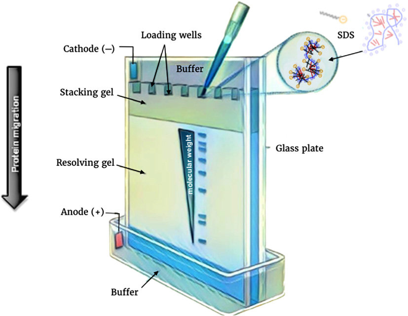 Figure: Principle of polyacrylamide gel electrophoresis, Image source: DOI: 10.1016/B978-0-12-803077-6.00012-6
In polyacrylamide gel electrophoresis, the sample (usually protein) is dissolved in a loading buffer containing denaturing agents (sodium dodecyl sulfate and β-mercaptoethanol), glycerol, and bromophenol blue. Sodium dodecyl sulfate is an anionic detergent. It denatures the protein and binds along the polypeptide chain at approximately one SDS molecule per two amino acid residues
Therefore, the negative charge of SDS results in a net negative charge in the protein sample. Similarly, β-mercaptoethanol also denatures the protein sample by cleaving the disulfide bond.
Glycerol increases the density of the sample so that it sinks to the bottom of the well during loading, rather than floating away into the running buffer. Bromophenol blue is a small, fast-migrating tracking dye. It does not stain the protein. It runs ahead of nearly all proteins as a visible dye front, so the operator can see how far the run has progressed and stop it before the smallest proteins run off the end of the gel.
When the electric current passes through the electrophoretic chamber, the protein-sodium dodecyl sulfate complex starts moving toward the anode. Gel percentage is chosen to match the size of the proteins being separated. A low-percentage gel (4 to 8% acrylamide) has large pores, so large proteins can enter it and spread out, while small proteins run through almost unimpeded and are poorly resolved. A high-percentage gel (12 to 20% acrylamide) has small pores that hold back large proteins near the top of the gel, and resolve small proteins sharply.
Materials Required for PAGE
Various materials are required to conduct polyacrylamide gel electrophoresis, which include;
- Polyacrylamide gel: It is the matrix that helps to separate proteins based on their size. It can be either prepared in the lab or can be purchased.
- Running buffer: It varies based on sample type. Its primary function is to allow the conduction of current across the gel.
- Protein or nucleic acid sample: The primary sample needed to separate based on its molecular weight.
- Staining and de-staining reagent: Coomassie stain solution is mainly helps to stain protein bands after electrophoresis. In contrast, the de-staining reagent (prepared by mixing methanol, acetic acid, and water) helps to de-stain the gel.
Essentials of electrophoresis:
- Gel plate: It is the plate that holds the polymerized gel in an electrophoresis chamber.
- Comb: A toothed plate inserted into the liquid stacking gel before it sets. Once the gel polymerizes, the comb is removed, leaving behind the wells into which samples are loaded.
- Electrophoresis chamber: Polyacrylamide gel is packed within a running buffer, and electrophoresis is carried out.
- Protein ladder: It is a reference protein ladder with known size. It helps to confirm the molecular weight of the protein of interest.
- Power supply: It supports converting AC to DC, which is essential to create an electric field.
Procedure involved in PAGE
The procedure involved while operating polyacrylamide gel electrophoresis is;
Sample preparation
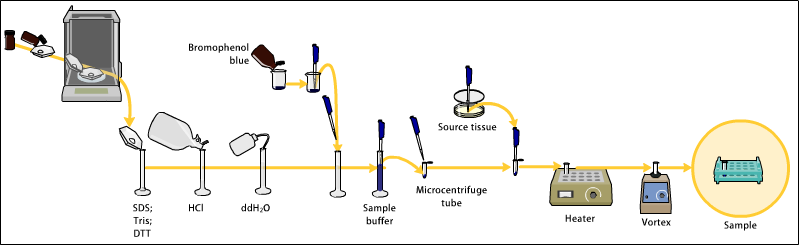 Figure: Sample preparation
- The sample can be either protein or nucleic acid.
- The sample is mixed with a loading buffer containing denaturing agents (SDS and β-mercaptoethanol), glycerol, and bromophenol blue. For nucleic acids, urea is used as the denaturing agent instead of SDS.
- Heating the samples with denaturing agents and mercaptoethanol for 5-10 minutes further enhances the denaturation.
Preparation of polyacrylamide gel
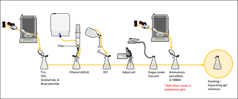 Figure: Preparing of the polyacrylamide gel
- The gel mixture comprises acrylamide, bis-acrylamide, a buffer at the appropriate pH, and an optional denaturant (SDS for protein, urea for nucleic acid).
- Polymerization requires two further reagents. Ammonium persulfate (APS) is the free-radical initiator, and TEMED (N,N,N',N'-tetramethylethylenediamine) is the catalyst that accelerates radical formation. Both are added last, immediately before pouring, because the gel begins to set as soon as they are mixed in.
- Two gels are cast. The separating (resolving) gel at pH 8.8 is poured first, between the glass plates. Water-saturated isobutanol is gently layered on top of it. This gives a flat gel surface and, importantly, excludes atmospheric oxygen, which inhibits acrylamide polymerization.
- Once the separating gel has set, the isobutanol is poured off and the stacking gel at pH 6.8 is poured on top. The comb is inserted into the still-liquid stacking gel.
- After the stacking gel polymerizes, the comb is withdrawn, leaving wells. The assembled unit is now called a gel cassette.
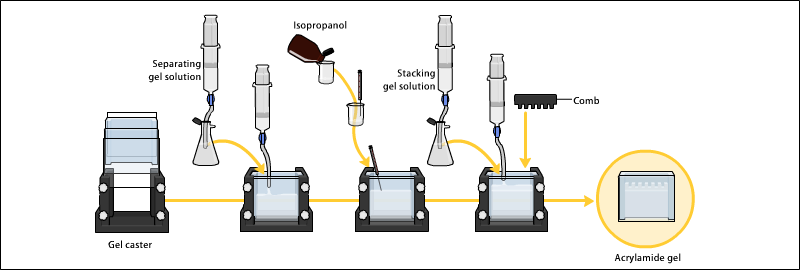 Figure: Preparation of gel cassette
Electrophoresis
- Depending on the sample type, the use of different buffer systems can help to run polyacrylamide gel electrophoresis.
- The buffer used at the cathode or anode might be the same or different.
- The gel cassette is removed from the casting stand, placed in the electrode assembly, and secured in the clamp stand.
- Then, 1X running buffer is poured into the electrophoresis chamber.
- Each well is then loaded with a protein sample. Similarly, marker protein is also loaded into a single well of gel.
- The tank is then covered with a lid, and the sample can run at 30mA for about 1 hour.
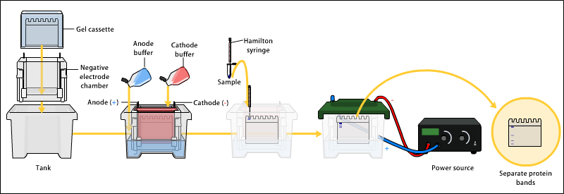 Figure: Running of polyacrylamide gel electrophoresis
Detection
Following electrophoresis, the gel is stained so the bands become visible and can be compared against the ladder. Coomassie brilliant blue is used for proteins. Ethidium bromide has traditionally been used for nucleic acids, but it is a mutagen, and safer intercalating stains such as SYBR Gold or GelRed are now preferred where available.
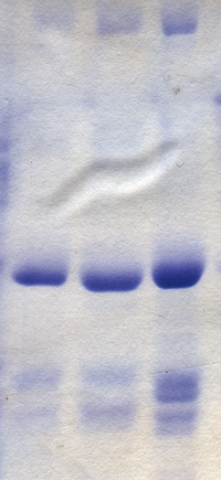 Figure: Polyacrylamide gel after staining with Coomassie blue
Applications of Polyacrylamide Gel Electrophoresis (PAGE)
Polyacrylamide gel electrophoresis is applicable in a wide range of that includes;
- It helps to determine the purity of samples.
- It is also helpful in determining the molecular weight of protein.
- PAGE helps to quantify the proteins.
- It helps in monitoring changes in protein in body fluids.
- PAGE is useful in peptide mapping.
- It is useful to estimate the purity of the protein and nucleic acid.
- SDS-PAGE is the first step of every Western blot. Proteins are separated by size on the gel, transferred onto a membrane, and then probed with a specific antibody. The band's position on the gel is what identifies the protein.
- It is useful for the detection of protein ubiquitination.
- PAGE is helpful to analyze the size and number of polypeptide subunits.
- It underpins Western blot serology. In the historical HIV confirmatory Western blot, patient antibodies were detected against viral proteins separated by SDS-PAGE, and the proteins are named for the molecular weights at which they resolve: p24 (24 kDa capsid protein), gp41 and gp120 (41 and 120 kDa envelope glycoproteins). The HIV algorithm has since replaced Western blot with an antigen/antibody immunoassay followed by an HIV-1/HIV-2 differentiation assay, but Western blot remains standard in the two-tier serology of Lyme disease, and SDS-PAGE remains its first step.
Advantages of Polyacrylamide Gel Electrophoresis (PAGE)
Polyacrylamide gel electrophoresis consists of the following benefits:
- It helps to determine the molecular weight, as migration is directly proportional to the molecular weight.
- It is a sensitive technique, capable of detecting small quantities of protein.
- It can provide results even with a small amount of sample.
- It consists of chemically crosslinked stable gel.
- The sample recovered from the gel is exceptionally pure.
- It is best for separating proteins of low molecular weight.
Disadvantages of Polyacrylamide Gel Electrophoresis (PAGE)
Despite the benefits, polyacrylamide gel electrophoresis has some drawbacks which are as follows:
- Unpolymerized acrylamide monomer is a potent neurotoxin and a probable human carcinogen. It is absorbed through the skin, so it must be handled with gloves, and powder must be weighed with care to avoid inhaling dust.
- Preparation of gel is time-consuming, and electrophoresis also takes a longer time.
- It requires a higher budget to operate.
- The preparation of a new gel for each test is necessary.
Precautions of Polyacrylamide Gel Electrophoresis (PAGE)
While performing polyacrylamide gel electrophoresis one should consider following precautionary measure:
- Routine care should be exercised in handling buffers and samples to avoid accidental ingestion, needle stick, etc.
- Acrylamide and bisacrylamide are highly toxic monomers of polyacrylamide gel. Therefore, the use of gloves for handling polyacrylamide gel.
- A precise amount of polyacrylamide monomers should be weighed to obtain desired pore size.
- Always wear gloves, goggles, and a lab coat while handling samples and buffers.
Differences Between Agarose Gel and Polyacrylamide Gel Electrophoresis (PAGE)
Although both techniques are used for separation purposes, they have some differences. The differences between agarose gel electrophoresis and polyacrylamide gel electrophoresis are given below;
| Polyacrylamide gel electrophoresis (PAGE) | Agarose gel electrophoresis |
|---|---|
| In polyacrylamide gel electrophoresis, polyacrylamide gel separates macromolecules, i.e., proteins of size five kDa to 250 kDa. Similarly, it can also isolate DNA of 5- 500 bp size. | In agarose gel electrophoresis, agarose gel separates DNA, RNA, and protein. It can isolate DNA about 50-20,000 bp in size. |
| The run configuration of polyacrylamide gel electrophoresis is vertical. | The run configuration of agarose gel electrophoresis is horizontal. |
| Unpolymerized acrylamide monomer is a potent neurotoxin and probable carcinogen. The polymerized gel is far safer, but may contain residual monomer, so gloves are worn. | Agarose is non-toxic in both powder and gel form. |
| It gives better resolution than agarose gel. | It gives poor resolution than polyacrylamide gel. |
| Gels must be cast between glass plates by chemical polymerization, which takes time, and the finished gel is thin and fragile. | Gels are poured from melted agarose in minutes, are physically robust, and the agarose can be re-melted and recast. |
| It requires two layers of gel, i.e., staking and separating gel. | It only requires a single layer of agarose gel. |
References
- Laemmli UK. Cleavage of structural proteins during the assembly of the head of bacteriophage T4. Nature. 1970;227(5259):680-685. doi:10.1038/227680a0
- Wilson K, Walker J. Principles and Techniques of Biochemistry and Molecular Biology. 8th ed. Cambridge: Cambridge University Press; 2018. Chapter: Electrophoretic Techniques.
- Westermeier R. Electrophoresis in Practice: A Guide to Methods and Applications of DNA and Protein Separations. 5th ed. Weinheim: Wiley-VCH; 2016.
- Ornstein L. Disc electrophoresis I: Background and theory. Annals of the New York Academy of Sciences. 1964;121(2):321-349.
- Davis BJ. Disc electrophoresis II: Method and application to human serum proteins. Annals of the New York Academy of Sciences. 1964;121(2):404-427.
Frequently Asked Questions
Why is SDS added in SDS-PAGE?
Why does an SDS-PAGE gel have two layers?
What is the difference between native PAGE and SDS-PAGE?
How do I choose the acrylamide percentage?
Which direction do proteins move in SDS-PAGE, and why?
What is the role of β-mercaptoethanol, and how is it different from SDS?
What do APS and TEMED do?
Is polyacrylamide gel toxic?
What is the tracking dye in SDS-PAGE?
How is SDS-PAGE related to the Western blot?

Tankeshwar Acharya, MSc (Medical Microbiology)
Tankeshwar Acharya is an Assistant Professor in the Department of Microbiology at Patan Academy of Health Sciences (PAHS), Nepal, where he has been teaching and practicing clinical microbiology for over 14 years. He is the founder of Microbe Online, one of the leading free microbiology education resources on the web, covering bacteriology, mycology, parasitology, immunology, and clinical laboratory diagnostics written from direct experience in both the classroom and the diagnostic laboratory.