Phenotypic Methods for the Detection of Carbapenemases
Phenotypic Methods for the Detection of Carbapenemases
The enzyme carbapenemase can be detected by various phenotypic and molecular methods. Phenotypic methods for carbapenemases detection are preferred over molecular techniques because they are rapid, affordable, accurate, and feasible for implementation in clinical microbiology laboratories of all sizes.
Screening for carbapenem resistant organism (CRO)
For detecting the presence of carbapenemase enzyme, first, we need to know whether the isolate is a suspected carbapenemase producer or not. An organism is defined as a suspected carbapenemase producer if it shows reduced susceptibility to carbapenems either by disc diffusion or MIC. CLSI defines isolates with MICs of ≤1 μg/ml for meropenem or imipenem, and MICs of ≤0.5 μg/ml for ertapenem as carbapenem-resistant organism.
Overview of phenotypic assays
Phenotypic assays currently used in clinical practice consist of the following:
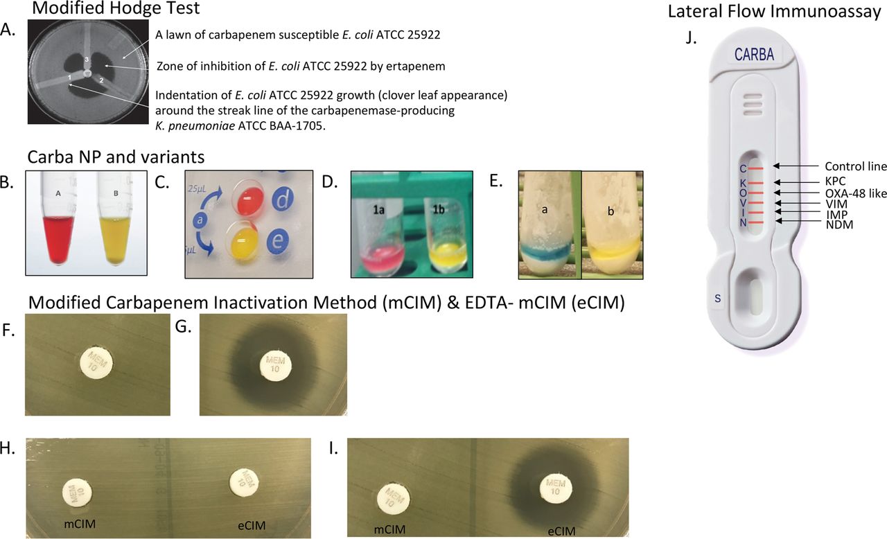
- Growth-based assays which measure resistance based on growth in the presence of an antibiotic (e.g., modified Hodge test [MHT]) and modified carbapenem inactivation method [mCIM])
- Hydrolysis methods which detect the product of hydrolysis that is catalyzed by carbapenemase enzymes (e.g., Carba NP, targeted carbapenemase assays, and matrix-assisted laser desorption–ionization-time of flight mass spectrometry [MALDI-TOF MS] methods); and
- Lateral flow immunoassays detect carbapenemase enzymes through the use of specific antibodies.
Modified Hodge test (MHT)
The modified Hodge test (MHT) is probably the most widely used approach for carbapenemase detection. It involves streaking of a clinical isolate in a line away from an ertapenem or meropenem disk which placed previously on an agar plate inoculated with a lawn of a carbapenem-susceptible Escherichia coli strain.
The MHT relies on the ability of carbapenemase producers to decrease the local concentration of carbapenem antibiotics, enabling the carbapenem-susceptible E. coli to grow uninhibited around the streak line near the carbapenem disk, producing a cloverleaf appearance.
Carbapenem inactivation method
Meropenem disk (10-µg) is placed on a suspension (containing 10 µL loop full of test organism suspected of producing carbapenemase) and incubated for 2 hours at 35°C. If the test organism produces carbapenemase, the meropenem will be hydrolyzed. Alternatively, if the organism does not produce a carbapenemase, the meropenem disc retains its activity.
The meropenem disk is then removed and placed on a Mueller-Hinton agar plate streaked with a susceptible laboratory strain of E. coliand incubated overnight.
Following incubation, the isolate is considered as a carbapenemase producer if there is no zone of inhibition (i.e. the meropenem was hydrolyzed). In contrast, the presence of a zone of inhibition indicates that the meropenem in the disk has preserved its activity and the isolate is not producing a carbapenemase.
Carba NP test
The Carba NP test detects carbapenemases by measuring the in vitro hydrolysis of imipenem in bacterial extracts and produces color changes within approximately 2hour. Imipenem hydrolysis results in a carboxylic derivative, which in turn decreases the pH, producing a resultant color shift of a phenol red indicator from red to yellow.
Targeted carbapenemase assays
Targeted phenotypic carbapenemase assays compare carbapenem activity with and without the presence of inhibitors. Certain variations include
- Synergism with phenylboronic acid [PBA]: detect KPC (or other class A serine carbapenemases)
- Synergism with phenylboronic acid [PBA] and cloxacillin: detect AmpC carbapenemase
- Synergism with EDTA: for detection of Metallo beta-lactamases (MBLs)
Targeted carbapenemase assays can be valuable during outbreaks or when considering treatment options with activity against select carbapenemases.
MALDI-TOF MS method
Two major applications of MALDI-TOF MS for the rapid identification of carbapenemase production are being pursued.
- Hydrolysis approach: It detects carbapenem degradation products when bacterial protein extracts are incubated with a carbapenem substrate.
- Plasmid-associated peak approach: It involves the detection of a known carbapenemase-bearing plasmid-associated protein peak.
Lateral flow immunoassay
Lateral flow immunoassays (LFIAs) are antibody-based methods to identify the presence of carbapenemases. A number of LFIAs have been recently developed but generally enable the detection of one or a few of the most epidemiologically important carbapenemases family such as KPC, NDM, VIM, IMP, and OXA-48-like carbapenemases. Available data suggest that LFIAs produce accurate results from cultured isolates within 15 min.
The procedure involves suspending a single bacterial colony from a Mueller-Hinton agar plate in 150 µl of extraction buffer. 100 µl of this extract is loaded on a cassette, and results are read within 15 min of migration, based on the presence of visible lines indicating a positive test.
Factors influencing the choice of methods
The selection of a carbapenemase detection test is dependent upon several factors, including Local carbapenemase prevalence
- Regional molecular epidemiology
- Diagnostic performance characteristics
- Labor intensity (ease of use, workflow, and reagent preparation requirements)
- Availability of necessary equipment such as MALDI TOF
- Cost
- Turnaround time (TAT) of the test: The TAT is important both for therapeutic decision-making and infection control purposes, with same-day results being ideal.
- Organisms to be tested (i.e., Enterobacteriaceaeand/or glucose-non fermenting Gram-negatives)
Unfortunately, no single assay has a favorable profile for all of the criteria listed above. Laboratory has to choose and implement method that best suits their needs.
References and further readings:
- Tamma PD, Simner PJ. 2018. Phenotypic detection of carbapenemase producing organisms from clinical isolates. J Clin Microbiol 56:e01140-18. https://doi.org/10.1128/JCM.01140-18.
- Workneh M, Rebecca Yee R, Simner P J 2019.Phenotypic Methods for Detection of Carbapenemase Production in Carbapenem-Resistant Organisms: What Method Should Your Laboratory Choose? Clinical Microbiology Newsletter Volume 41, Issue 2,15 January 2019, Pages 11-22 https://doi.org/10.1016/j.clinmicnews.2019.01.001
- Sun K, Xu X, Yan J, Zhang L. Evaluation of Six Phenotypic Methods for the Detection of Carbapenemases in Gram-Negative Bacteria with Characterized Resistance Mechanisms. Ann Lab Med. 2017 Jul; 37(4):305-312. doi: 10.3343/alm.2017.37.4.305. PMID: 28445009; PMCID: PMC5409027
- Clinical and Laboratory Standards Institute. 2020. Performance standards for antimicrobial susceptibility testing, 30th ed. CLSI supplement M100. Clinical and Laboratory Standards Institute, Wayne, PA

Tankeshwar Acharya, MSc (Medical Microbiology)
Tankeshwar Acharya is an Assistant Professor in the Department of Microbiology at Patan Academy of Health Sciences (PAHS), Nepal, where he has been teaching and practicing clinical microbiology for over 14 years. He is the founder of Microbe Online, one of the leading free microbiology education resources on the web, covering bacteriology, mycology, parasitology, immunology, and clinical laboratory diagnostics written from direct experience in both the classroom and the diagnostic laboratory.