
Kato-Katz Technique: Principle, Procedure, EPG Calculation, and Results
How to perform the Kato-Katz technique for helminth diagnosis: principle, step-by-step procedure, egg-per-gram (EPG) calculation, WHO infection intensity thresholds, and when to use it over formal-ether concentration.

On this page
A public health officer in a rural district of Bangladesh needs to know whether the school deworming program is working. Before treatment, surveys showed 65% of children infected with Ascaris lumbricoides. Six months after mass albendazole administration, she needs objective data: not just "are children still infected?" but "how intensely infected are they?" Did treatment reduce worm burden or merely clear some infections?
The Kato-Katz technique answers this second question. It is not simply another method for detecting parasites. It is the only field-suitable technique that quantifies infection intensity, expressing results as eggs per gram (EPG) of stool. This makes it the backbone of WHO soil-transmitted helminth surveillance and the standard tool for assessing treatment program effectiveness globally.

Kato Katz technique is used for qualitative and semi-quantitative diagnosis of intestinal helminthic infestations; caused by Ascaris lumbricoides, Trichuris trichiura, hookworm, and especially Schistosoma spp.
WHO has recommended the Kato Katz technique in areas with moderate to high transmission rates of soil-transmitted helminths (i.e. where the proportion of infected individuals is >20– >50%) or intestinal schistosomiasis (>10–50%). Where the prevalence of soil-transmitted helminths (STH) is <20%, the specificity of this technique makes it less appropriate, and more sensitive tools should be used.
Direct wet mount is the commonly used method for the diagnosis of intestinal parasitic infections but its sensitivity is low. Formal ether concentration (FEC) technique is used to increase the sensitivity of the microscopic detection methods.
WHO Infection Intensity Thresholds (Eggs per Gram of Stool)
The Kato-Katz EPG count classifies infection intensity according to WHO criteria. This classification determines treatment decisions in endemic areas:
| Species | Light infection (EPG) | Moderate infection (EPG) | Heavy infection (EPG) |
|---|---|---|---|
| Ascaris lumbricoides | 1–4,999 | 5,000–49,999 | ≥50,000 |
| Trichuris trichiura | 1–999 | 1,000–9,999 | ≥10,000 |
| Hookworm (Ancylostoma/Necator) | 1–1,999 | 2,000–3,999 | ≥4,000 |
| Schistosoma mansoni | 1–99 | 100–399 | ≥400 |
| Schistosoma japonicum | 1–99 | 100–399 | ≥400 |
Clinical relevance: Heavy infections are associated with clinically significant disease: iron-deficiency anemia and protein malnutrition from heavy hookworm, bowel obstruction from heavy Ascaris, and rectal prolapse from heavy Trichuris. Light infections are usually asymptomatic. This is why EPG is not just academic. It identifies the children who need urgent treatment and nutritional support.
Principle
People infected with STH or intestinal schistosomes pass the eggs of the worms through their feces. In the Kato-Katz technique, feces are pressed through a mesh screen to remove large particles. A portion of the sieved sample is then transferred to the hole of a template on a slide. After filling the hole, the template is removed and the remaining sample is covered with a piece of cellophane soaked in glycerol. The glycerol clears the fecal material from around the eggs. The eggs are then counted and the number is calculated per gram of feces.
Materials
- Kato-set (template with hole, screen, nylon or plastic, plastic spatula)
- Newspaper or glazed tile
- Microscope slides
- Cellophane as coverslip, soaked in a glycerol-malachite green or glycerol-methylene blue solution.
- Fresh stool
- Gloves
Procedure of Kato-Katz Technique
- Label a glass slide with the sample number and then place a plastic template on top of it.
- Place a small amount of the fecal sample on a newspaper and press a piece of nylon screen on top. Using a spatula, scrape the sieved fecal material through the screen so that only the debris remains.
- Scrape up some of the sieved feces to fill the hole in the template, avoiding air bubbles and leveling the feces off to remove any excess.
- Carefully lift off the template and place it in a bucket of water mixed with concentrated detergent so that it can be reused.
- Place one piece of the cellophane, which has been soaked overnight in methylene blue glycerol solution, over the fecal sample.
- Place a clean slide over the top and press it evenly downwards to spread the feces in a circle. Carefully remove the slide by gently sliding it sideways to avoid separating the cellophane strip. If done well, it should be possible to read newspaper print through the stool smear. Place the slide with the cellophane upwards.
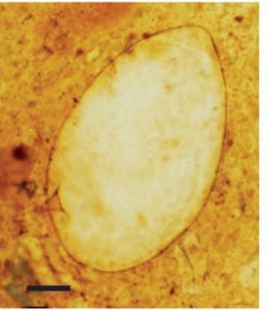 Figure: S mansoni egg in Kato-Katz Preparation
⚠️ Critical time limit for hookworm: If hookworm is suspected, examine Kato-Katz slides within 30–60 minutes of preparation. Hookworm eggs are thin-shelled and the glycerol-cellophane clearing process dissolves their shell contents over time, making them unrecognizable after 1 hour. Slides prepared and left overnight will give false-negative results for hookworm specifically. Ascaris, Trichuris, and Schistosoma eggs are stable and can be read the following day.
Examination and Results
- Place the slide under a microscope and examine the whole area in a systematic zigzag pattern.
- Record the number and the type of each egg of each species on a recording form alongside the sample number.
- Finally, multiply the number of eggs by the appropriate number (see inlet-information of the kato-set) to give the number of eggs per gram (epg) – the standard measurement to assess the intensity of infection.

EPG Calculation: Converting Egg Count to Eggs Per Gram
The Kato-Katz template holds a standardized volume of stool equivalent to 41.7 mg.
Formula:
EPG = Eggs counted on slide × 24
(The multiplier 24 = 1000 mg ÷ 41.7 mg, converting from "eggs per 41.7 mg" to "eggs per gram)
Worked example:
- You count 35 Ascaris eggs on a Kato-Katz slide
- EPG = 35 × 24 = 840 EPG
- WHO classification: light infection (1–4,999 EPG for Ascaris)
- Clinical interpretation: likely asymptomatic; eligible for mass drug administration program but not urgent individual treatment
If using a commercial kit: The kit insert specifies the exact multiplier for that template size; always check, as some kits use templates of slightly different volumes (e.g., 50 mg templates use a multiplier of 20).
Where Students Actually Get Confused
1. "Kato-Katz detects protozoa." Kato-Katz is designed for helminth eggs only. The glycerol-malachite green cellophane technique is optimized for clearing and staining eggs. Protozoan cysts are not visible. Never use Kato-Katz as the sole method when Giardia, Entamoeba, or other protozoa are suspected.
2. "Any stool can be used for Kato-Katz." The technique requires fresh formed or semi-formed stool. Liquid stool cannot be pressed through the mesh screen correctly and the template cannot be filled properly. Preserved (formalin or PVA-fixed) stool is also unsuitable; the eggs may be distorted and EPG quantification is unreliable. Fresh stool is mandatory for valid EPG counts.
3. "The slide can be read at any time." Only for Ascaris, Trichuris, and Schistosoma. For hookworm, slides must be read within 30–60 minutes.
4. "EPG from one slide gives the true worm burden." EPG is an estimate, not a direct worm count. Egg output varies with sex (female worms produce eggs; male worms don't), worm age, and stool consistency. For population surveys, duplicate Kato-Katz slides from each specimen are recommended to improve precision. For individual diagnosis, a single slide is sufficient.
5. "Kato-Katz can be used in any endemic area." WHO specifically recommends Kato-Katz where STH prevalence is >20% or schistosomiasis prevalence is >10%. Below these thresholds, the specificity-sensitivity balance favors other methods. In very low prevalence settings, multiple Kato-Katz slides or formal-ether concentration gives better yield per effort.
Key Exam Facts
| Fact | Detail | Memory hook |
|---|---|---|
| Template volume | 41.7 mg of stool | Standardized, enables EPG calculation |
| EPG multiplier | ×24 (for standard 41.7 mg template) | 1000 ÷ 41.7 = 24 |
| Stain/clearing agent | Glycerol-malachite green (or methylene blue) cellophane | Glycerol clears fecal debris; dye stains background |
| Detects | Helminth eggs only | NOT protozoa |
| Hookworm reading time | Within 30–60 minutes | Thin-shelled eggs clear and disappear |
| Ascaris/Trichuris/Schistosoma | Can read next day | Thick-shelled eggs stable |
| WHO use threshold (STH) | Prevalence >20% in a community | Below 20%, other methods preferred |
| WHO use threshold (schistosomiasis) | Prevalence >10% | |
| Heavy hookworm EPG | ≥4,000 EPG | Anemia risk |
| Heavy Ascaris EPG | ≥50,000 EPG | Bowel obstruction risk |
| Specimen type | Fresh stool only | Preserved specimens give unreliable EPG |
| Screens/templates | Reusable after cleaning | Cost-effective for field surveys |
Frequently Asked Questions
What is the Kato-Katz technique used for?
The Kato-Katz technique is used for qualitative and quantitative diagnosis of intestinal helminthic infections, specifically soil-transmitted helminths (Ascaris lumbricoides, Trichuris trichiura, hookworm) and Schistosoma species.
It presses a standardized volume of stool (41.7 mg) through a mesh screen onto a slide, covers it with glycerol-malachite green cellophane, and allows microscopic identification and counting of helminth eggs. The egg count is multiplied by 24 to give eggs per gram (EPG) of stool, a measure of infection intensity.
Why must Kato-Katz slides for hookworm be read within 30-60 minutes?
Hookworm eggs have thin shells that are progressively dissolved by the glycerol in the cellophane during the clearing process. After 60 minutes, the shell contents become unrecognizable, and the eggs appear as empty outlines or disappear entirely.
Ascaris, Trichuris, and Schistosoma eggs have thicker shells and are stable for up to 24 hours, but hookworm diagnosis requires immediate slide reading.
How is EPG calculated from a Kato-Katz slide?
EPG (eggs per gram) = number of eggs counted on the slide × 24. The multiplier 24 comes from dividing 1,000 mg (1 gram) by the template volume of 41.7 mg. For example, if you count 50 Ascaris eggs, EPG = 50 × 24 = 1,200 EPG, which classifies as a light infection (WHO threshold: light = 1–4,999 EPG for Ascaris). Always check the kit insert, some templates use slightly different volumes.
Reference
- World Health Organization. (2012). Bench aids for the diagnosis of intestinal parasites (2nd ed.). WHO. https://apps.who.int/iris/bitstream/handle/10665/37323/9789241544764_eng.pdf
- World Health Organization. (2011). Helminth control in school-age children: a guide for managers of control programmes (2nd ed.). WHO. https://www.who.int/publications/i/item/9789241548267
- Katz, N., Chaves, A., & Pellegrino, J. (1972). A simple device for quantitative stool thick-smear technique in schistosomiasis mansoni. Revista do Instituto de Medicina Tropical de São Paulo, 14(6), 397–400.
- Garcia, L. S. (2016). Diagnostic Medical Parasitology (6th ed.). ASM Press.

Tankeshwar Acharya, MSc (Medical Microbiology)
Tankeshwar Acharya is an Assistant Professor in the Department of Microbiology at Patan Academy of Health Sciences (PAHS), Nepal, where he has been teaching and practicing clinical microbiology for over 14 years. He is the founder of Microbe Online, one of the leading free microbiology education resources on the web, covering bacteriology, mycology, parasitology, immunology, and clinical laboratory diagnostics written from direct experience in both the classroom and the diagnostic laboratory.
Comments
No comments yet. Be the first to share your thoughts.
Leave a comment
All comments are reviewed before they appear.