Cell Biology
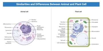
Similarities and Differences Between Plant and Animal Cells
Similarities and Differences Between Plant and Animal Cells

Posts related to cell biology
This page contains all posts in the Cell Biology category.
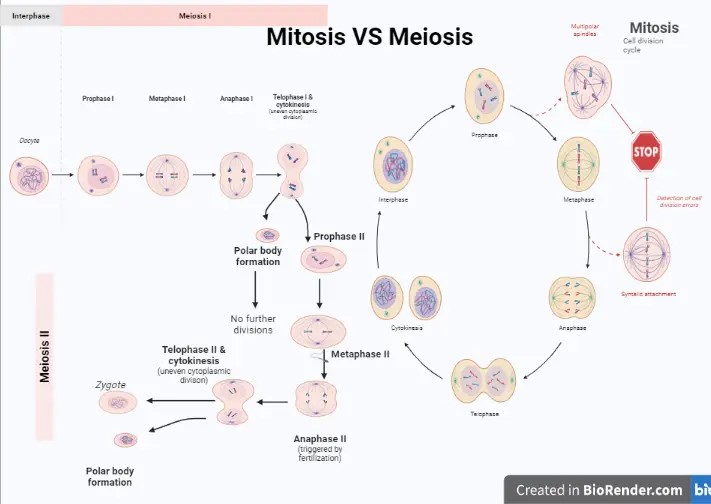
Cell Division: Mitosis and Meiosis
Similarities and Differences Between Plant and Animal Cells
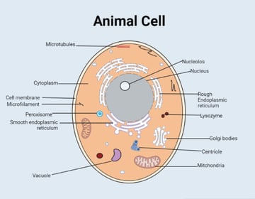
Animal Cell: Structural Components and Types
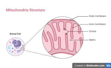
Mitochondria, Structure, Functions, and Location