
How a Colorimeter Measures Concentration: Principle, Parts, Types, and Filter Selection

On this page
A student sets up a colorimeter to measure a blue copper sulfate solution, loads a blue filter because the solution is blue, and gets an absorbance reading so low it barely lifts off the blank. The dilution series that should climb steadily stays almost flat. Nothing is wrong with the instrument or the samples. The filter is the error. A blue solution absorbs least where it looks blue and most in the red-orange band it is subtracting from the light. Reading a colored solution is not about matching the filter to the color you see. It is about measuring where the solution actually absorbs, and that single decision, made before any sample goes in, determines whether the numbers mean anything.
A colorimeter is a laboratory instrument that measures how much light of a selected color a solution absorbs, and uses that absorbance to determine the concentration of the colored compound. A more concentrated solution absorbs more light, and within a working range absorbance rises in direct proportion to concentration. This proportionality is the basis of colorimetry, described by the Beer-Lambert law.
A colorimeter is a photometric device that measures light in the visible range of the electromagnetic spectrum. It suits applications that do not need fine spectral resolution, because it works across only a few broad bands of the visible range, unlike more precise photometric instruments. Common uses include measuring ink color in forensic science, beverage color in the food industry, and the growth density of yeast and bacterial cultures in the microbiology laboratory.
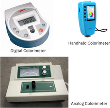 Figure: Different types of colorimeter
Principle of Colorimeter
The principle is colorimetry: a colored solution is analyzed by how much visible light it absorbs. Light of a chosen color passes first through a filter, then through the solution held in a cuvette. The light leaving the solution is less intense than the light that entered, because the colored compound absorbs part of it. The more concentrated the solution, the more light is absorbed, and within a working range this absorbance is directly proportional to concentration. That direct proportionality is what allows a color reading to stand in for a concentration.
The Beer-Lambert law is the theory behind this. It states that the absorbance of a solution is directly proportional to the concentration of the absorbing compound and to the path length the light travels through it. Transmittance, the fraction of light that passes through, falls as concentration rises, which is why absorbance and transmittance run in opposite directions.
Instruments often read percent transmittance (%T). This is converted to absorbance, also called optical density, so that measurements relate directly and linearly to concentration:
Absorbance (A) = log (100 / %T)
Absorbance is directly proportional to both the concentration of the solution (C) and the path length the light travels (L):
A ∝ CL, or A = εCL
where ε is the molar absorptivity, a constant for a given compound at a given wavelength.
Because the cuvette path length is fixed and molar absorptivity is constant for a given compound, absorbance depends only on concentration. In practice you do not calculate concentration from ε directly. You read the unknown off a standard curve of absorbance against known concentrations, run on the same instrument with the same filter and cuvette. Two limits matter: the proportionality holds only over a working range, so the standard curve bends at high concentration, and it assumes a constant path length, which is why cuvettes must be filled and matched consistently.
Choosing the right filter
A colorimeter measures the light a solution removes, not the light it lets through, so the filter you choose must pass the color the solution absorbs. That color is the complement of the color the solution appears. A blue solution is read through a red or orange filter, a red solution through a green filter, a yellow solution through a blue filter. Choosing a filter that matches the solution's own color sends in light the solution barely absorbs, which produces almost no signal. This is the flat, meaningless reading in the copper sulfate example above.
| Solution appears | Color it absorbs | Filter to use |
|---|---|---|
| Blue | Red-orange | Red or orange |
| Green | Red | Red |
| Yellow | Blue | Blue |
| Red | Green | Green |
| Violet | Yellow-green | Yellow-green |
Parts of Colorimeter
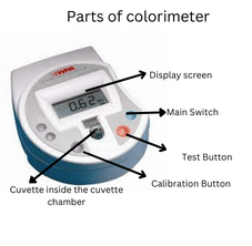 Figure: Parts of colorimeter
The essential parts of the colorimeter include a light source, cuvette chamber or sample container, cuvette, filter, detector, and galvanometer.
- Light source: In the colorimeter, it produces light energy of the required intensity throughout the visible spectrum, 380-780 nm. The light source used in the laboratory equipment is a simple tungsten lamp that provides light in the visible range.
- Cuvette chamber or sample container: It is the area in the equipment where cuvettes or container is held. It is present at the topmost part of the equipment.
- Cuvette: The sample is held in a transparent container called a cuvette, made of optical glass or clear plastic. Cuvettes are rectangular or square cells with an exact, known path length, usually 10 or 20 mm, though other path lengths are available. The fixed path length is what makes measurements precise and reproducible. A common cuvette is about 45 mm high and holds roughly 4 mL. Test tubes are a cheaper alternative but lack the accuracy and precision of a matched cuvette, since their path length is neither exact nor consistent.
- Filter: The filter selects the band of visible light that reaches the solution, and it is what defines an instrument as a colorimeter rather than a spectrophotometer. A colorimeter uses fixed optical filters that pass a fairly broad band of wavelengths, chosen to match the color the sample absorbs. The common filter types are gelatin, glass, and interference filters. A spectrophotometer, by contrast, replaces the filter with a monochromator (a prism or diffraction grating) that selects a narrow, tunable wavelength, which is why it is more precise and can scan across the spectrum.
- Gelatin filters: It is formed by sandwiching a thin layer of colored gelatin between two thin glass plates. These are cost-effective filters but can absorb 30-40% of all incident radiation, decreasing the detectors' energy throughput.
- Glass filters: Another type of filter is colored glass filters with wide band passes up to 150 nm. Specific wavelengths are achieved by combining different glass filters.
- Interference filter: It comprises many reflecting but semi-transmitting films of silver separated by thin layers of transparent dielectric material. When white light passes through the dielectric layers, multiple reflections occur between the semi-transparent mirrors. Some energy passes straight through the filter as the desired wavelength for analysis. The thickness of the dielectric layer determines the resulting wavelength.
- Detector: The detector, also called a photocell, converts the transmitted light leaving the sample into an electrical signal. The type depends on the material used. The common detectors are the selenium photocell, phototube, and silicon photocell.
- Selenium photocell: It is the simplest type of detector and does not require any power supplies for functioning.
- Phototube: It is made up of a glass bulb coated with photosensitive materials like cesium or potassium.
- Silicon photocell: It generates electrons when a photon of light strikes the semi-conductive surface of the silicon photocell.
- Galvanometer: It measures the electric signal generated by the detectors and displays the value in the display area.
- Display: It can be analog or digital. The analog appears like meters and is calibrated in absorbance along with a supplementary percent transmission scale. The digital display indicates an absorbance value identical to the resolution despite the change in the magnitude of data.
Types of Colorimeter
Colorimeters are divided into various types based on its size and the filters used in the instrument. On the basis of size colorimeters are of two types: benchtop and portable colorimeters. Whereas based on the filters used, it is of two types: tristimulus and densitometer.
Based on size
- Benchtop colorimeter: It is slightly larger and requires a benchtop for operating. It comes in the wavelength range 420-660 nm. It is highly accurate and consumes only 1.5 mL of reagent. It is suitable in analysis of compounds in different laboratories.
- Portable/handheld colorimeter: It is a compact device that can be easily carried outside laboratory setting. It is useful in food analysis and water analysis at outdoor settings. The wavelength range offered by most manufacturers is 420 to 660 nm.
Based on filters
- Tristimulus colorimeter: Uses three filters to measure the intensity of the three primary colors, red, green, and blue (RGB). This is the most common filter arrangement for measuring perceived color, as in paint, textile, and food color matching.
- Densitometer colorimeter: Uses a single filter to measure the intensity of one color. It is useful for measuring the density of bacterial and yeast growth in microbiology.
A spectrophotometer is sometimes grouped alongside colorimeters, but it is a distinct instrument: instead of fixed filters it uses a monochromator (prism or grating) to select a narrow, tunable wavelength, which makes it more precise and able to scan across the spectrum. Read more about spectrophotometer here
Based on the display
- Analog colorimeter: The display area of the analog colorimeter has scale. The upper number scale denotes the transmittance, whereas the lower scale represents the absorbance. The change in the arrow head’s placement indicates the absorbance and transmittance.
- Digital colorimeter: The digital colorimeter displays on an LED screen. The absorbance and % transmittance displays in digits. The digital colorimeter is quickly replacing analog colorimeters.
Operating a Colorimeter
Steps of operating colorimeter
- Select the required filter.
- Calibrate the colorimeter.
- Fill two-thirds of the cuvette with the desired sample solution.
- Slide the lid of the cuvette chamber and place the cuvette with the sample inside it.
- Press the T button or test button to start testing the sample solution.
- Observe the absorbance in the display area.
How to calibrate a colorimeter?
- Fill two-thirds of the cuvette with distilled water.
- Slide the lid of the cuvette chamber and place the cuvette inside it.
- Press the CAL button (labeled R on some instruments) in the colorimeter until the LED light flashes.
- When the LED light stops flashing, the calibration completes, and the absorbance shown in the display should be 0.00 (100% transmittance).
- Now, remove the cuvette and start analyzing test samples.
Things to consider
- Pre-heat the instrument for about 5 minutes before use.
- Calibration after every filter change is a must.
- Fill the cuvette two-thirds to three-fourths for good transmittance.
- Placing the cuvette in the right way is also essential.
- Cover the cuvette with a lid before the run to reduce the risk of spillage.
- Match the filter to the color the solution absorbs, which is the complement of the color it appears, not the color itself.
Uses of Colorimeter
The colorimeter is used in various fields of science as well as non-science for measuring the concentration of solutions or density of the solution. The following are the uses of colorimeter based on the areas:
- In the clinical laboratory, a colorimeter is used to analyze urine, plasma, serum, and cerebrospinal fluids for biochemical studies.
- Studying the growth density of bacterial and yeast cultures using a densitometer colorimeter is very helpful in the microbiology laboratory.
- The concentration of food preservatives and harmful toxins in food industries is analyzed using colorimeters.
- Quality control in various laboratories, like water quality in the water supply area quality of drugs produced by pharmaceutical industries, is analyzed using a colorimeter.
- In the textile and paint industries, to analyze different colors.
- In forensic science, a colorimeter helps in analyzing different samples.
Advantages of Colorimeter
- It is low-cost equipment.
- A colorimeter is easy to repair and maintain.
- It is a simple instrument to use.
- The handheld colorimeter is very helpful for on-site analysis.
Disadvantages of Colorimeter
- Some surfaces can reflect the light hindering the specificity of the equipment.
- It does not work in ultraviolet and infrared rays.
- The colorimeter is not applicable for colorless substances.
How to Remember
Complementary filter. The filter fights the color. You do not match the solution's color, you oppose it. Ask yourself the one-line check: "What color does this solution remove from white light?" That removed color is your filter. Blue solution removes red-orange, so a red filter goes in.
Beer-Lambert direction. More stuff, more blocked, higher absorbance. Concentration and absorbance move together; concentration and transmitted light move opposite. If someone quotes transmittance, remember it runs backward.
Colorimeter vs. spectrophotometer. Filter versus grating. A colorimeter picks a color with a filter (crude, fixed). A spectrophotometer picks a wavelength with a grating (fine, tunable). Filter = fewer choices; grating = a continuous dial.
Key exam facts in one table
| Concept | Fact to remember |
|---|---|
| Working principle | Beer-Lambert law: within range, absorbance is directly proportional to concentration at a fixed path length |
| What is measured | Absorbance (light removed by the colored solution), not the color you see |
| Filter selection | Filter is complementary to the solution's color (blue solution, red or orange filter) |
| Light source | Tungsten filament lamp for the visible range |
| Wavelength selector | Optical filter (broad band), unlike a spectrophotometer's monochromator |
| Detector | Photocell or photodiode converting transmitted light to current |
| Filter-based types | Tristimulus (three RGB filters) and densitometer (single filter); a spectrophotometer is a separate instrument, not a colorimeter type |
| Colorimeter vs. spectrophotometer | Filter (broad band, fixed) versus monochromator (narrow band, tunable); the spectrophotometer is more precise and can scan wavelengths |
| Range limitation | Linearity fails at high concentration; the standard curve bends |
| Path length | Must stay constant; use matched cuvettes filled consistently |
Where Students Get Confused
"Match the filter to the solution color." The single most common error. You choose the complementary color, because the instrument measures absorbed light, not transmitted light. Matching the color sends in light the solution ignores.
Absorbance versus transmittance. They are inversely related. High absorbance means low transmittance. A concentrated solution gives high absorbance and low transmittance; students often flip this.
"A colorimeter and a spectrophotometer are the same thing." They share the Beer-Lambert basis, but wavelength selection differs (filter versus monochromator), and that difference sets precision, range, and whether you can scan a spectrum.
Why the standard curve must be run every time. Absorbance is only proportional to concentration within a range and for a given setup. You read unknowns off a curve built from known standards on the same instrument with the same filter and cuvette, not from a memorized value.
The blank is not optional. Zeroing on a blank (reagent without the analyte) subtracts absorbance from the solvent, reagents, and cuvette so the reading reflects only the analyte. Skipping it shifts every value.
References
Skoog, D. A., Holler, F. J., & Crouch, S. R. (2018). Principles of Instrumental Analysis (7th ed.). Cengage Learning.
Sherwood Scientific Ltd. (n.d.). A guide to colorimetry. Retrieved from https://archive-resources.coleparmer.com/Manual_pdfs/Sherwood/Chroma%20Colorimeters/Manuals/A%20Guide%20to%20Colorimetry.pdf
Vernier Science Education. (n.d.). Colorimeter (COL-BTA) user manual. Retrieved from https://www.vernier.com/manuals/col-bta/
Frequently Asked Questions
Why does a colorimeter use a filter complementary to the solution's color?
Why does a colorimeter use a filter complementary to the solution's color?
Because it measures the light the solution absorbs, not the light it transmits. A solution absorbs most strongly in the color complementary to the one it appears, so a filter passing that complementary color gives the strongest, most useful signal. A blue solution is read through a red or orange filter.
What is the principle of a colorimeter?
What is the principle of a colorimeter?
It is based on the Beer-Lambert law. Within a working range and at a fixed path length, the absorbance of a colored solution is directly proportional to the concentration of the absorbing compound. The colorimeter measures absorbance and converts it to concentration using a standard curve.
What is the difference between a colorimeter and a spectrophotometer?
What is the difference between a colorimeter and a spectrophotometer?
Both apply the Beer-Lambert law, but they select wavelength differently. A colorimeter uses fixed optical filters that pass a broad band of light; a spectrophotometer uses a monochromator (prism or grating) that selects a narrow, tunable wavelength. The spectrophotometer is more precise, can work in the UV range, and can scan across wavelengths, while a colorimeter is limited to its filters and the visible range.
Why do you need to zero the colorimeter with a blank?
Why do you need to zero the colorimeter with a blank?
The blank contains everything except the analyte. Zeroing on it subtracts the absorbance contributed by the solvent, reagents, and cuvette, so the final reading reflects only the compound being measured.
Can a colorimeter measure a colorless solution?
Can a colorimeter measure a colorless solution?
Not directly. The analyte must absorb visible light. Colorless compounds are measured after a reagent converts them into a colored product, then read at the appropriate complementary filter.
Why does the standard curve bend at high concentration?
Why does the standard curve bend at high concentration?
The Beer-Lambert law holds only over a limited range. At high concentration, absorbance stops rising in proportion (from molecular interactions and stray light effects), so the line flattens and readings there are unreliable. Dilute the sample back into the linear range.

Tankeshwar Acharya, MSc (Medical Microbiology)
Tankeshwar Acharya is an Assistant Professor in the Department of Microbiology at Patan Academy of Health Sciences (PAHS), Nepal, where he has been teaching and practicing clinical microbiology for over 14 years. He is the founder of Microbe Online, one of the leading free microbiology education resources on the web, covering bacteriology, mycology, parasitology, immunology, and clinical laboratory diagnostics written from direct experience in both the classroom and the diagnostic laboratory.
Comments
No comments yet. Be the first to share your thoughts.
Leave a comment
All comments are reviewed before they appear.