Blood cells: Types and Their Functions
Blood cells: Types and Their Functions
Blood is a fluid connective tissue that transports oxygen and nutrients to the cells and carries away carbon dioxide and other waste products. It is composed of a clear, straw-colored watery fluid known as plasma in which different types of blood cells are suspended.
Blood cells are different types of cells found in the blood but formed in bone marrow by a process known as hematopoiesis. These constitutes about 45% of the volume of blood.
Types of blood cells
Blood cells arise from the same bone marrow stem cells. Later, the immortal, undifferentiated, pluripotent stem cells differentiate into blood cells, i.e., red blood cells, white blood cells, and platelets.
Red blood cells (RBCs)
Red blood cells are red colored and non-nucleated elements in the blood. These cells are red due to the presence of coloring substance known as hemoglobin. The location of production of red blood cells are in bone marrow. The hormone erythropoietin controls the production of RBCs. The other terms for red blood cells are erythrocytes, red cells, red blood corpuscles, haematids, or erythroid cells.
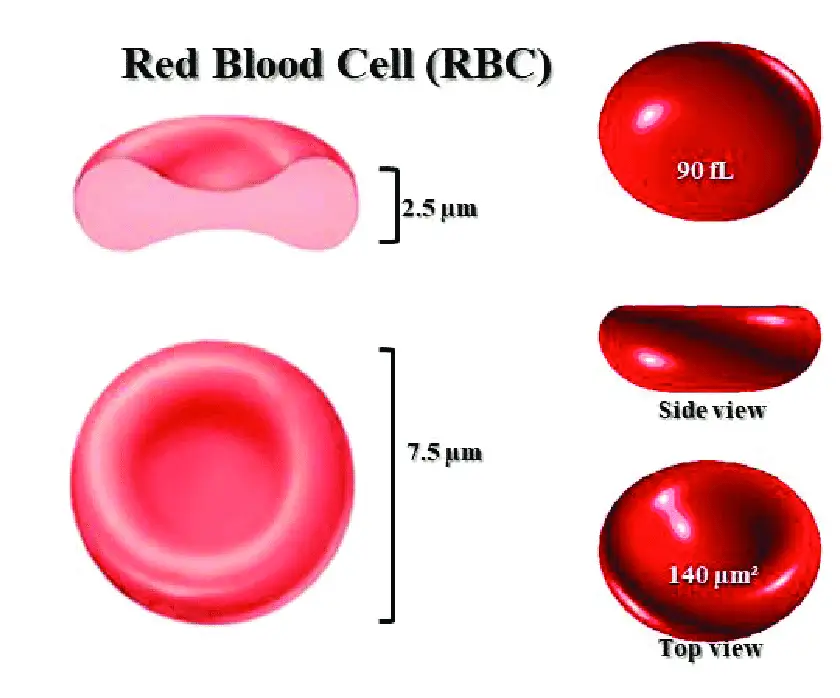
Morphology (structure) of Red blood cells
- Shaped: Normally, disc-shaped and biconcave (dumb-bell shaped).
- Size: 7.5 µm in diameter.
- Thickness: At the periphery, it is thicker with 2.5µm, and it is thinner at the center at 1µm. This difference in thickness is because of the biconcave shape.
- Surface area: 120 square µ.
- Volume: 85-90 cubic u.
- Composition: They are non-nucleated and covered with a membrane composed of protein and lipids. DNA and other cell organelles such as mitochondria and Golgi body are also absent in RBC.
Normal value of red blood cells
- Average red blood cell count is 4.5-6 million per cubic millimeter of blood.
- In adults male: 5-6 million per cubic millimeter of blood.
- In adult females:4.2-5.5 million per cubic millimeter of blood.
Life span of red blood cells
- The average life span of RBCs is about 120 days.
- After the lifespan, the old RBCs breakdown or hemolyse, carried out by macrophages in the spleen, bone marrow, and liver.
- Iron released by hemolysis returns to bonemarrow to form new hemoglobin molecules.
Functions of Red Blood Cells
- Transport of oxygen from the lungs to the tissues: Haemoglobin in the RBCs binds with oxygen to form oxyhemoglobin. About 97% of oxygen transports in the blood in the form of oxyhemoglobin.
- Transport of carbon dioxide from the tissues to the lungs: Haemoglobin combines with carbon dioxide to form carboxyhemoglobin. About 30% of carbon dioxide is transported in this form.
- Buffer function: Haemoglobin in red blood cells acts as a buffer by regulating hydrogen ion concentration and thereby plays a role in maintaining acid base balance.
- Blood group determination: Red blood cells carry the blood group antigens like A antigen, B antigen, and Rh factor. This helps in the determination of blood groups and enables the prevention of reactions due to incompatible blood transfusion.
White Blood Cells
White blood cells are the colorless and nucleated type of blood cell made in bone marrow but found in blood and lymph tissue. It is also known as leukocytes. WBCs are an essential part of the body’s immune system.
| Types of white blood cells | Normal count (per cumm of blood) | Percentage (%) |
|---|---|---|
| Neutrophils | 3,000-6,000 | 50-70 |
| Eosinophils | 150-450 | 2-4 |
| Basophils | 0-100 | 0-1 |
| Monocytes | 200-600 | 2-6 |
| Lymphocytes | 1500-2700 | 20-30 |
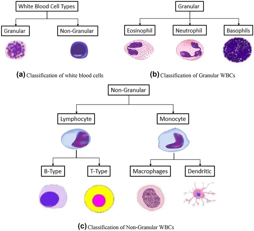
Classification of white blood cells
Based on the presence or absence of granules in the cytoplasm, the WBCs are classified into two groups: Granulocytes and Agranulocytes.
- Granulocytes: The presence of granules in the cytoplasm when stained and viewed under a microscope, granulocytes. It is further classified into three types; Neutrophils, Eosinophils, and Basophils.
- Agranulocytes: The absence of granules in the cytoplasm when stained and viewed under a microscope, known as agranulocytes. It is further classified into Monocytesand Lymphocytes.
Lymphocytes are further subdivided into T-lymphocytes and B-lymphocytes.
Morphology of white blood cells
- Neutrophils
They are mostly circular, but their shape changes into ameboid form once activated. They are the smallest among all granulocytes with a diameter of about 10-12µ. They have fine or small granules in the cytoplasm, which take acidic or basic stains. The nucleus is multilobed. The number of lobes varies. In the younger cells, the nucleus is not lobed, but the older one has 3-5 lobes.
- Eosinophils
The diameter of cells varies between 10-14 µ. They consist of larger granules in their cytoplasm, which stain red or pink with eosin. The nucleus is segmented or bilobed and spectacle shaped.
- Basophils
The diameter of the cell varies between 8-10µ. They also have larger granules in their cytoplasm, which stains purple-blue with methylene blue. The nucleus is bilobed and is ‘S-shaped.’
- Monocytes
They are the largest leukocytes, with a diameter of 14-18µ. They are motile and, on maturation, develop into macrophages and dendritic cells. The cytoplasm is clear without granules. The nucleus is round, oval, and horseshoe, bean, or kidney-shaped is placed at one side.
- Lymphocytes
They have clear cytoplasm without granules. The nucleus is oval, bean-shaped, or kidney-shaped, occupying the whole of the cytoplasm. These are the only blood cells with specific receptors for antigens. Thus they are key mediators of adaptive immunity. Although all types of lymphocytes are morphologically similar and rather unremarkable in appearance, they are extremely heterogeneous in lineage, function, and phenotype and are capable of complex biologic responses and activities. So, the use of panels of monoclonal antibodies specific to the surface proteins helps differentiate these by the surface proteins. The standard nomenclature for these proteins is the “CD” (cluster of differentiation) numerical designation, which helps to delineate surface proteins that define a particular cell type or stage of cell differentiation.
Normal count of white blood cells
- Total white blood count (TLC or TC): 4,000-11,000/cumm of blood.
- Differential white blood count is given in the table below
Life span of white blood cells
The lifespan of WBC varies, depending upon the demand of the body and its function. Lifespan can be as short as a few hours or maybe can last as long as 3 to 6 months. However, the normal lifespan of WBCs is given in the table below
Functions of white blood cells
WBCs play a crucial role in the defense mechanism of the body. Each type of WBCs functions in different ways;
- Neutrophils play a vital role in the defense mechanism of the body. Along with monocytes, it provides a first-line defense against invading microorganisms. Similarly, it also helps to boost the response of other cells.
- Eosinophils play an essential role in the host’s defense against parasites.
- Basophils play an essential role in allergy.
- Monocytes help the body’s defense mechanism by a process known as phagocytosis.
- Lymphocytesplay a major role in immunity. B lymphocytes are the only cells capable of producing antibodies; therefore, they are the cells that mediate humoral immunity. T-Lymphocytes are the cells of cell-mediated immunity. The antigen receptors of T lymphocytes only recognize peptide fragments of protein antigens bound to major histocompatibility complex (MHC) molecules, on the surface antigen-presenting cells (APCs)
Platelets
Platelets are small, colorless, non-nucleated, and moderately refractive bodies. They are produced when large cell known as megakaryocytes breaks into pieces of 2000-3000 in numbers.
Morphology of platelets
- Diameter: 2-4 µ
- Volume: 7.5 cu µ
- Shape: They are of several shapes, spherical or rod-shaped, and become oval and disk-shaped when inactivated. Sometimes, the platelets are of dumbell shape, comma shape, cigar shape, or any other unusual form.
Lifespan of platelets
- The average lifespan of platelets is 8-11 days.
- The tissue macrophage system destroys platelets in the spleen. So, splenomegaly (enlargement of the spleen) decreases platelet count, and splenectomy (removal of the spleen) increases the platelet count.
Normal count of platelets
- The normal platelet count in blood is 2,50,000/cu mm.
- Its value ranges from 1,50,000-4,00,000/cu mm in the blood.
Functions of platelets
- It helps to prevent blood loss (hemostasis) in three ways;
Platelets secrete 5HT, which causes constriction of blood vessels. The adhesive property of platelets helps to seal damaged blood vessels such as; capillaries. Platelets help seal the damaged part of the blood vessel by forming a temporary plug.
- The platelets are responsible for forming an intrinsic prothrombin activator responsible for the onset of blood clotting.
- The platelets are useful for repairing the endothelium and other structure of blood vessels.
- By the property of agglutination, platelets surround the foreign bodies and kill them through phagocytosis.
- The cytoplasm of platelets contains contractile proteins, namely actin, myosin, and thrombosthenin. These contractile proteins are responsible for clot retraction.
References
- Peter J. Delves, Seamus J. Martin, Dennis R. Burton, and Ivan M. Roitt(2017). Roitt’s Essential Immunology, Thirteenth Edition. John Wiley & Sons, Ltd.
- White blood cells. National Cancer Institute. Retrieved on 14th September 2022. Retrieved from https://www.cancer.gov/publications/dictionaries/cancer-terms/def/white-blood-cell
- Image 1 Source: Yeği̇n, Ecem & Yegin, Mehmet & Kosova, Buket. (2021). A CHRONOLOGICAL REVIEW OF BIOINFORMATICS SCIENCE IN PLASTIC SURGERY.

Tankeshwar Acharya, MSc (Medical Microbiology)
Tankeshwar Acharya is an Assistant Professor in the Department of Microbiology at Patan Academy of Health Sciences (PAHS), Nepal, where he has been teaching and practicing clinical microbiology for over 14 years. He is the founder of Microbe Online, one of the leading free microbiology education resources on the web, covering bacteriology, mycology, parasitology, immunology, and clinical laboratory diagnostics written from direct experience in both the classroom and the diagnostic laboratory.