
Yeast: Structure, Reproduction, and Uses
Yeast structure, reproduction (budding, fission, sexual life cycles), and clinical relevance. How Candida and other pathogenic yeasts relate to general yeast biology, pseudohyphae formation, and why understanding budding matters for diagnosis.

On this page
Yeast is a unicellular eukaryotic cell. It is a saprophytic fungus and is found in sugary mediums like the juice of sugarcane and sweet fruits, the nectar of flowers, etc. Yeast is larger than most bacteria. It is non-motile and does not consist of flagella or any other organ of locomotion.
Why yeast biology matters beyond baking and brewing
Saccharomyces cerevisiae is the textbook model organism for yeast structure and reproduction, and is also genuinely important industrially (baking, brewing, biofuel, as covered below). But for clinical microbiology students, understanding general yeast biology serves a more specific purpose: everything described in this article about yeast structure and budding directly explains the behavior of pathogenic yeasts you will encounter clinically, most importantly Candida albicans, the most common fungal pathogen in humans.
Candida, Cryptococcus neoformans, and other medically important yeasts share the same fundamental structure (cell wall, plasma membrane, cytoplasmic organelles) and the same primary reproductive mechanism (budding) described in this article for Saccharomyces. Understanding the general biology here is the foundation for understanding why Candida forms the structures it does under the microscope, and why those structures matter diagnostically.
Yeast reproduces by vegetative and sexual methods of reproduction. Vegetative reproduction occurs in yeast by budding or fission method. Sexual reproduction occurs in yeast by haplobiontic, diplobiontic, and haplodiplobiontic life cycle. Yeast is used mainly in the baking and brewing industry. Besides, it is used as a source of protein, biofuel, and commercially for producing different enzymes and organic compounds.
Classification
Kingdom: Fungi
Phylum: Ascomycota
Class: Saccharomycetes
Order: Saccharomycetales
Family: Saccharomycetaceae
Genus: Saccharomyces
Structure of Yeast
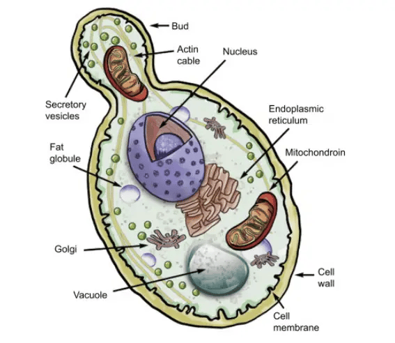 Figure: Structure of yeast (Image source: Sciencedirect)
The structure of yeast is very simple since it is a unicellular cell. Sometimes this single cell form the pseudomycelium, where the cluster of yeast cells are seen in a chain. The shape of a yeast cell is elliptical, round, or spherical. Its size is 3-15 μm in length and 2-8 μm in diameter. It is in hyaline (pearl grey) color.
It is surrounded by the cell wall, which is made up mainly of beta-glucans and mannoproteins, with chitin as a minor component concentrated at the bud scars. Just beneath the cell wall, a cytoplasmic membrane is present in the yeast cell. This membrane surrounds the cytoplasm.
The cytoplasm consists of different cell organelles like the Golgi apparatus, ribosomes, endoplasmic reticulum, mitochondria, spherosome, nucleus, and vacuole. Volutin granules are also present in the cytoplasm, but chloroplast is absent in the yeast cell. Reserve food material is present in the form of glycogen or fats.
The cell wall is worth pausing on, because it explains two things you will meet later.
- First, it is made of chitin and beta-glucans with an outer layer of mannoproteins, none of which human cells make. That difference is precisely what antifungal drugs exploit: echinocandins block beta-glucan synthesis, so they damage the fungal wall without a human target to hit.
- Second, the chitin in the wall is what binds fluorescent stains like calcofluor white and takes up the KOH preparation contrast, which is why a yeast cell wall shows up so clearly on a direct mount. The simple structure of a yeast cell, in other words, is also the reason it is stainable and druggable.
Clinically important yeasts that share this structure
| Organism | Clinical significance | Structural note |
|---|---|---|
| Candida albicans | Most common cause of candidiasis (thrush, vaginal candidiasis, invasive candidiasis) | Forms true hyphae AND pseudohyphae, unlike Saccharomyces |
| Cryptococcus neoformans | Cryptococcal meningitis, especially in HIV/AIDS | Surrounded by a prominent polysaccharide capsule which can be visualized by India ink |
| Candida auris | Emerging multidrug-resistant nosocomial pathogen | Often misidentified by automated systems; requires specific identification methods |
| Saccharomyces cerevisiae | Generally non-pathogenic; rare opportunistic infections in severely immunocompromised patients | The model organism described in this article |
Reproduction
In short: yeast reproduces mainly by budding (an asexual, vegetative process where a small outgrowth pinches off the mother cell), and some yeasts also divide by fission. Under stress, yeast can reproduce sexually by forming ascospores. The rest of this section walks through each mechanism and how it looks under the microscope.
Vegetative reproduction
Vegetative reproduction occurs in the yeast cell in the favorable condition where there is sufficient nutrition, as in a sugary solution. It takes place either by budding or fission method.
Budding (Gemmation)
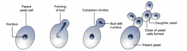 Figure: Budding in yeast cell
The most common method of reproduction in yeast is budding. During the favorable condition, it produces the bud or the small outgrowth at its end. It enlarges gradually. Then the nucleus divides into two. One nucleus is present in the mother cell, and another moves to the bud. Then constriction appears between the mother cell and bud, separating them by a transverse wall. Sometimes pseudomycelium is also formed. Pseudomycelium is the chain of the temporary bud, which is not separated from the mother cell. It looks like the mycelium, which is long and unbranched. These cells of pseudomycelium later separate and form many vegetative cells.
Why understanding budding matters for diagnosing Candida infections
The budding process described above for Saccharomyces is exactly what a microbiologist looks for when examining a wet mount, gram stain, or KOH preparation for suspected candidiasis.
The germ tube test exploits this exact biology. When Candida albicans (and C. dubliniensis) is incubated in serum at 37°C for 2–3 hours, it produces a germ tube, a true hyphal extension growing directly from the yeast cell without any constriction at the point of origin. This is structurally distinct from a bud, which always shows a visible constriction (narrowing) at the junction between mother cell and daughter cell, exactly as described in the budding section above. The presence or absence of this constriction is literally the diagnostic feature being assessed in the germ tube test.
Pseudohyphae formation is the same pseudomycelium process described above, applied clinically. When budding occurs repeatedly but daughter cells fail to fully separate from the mother cell, Candida forms pseudohyphae: chains of elongated, constricted yeast cells that resemble true hyphae but retain visible constrictions at each junction. This is a key microscopic feature used to identify Candida species in tissue sections, blood cultures, and direct smears, and it is mechanistically identical to the pseudomycelium formation described for Saccharomyces in this article.
The clinical distinction that matters:
| Structure | Constriction at junction? | Organism example | Clinical significance |
|---|---|---|---|
| Bud | Yes, clearly visible | Saccharomyces, Candida, Cryptococcus | Normal vegetative reproduction |
| Pseudohyphae | Yes, at each segment junction | Candida albicans, C. tropicalis | Tissue invasion; visible in histopathology |
| True germ tube/hyphae | No constriction | Candida albicans, C. dubliniensis ONLY | Diagnostic for these two species, basis of germ tube test |
See more: Germ Tube Test: Principle, Procedure, Results
Fission
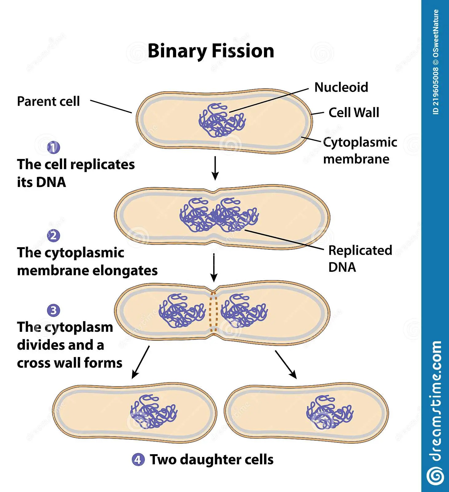 Figure: Binary fission in yeast cell
Fission is a common method of vegetative reproduction in Schizosaccharomyces (fission yeast). During fission, the parental yeast cell elongates, and its nucleus divides into two same-sized daughter nuclei. A transverse wall is developed in the middle of the cell. Then the parent cell divides into the two daughter cells.
Budding vs fission: which yeasts use which, and how to tell them apart
Most yeasts, including Saccharomyces cerevisiae, reproduce by budding. Fission is the exception, used mainly by Schizosaccharomyces (the fission yeasts). Both are asexual vegetative methods, but they look different under the microscope and produce different daughter cells.
| Feature | Budding | Fission |
|---|---|---|
| Which yeasts | Most yeasts (Saccharomyces, Candida, Cryptococcus) | Schizosaccharomyces only |
| How it starts | A small outgrowth (bud) appears at one point on the mother cell | The whole cell elongates |
| Daughter cell size | Smaller than the mother at first | Equal to the mother |
| Division plane | Localized at the bud site | A transverse wall forms across the middle |
| Microscope tell | One large cell with a small cell attached at a constriction | Two equal cells splitting end to end |
The quickest way to decide which you are looking at: a bud is a small daughter attached to a larger mother at a narrow neck. Fission gives two daughters of equal size, like a rod pinching in the middle. If the two cells are the same size, it is fission; if one is clearly the parent and one is a smaller offshoot, it is budding.
Sexual reproduction
Sexual reproduction occurs in yeast cells by the ascospores formation. During unfavorable conditions, when there is high stress, the diploid cell can go through sporulation. During this process, meiosis division takes place, forming various haploid spores. These cells conjugate and again forms the diploid cells.
Based on the types of vegetative cells involved in sexual reproduction and the dominant phase in the life cycle (haploid or diploid phase), Guilliermond (1949) classified the life cycle of yeast into three types, i.e., haplobiontic, diplobiontic, and haplodiplobiontic lifecycle.
Yeast life cycle: haplobiontic, diplobiontic, and haplodiplobiontic
Haplobiontic lifecycle
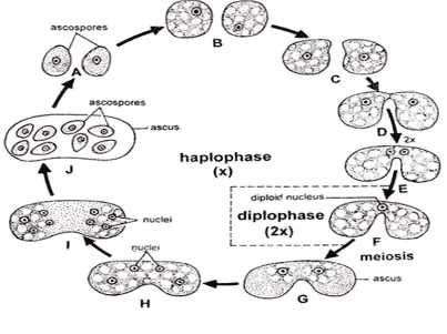 Figure: Haplobiontic lifecycle
It occurs in fission yeast (Schizosaccharomyces octosporus). In this type of life cycle, a vegetative cell is only haploid, and the dominant phase in the life cycle is haploid. During unfavorable conditions, the two opposite strains (haploid vegetative) cells come in contact. Then it behaves as the gametangia. Then these strains develop short beak-like protuberances, which elongate gradually and come in contact with each other. It then forms a single conjugation tube by dissolving the contact wall between them. Then their nuclei fuse, forming the zygote. It grows and develops the ascus.
The diploid nucleus is present in the ascus. It divides and forms eight haploid nuclei. From the eight haploid nuclei, eight ascospores are formed. These ascospores have their cytoplasm and cell membrane. On maturation, there is a release of the ascospores from the ascus. Out of eight ascospores, four become the positive vegetative cell, and four become the negative vegetative cell. These vegetative cells also multiply by fission and produce many haploid vegetative cells.
Diplobiontic lifecycle
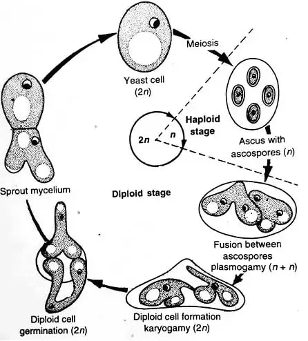 Figure: Diplobiontic lifecycle
It occurs in helobial yeast (Saccharomycodes ludwigii). In this type of life cycle, a vegetative cell is only diploid, and the dominant phase in the life cycle is diploid.
During unfavorable conditions, the diploid vegetative cell acts as an ascus. Ascus consists of the diploid nucleus. Then by the process of meiosis, it forms four haploid nuclei. From the four nuclei, four ascospores are formed in the ascus. They have their cytoplasm and cell membrane.
Inside the ascus, the opposite strain of ascospores develops the conjugation tube. The nuclei of opposite strain ascospores fuse at the conjugation tube. Thus, two diploid zygotes are formed within a single ascus.
Each diploid zygote develops a germination tube that pierces through the ascus wall and divides to form a short sprout mycelium consisting of many bud cells. These bud cells separate later and form many diploid vegetative cells.
Haplodiplobiontic lifecycle
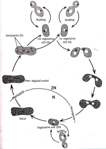 Figure: Haplodiplobiontic lifecycle
It occurs in budding yeast (Saccharomyces cerevisiae). In this type of life cycle, a vegetative cell is both haploid and diploid, and both phases are equally dominant in the life cycle.
During the unfavorable condition, two opposite strains (haploid vegetative cells) come near each other, behaving as gametangia. From each cell, protuberances develop, which resemble a beak. Then these protuberances elongate and come in contact with each other. Then the conjugation tube is formed after dissolving the protuberances in between them. In the conjugation tube, two nuclei fuse, forming the zygote. These diploid zygotes directly change into diploid vegetative cells.
During favorable conditions, there is the multiplication of diploid vegetative cells by the budding process. During unfavorable conditions, the diploid vegetative cell directly changes into an ascus. The diploid nucleus of the ascus undergoes meiosis and forms four haploid nuclei. From the four haploid nuclei, four ascospores are formed in the ascus. These ascospores have their cytoplasm and cell membrane. After maturation, ascospores break the wall of the ascus and releases from it. Among these four ascospores, two ascospores form positive vegetative cells, and two ascospores later form negative vegetative cells. These vegetative cells also multiply by budding and produce many haploid vegetative cells.
Uses of Yeast
Among the various microorganisms, yeast has proven to be the most useful microorganisms to humankind. Its uses range from food industry to fuel industry. Some of its uses are as follows:
- Alcohol or brewery industry: Different species of yeasts like Saccharomyces cerevisiae (including the wine strain formerly called S. ellipsoideus) is used for making alcoholic products like beer, wine, whisky, rum, gin, vodka, brandy, etc.
- Baking industry: The yeast used in baking is Saccharomyces cerevisiae which is used for making bread, doughnut, cakes, etc.
- Flavoring agent: Yeasts ferment the cocoa, which is used in making the chocolates. After the fermentation, it gives the flavor.
- Biofuel: Yeast is used to produce biofuel. It utilizes the sugar substrates and converts them into ethanol. It can be used in vehicles as a source of fuel. Saccharomyces cerevisiae has a high ethanol production capacity and can ferment a wide range of sugars. The use of biofuel helps in the reduction of crude oil consumption.
- Source of protein: Yeast is used as a source of protein, most as single cell protein. It supplements the protein in the diet like as milk and meat.
- Enzyme production : Saccharomyces cerevisiae helps in the production of the enzyme invertase on a commercial scale.
- Production of organic compounds: Saccharomyces cerevisiae helps in the production of different types of organic compounds like acetic acid, lactic acid, and glycerol.
How to Remember
The one distinction to lock in: bud vs. pseudohypha vs. true hypha. All three come from the same budding biology; what separates them is the constriction. A bud has a clear constriction where it meets the mother cell. Pseudohyphae are chains of buds that failed to separate, still showing a constriction at each junction. A true hypha (germ tube) shows no constriction at its point of origin at all.
The single sentence that captures the clinical payoff: every constriction you see in a budding yeast under the microscope tells a story, and the one organism whose germ tubes show no constriction at all is the clue that identifies Candida albicans.
Budding vs fission, by size: if the two cells are the same size, it is fission (Schizosaccharomyces splitting like a rod). If one cell is clearly the mother and one is a smaller offshoot at a neck, it is budding (everything else). Same size = fission; mother-plus-baby = budding.
Where Students Get Confused
All yeasts reproduce by budding. Most do, but not all. Schizosaccharomyces reproduces by fission, splitting into two equal daughter cells rather than budding off a smaller one. When an exam asks for the yeast that does not bud, the answer is the fission yeast.
Budding and binary fission are the same thing. They are not. Binary fission (as in bacteria) splits one cell into two equal halves. Budding produces an unequal daughter that grows from the parent at a localized point. Yeast fission resembles binary fission; yeast budding does not.
A germ tube is just a big bud. No. A bud shows a constriction where it meets the mother cell; a true germ tube grows out with no constriction at its origin. That single difference is the whole basis of the germ tube test for Candida albicans.
Pseudohyphae are true hyphae. They are not. Pseudohyphae are chains of buds that did not fully separate, so they keep a visible constriction at every junction. True hyphae have parallel walls and no constrictions. Telling them apart is a routine identification step for Candida.
Yeast has chloroplasts because it is a "plant-like" microbe. Yeast is a fungus, not a plant. It has no chloroplasts and cannot photosynthesize; it is a saprophyte that lives on preformed sugars. The reserve food is glycogen, not starch.
Key exam facts
| Question | Answer |
|---|---|
| What is the most common method of yeast reproduction? | Budding (gemmation) |
| What is pseudomycelium? | A chain of incompletely separated buds resembling mycelium |
| What is the clinical equivalent of pseudomycelium in Candida? | Pseudohyphae |
| What distinguishes a true germ tube from a bud? | No constriction at the point of origin (germ tube); visible constriction (bud) |
| Which two Candida species are germ tube positive? | C. albicans and C. dubliniensis |
| What is the yeast cell wall composed of? | Mainly beta-glucans and mannoproteins, with a smaller amount of chitin |
| Does yeast have flagella? | No, yeast is non-motile |
| What reproductive method is used by Schizosaccharomyces? | Fission (not budding) |
| How does yeast reproduce? | Asexually by budding (most yeasts) or fission (Schizosaccharomyces); sexually by ascospore formation under stress |
| What is the mode of reproduction in yeast? | |
| Which yeast reproduces by fission instead of budding? | Schizosaccharomyces (fission yeast) |
| How many ascospores form in the haplobiontic life cycle? | Eight (four positive, four negative mating type) |
Frequently Asked Questions
How does yeast reproduce?
How does yeast reproduce?
Yeast reproduces mainly asexually. Most yeasts bud: a small daughter cell grows out from the mother and pinches off. Some, like Schizosaccharomyces, divide by fission into two equal cells instead. Under stress, yeast can also reproduce sexually by forming ascospores.
What is the most common mode of reproduction in yeast?
What is the most common mode of reproduction in yeast?
Budding (also called gemmation). It is the primary method in Saccharomyces cerevisiae and in the medically important yeasts such as Candida and Cryptococcus.
Does yeast reproduce by budding or fission?
Does yeast reproduce by budding or fission?
Both occur, but in different yeasts. Most yeasts bud. Fission is used mainly by Schizosaccharomyces, the fission yeast. If the two cells are equal in size it is fission; if one is a smaller offshoot of a larger mother, it is budding.
What is the structure of a yeast cell?
What is the structure of a yeast cell?
A yeast cell is a single eukaryotic cell, 3 to 15 μm long, surrounded by a wall of beta-glucans, mannoproteins, and a little chitin. Beneath the wall is the plasma membrane and cytoplasm containing a nucleus, mitochondria, endoplasmic reticulum, Golgi apparatus, ribosomes, and a vacuole. Yeast has no chloroplasts and no flagella.
What are the three types of yeast life cycle?
What are the three types of yeast life cycle?
Haplobiontic (the dominant phase is haploid, as in Schizosaccharomyces), diplobiontic (the dominant phase is diploid, as in Saccharomycodes ludwigii), and haplodiplobiontic (both phases are prominent, as in Saccharomyces cerevisiae).
Why does yeast reproduction matter clinically?
Why does yeast reproduction matter clinically?
Because the same budding biology explains what pathogenic yeasts look like under the microscope. The presence or absence of a constriction distinguishes a bud from a pseudohypha from a true germ tube, and that distinction is the basis of the germ tube test used to identify Candida albicans.
References
- Jach, M. E., Serefko, A., Ziaja, M., & Kieliszek, M. (2022). Yeast Protein as an Easily Accessible Food Source. Metabolites, 12(1). https://doi.org/10.3390/metabo12010063
- Mohd Azhar, S. H., Abdulla, R., Jambo, S. A., Marbawi, H., Gansau, J. A., Mohd Faik, A. A., & Rodrigues, K. F. (2017). Yeasts in sustainable bioethanol production: A review. Biochemistry and Biophysics Reports, 10, 52–61. https://doi.org/10.1016/j.bbrep.2017.03.003
- Sudbery, P. E. (2011). Growth of Candida albicans hyphae. Nature Reviews Microbiology, 9(10), 737–748. https://doi.org/10.1038/nrmicro2636
- Tille P. Bailey & Scott's Diagnostic Microbiology. 15th ed. St. Louis: Elsevier; 2022.
- Procop GW, Church DL, Hall GS, et al. Koneman's Color Atlas and Textbook of Diagnostic Microbiology. 7th ed. Philadelphia: Wolters Kluwer; 2017.

Tankeshwar Acharya, MSc (Medical Microbiology)
Tankeshwar Acharya is an Assistant Professor in the Department of Microbiology at Patan Academy of Health Sciences (PAHS), Nepal, where he has been teaching and practicing clinical microbiology for over 14 years. He is the founder of Microbe Online, one of the leading free microbiology education resources on the web, covering bacteriology, mycology, parasitology, immunology, and clinical laboratory diagnostics written from direct experience in both the classroom and the diagnostic laboratory.
Comments
No comments yet. Be the first to share your thoughts.
Leave a comment
All comments are reviewed before they appear.